

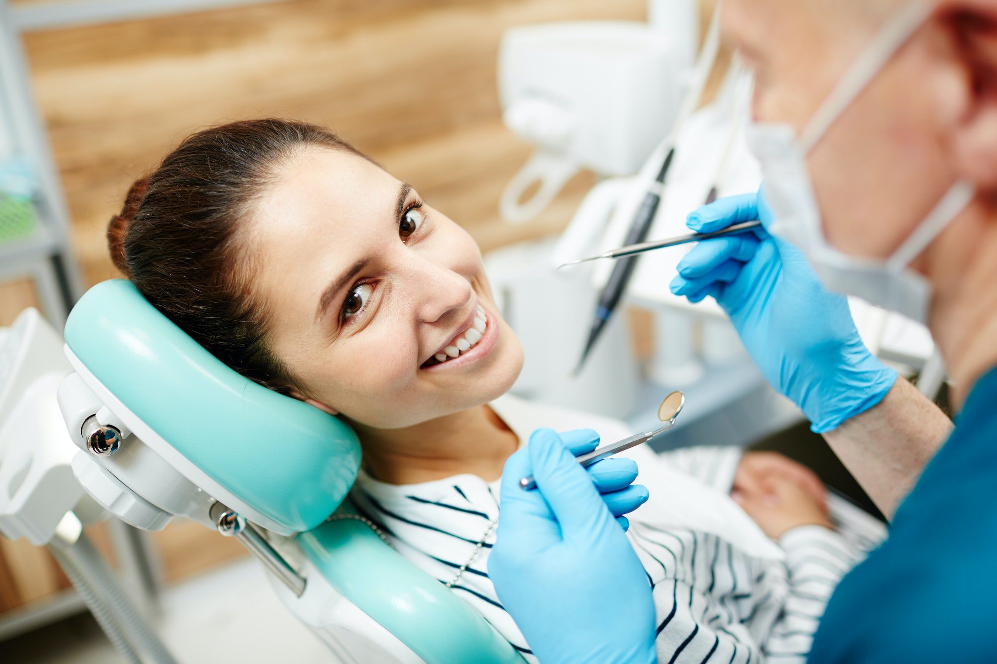
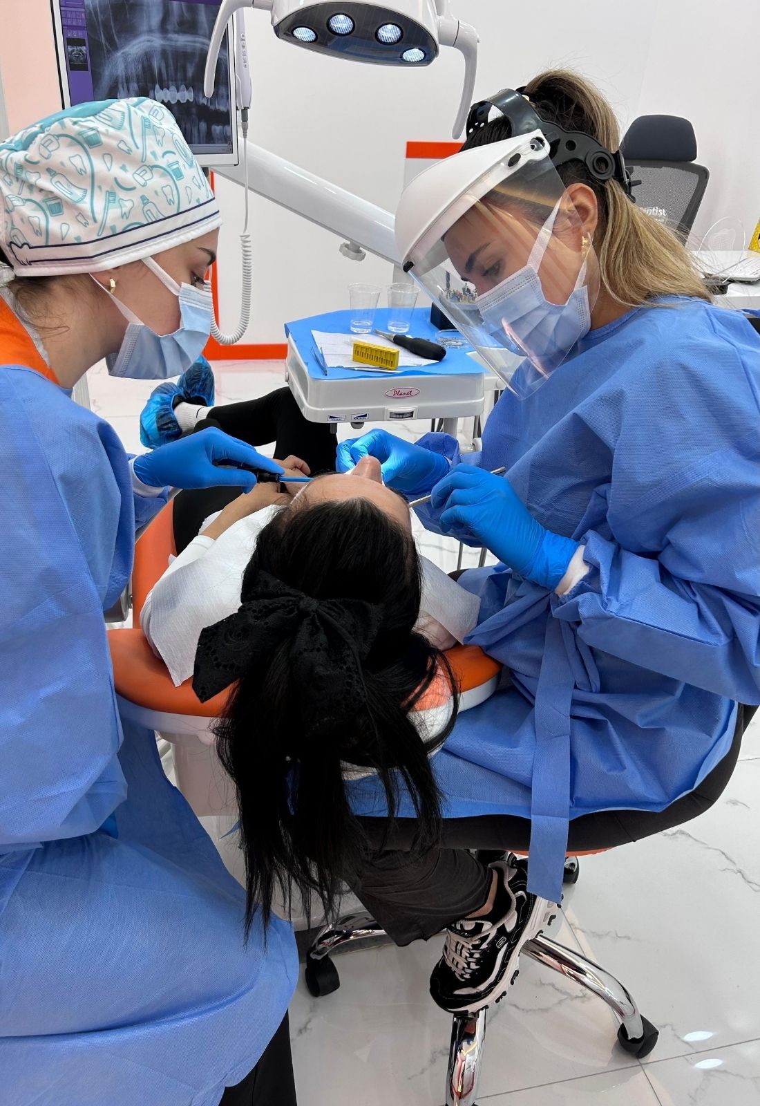
Oral Diagnosis, Radiology
Oral diagnosis and radiology are critical for detecting dental pathologies. Advanced imaging methods such as cone-beam computed tomography (CBCT), panoramic radiography, and intraoral imaging enable detailed examinations. CBCT, in particular, offers three-dimensional assessment in complex cases like impacted teeth and temporomandibular joint issues. Additionally, artificial intelligence-supported diagnostic tools provide high accuracy in detecting dental caries, oral cancer, and anomalies, improving the diagnostic process. These technological advancements reduce diagnostic errors and accelerate the treatment process, contributing to early detection.
| Treatment Purpose | Diagnose diseases in the oral, dental, and jaw regions and develop appropriate treatment plans. |
| Scope | – Detection of caries – Identification of abnormalities in dental and jaw structures – Diagnosis of tumors, cysts, or infections – Post-trauma assessment. |
| Methods Used | – Panoramic X-ray – Periapical X-ray – Cephalometric X-ray – Dental tomography (CBCT) – Intraoral camera imaging. |
| Application Process | 1. Clinical examination 2. Selection of necessary imaging methods 3. Imaging procedure and analysis 4. Diagnosis and treatment planning. |
| Suitable For | Individuals with oral and dental health issues, those requiring surgical or orthodontic treatment. |
| Risks and Complications | Radiation exposure (very low levels with modern devices), rarely allergic reactions (when contrast agents are used). |
| Advantages | – Detailed imaging for accurate diagnosis – Planning and monitoring of treatment processes – Quick results with minimal radiation risk. |
| Alternative Methods | Ultrasound (for soft tissues), MRI (magnetic resonance imaging; for evaluating non-bony structures). |
Open this in UX Builder to add and edit content
What is Oral Diagnosis and Why is it Important?
Oral diagnosis is a critical stage in dentistry that ensures the early detection and evaluation of oral and dental health issues. Effective treatment and preventive strategies involve taking patient history, clinical examination, and using diagnostic tools. In this process, the dentist evaluates various oral health conditions ranging from dental caries and periodontal diseases to complex issues like oral cancer.
Steps involved in the oral diagnosis process:
- Reviewing the patient’s history
- Clinical examination of the mouth and teeth
- Palpating any irregularities
- Probing for gum health
- Imaging with X-rays and intraoral cameras
Advanced diagnostic tools reveal conditions like impacted teeth, bone loss, and early-stage cavities that cannot be detected with the naked eye. Additionally, further tests such as biopsies or saliva tests play a significant role in identifying more complex pathologies like oral cancer and salivary gland disorders. This process enhances the accuracy of treatment planning and is crucial for protecting patient health and preventing the progression of oral diseases.
Recent advancements in digital imaging and artificial intelligence-supported diagnostic technologies have made oral diagnosis faster and more accurate. These technologies reduce the margin of error in the diagnostic process and ensure high precision in tailoring treatment plans to individual patients.
How Does Radiology Support Accurate Diagnosis in Oral Health?
Radiology improves diagnostic processes in oral health by utilizing advanced imaging techniques that facilitate accurate diagnosis. Methods like CBCT and MRI offer detailed anatomical structure analyses, providing effective pathways in complex cases.
- Three-dimensional imaging
- High-resolution details
- Soft tissue contrast
- Ability to diagnose without ionizing radiation
CBCT allows for detailed examination of hard tissues like teeth and jawbones, while MRI enhances diagnostic accuracy for soft tissues such as the temporomandibular joint. The high-resolution images provided by these methods simplify the treatment planning process, playing a significant role in patient care.
What are the Common Imaging Techniques Used in Oral Radiology?
Various imaging techniques are used in oral radiology to meet different diagnostic needs. These methods provide detailed information about dental structures and surrounding tissues and each has its unique advantages.
- X-rays (Intraoral Radiography)
- Panoramic Imaging (Orthopantomogram – OPG)
- Computed Tomography (CT)
- Magnetic Resonance Imaging (MRI)
Intraoral radiography is ideal for detecting local dental issues, providing high-resolution images. This method is effective in identifying caries, periodontal bone loss, and root abnormalities. Panoramic radiography offers a broad view, suitable for evaluating jaw fractures, impacted teeth, and dental alignments. CT, which provides three-dimensional imaging, is particularly useful in jaw trauma, tumor analyses, and implant planning, whereas MRI, which offers soft tissue detail, is preferred for examining structures like the temporomandibular joint.
How is Radiology Used in Detecting Oral Diseases and Cancer?
Radiology is a fundamental tool in the early diagnosis of oral diseases and cancers. Especially in individuals at high risk, radiological assessments can identify suspicious lesions before they become malignant. Advanced imaging techniques provide detailed and sensitive visualization of oral tissues, making it easier to detect potential risks.
- Cone-Beam Computed Tomography (CBCT)
- Digital Radiographs
- Panoramic Radiography
These methods can clearly visualize abnormal cell growths, non-healing ulcers, or pre-cancerous lesions like leukoplakia or erythroplakia. Routine use of these high-risk patient assessments allows for close monitoring of pre-cancerous changes and enables early intervention. This proactive approach facilitates early diagnosis, increasing survival rates and making treatment processes less invasive.
Additionally, radiological imaging is important during and after treatment. The progression or regression of diseases can be continuously monitored, and therapeutic effectiveness can be assessed. This allows for early detection of recurring conditions and facilitates comprehensive treatment planning by providing detailed mapping of oral tissues, including complex structures like osseointegration in tumors. These detailed assessments reduce the need for biopsies and help manage oral cancer risks by enabling the creation of comprehensive treatment plans.
What Role Does Oral Radiology Play in Treatment Planning?
Oral radiology is a critical element in modern dental treatment planning, enhancing accuracy, speed, and precision in patient diagnosis and treatment processes. Especially in orthodontics, implantology, and temporomandibular joint (TMJ) disorders, radiological imaging provides advanced assessments that shape the treatment process.
In orthodontic treatment, radiological evaluation analyzes growth patterns of teeth and skeletal systems, allowing for predictions of treatment impacts. CBCT enables the determination of the position of impacted teeth, risks of root resorption, and skeletal harmony, thereby improving both functional and aesthetic outcomes.
In implantology, CBCT provides essential information like bone density assessment and measurement of distances to sinus and nerve structures. This ensures precise implant placement with appropriate angulation and implant size, reducing the risk of complications.
In TMJ disorders, CBCT allows for clear visualization of joint morphology, evaluating condyle position and disc alignment. Accurate diagnosis of conditions like arthritis and disc displacement is facilitated, enabling appropriate treatment methods.
Digital radiology and CBCT technology integrate into clinical applications, enhancing diagnostic accuracy, predictability of treatments, and patient satisfaction in dentistry. This comprehensive imaging approach plays a significant role in ensuring the success of complex cases.
The Best Doctors Performing Oral Diagnosis, Radiology in Izmir
In Izmir, Oral Diagnosis, Radiology is not just about choosing a dentist; working with the best doctors directly impacts the success of your treatment. At AvrupaDent, we offer you the chance to choose from the best doctors performing Oral Diagnosis, Radiology in Izmir. Achieve a healthy and aesthetic smile with us.
Our Branches and Our Doctors
![]()
Health Group
As Avrupadent Health Group, we provide services in many districts of Izmir, especially in Buca and Gaziemir.
Frequently Asked Questions
Radiology plays a significant role in the diagnosis of dental diseases and its effectiveness varies depending on the imaging methods used and the specific condition. For instance, the diagnostic accuracy for detecting apical periodontitis (AP) lesions is 71% for periapical radiographs (PR) and 66% for panoramic radiographs (PAN). On the other hand, the sensitivity of radiographic evaluations for occlusal caries lesions ranges between 19% and 92%, while interproximal caries have a sensitivity between 39% and 94%. Artificial intelligence (AI)-supported systems enhance radiological diagnostic accuracy, achieving sensitivity comparable to experienced dentists in detecting impacted teeth, missing teeth, and caries on panoramic radiographs. AI models also provide over 80% accuracy in detecting conditions like implants and root canal treatments.
During a panoramic dental X-ray, lead aprons and thyroid protectors should be used to minimize radiation exposure. Metal accessories such as earrings, necklaces, and hearing aids should be removed to prevent interference with image quality. Pregnancy status must be reported to the dentist; in such cases, additional precautions may be taken or non-urgent X-rays postponed. Modern panoramic X-ray devices emit low levels of radiation, typically ranging between 3.85 and 30 microsieverts (μSv), equivalent to approximately 0.5 to 3.8 days of natural background radiation.
Digital radiography offers numerous advantages over traditional radiography, including up to 80% lower radiation exposure, faster image acquisition, higher image quality with adjustable contrast and brightness, and being more environmentally friendly by eliminating the need for chemical processing. Additionally, digital images are easily stored, shared, and accessed electronically, enabling better collaboration among healthcare providers.
Dental X-ray results are typically ready within a few minutes, as digital technology allows for instant image viewing. In urgent cases, preliminary results can be provided immediately for prompt intervention. In routine scenarios, such as standard dental X-rays, results are usually available within a few hours or by the next day at the latest. However, the turnaround time can vary depending on the type of X-ray, the equipment used, and the urgency of the procedure.
Oral examinations can reveal systemic health issues due to the strong connection between oral and overall health. For instance, individuals with inflammatory bowel disease (IBD) may exhibit oral signs like ulcers and gum diseases in up to 35% of cases, often before gastrointestinal symptoms appear. Periodontal disease is linked to cardiovascular diseases, diabetes, and adverse pregnancy outcomes. Additionally, oral signs can indicate nutritional deficiencies; for example, a red and smooth tongue may signal iron, folate, or vitamin B12 deficiencies. Regular dental check-ups are crucial for the early detection of such health issues, highlighting the holistic relationship between oral and general health.
![]()
Contact Us,
Renew Your Smile!
To schedule an appointment, please fill out the form below completely and get in touch with us.