Among the advanced imaging methods developed for the protection and treatment of oral and dental health, Dental Volumetric Tomography (often referred to as CBCT) has been attracting significant interest in recent years. Offering a three-dimensional view, this technology is invaluable when detailed examination of teeth and jawbones is required. In fact, it significantly helps dentists and patients make the most accurate treatment decisions when traditional two-dimensional X-rays are limited. By providing a high-resolution three-dimensional dataset in one go, Dental Volumetric Tomography opens the door to safer and more predictable dental treatments.
What is Dental Volumetric Tomography?
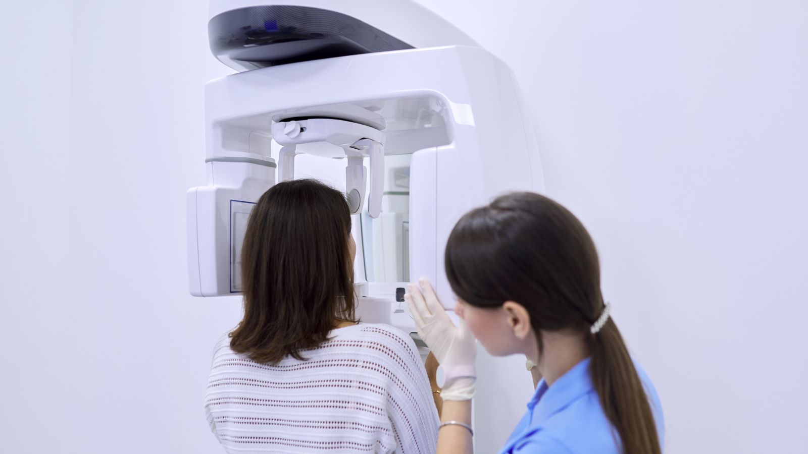
Dental Volumetric Tomography is an imaging technology that allows for three-dimensional examination of the mouth and jaw area. Unlike the flat images obtained with traditional X-rays, this system collects detailed three-dimensional data in a single scan. The technology works by rotating a cone-beam X-ray beam and a two-dimensional digital detector positioned opposite it around the patient. This allows raw images taken from dozens or hundreds of different angles to be processed by computer software to create a 3D map.
Dental Volumetric Tomography (DVT) can be performed on the patient in a sitting, standing, or, with some systems, lying position. With a single rotation, data on the entire anatomy of the jaw can be obtained. This three-dimensional view provides valuable information in many areas, from implant placement to the detection of pathologies such as cysts or tumors in the jawbones. Furthermore, the processing time is generally short, ranging from 5-20 seconds on most devices.
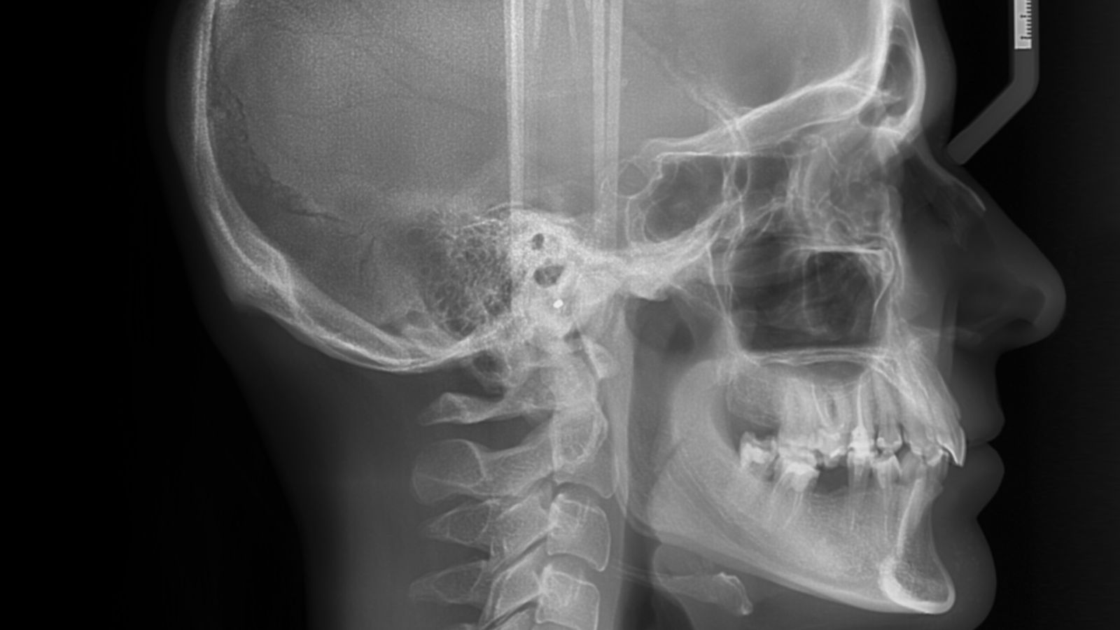
With DVT, the positions of teeth, bones, and joint surfaces are evaluated with accurate measurements. In traditional two-dimensional radiographs, overlapping structures can sometimes be difficult to distinguish clearly due to jaw curvature or other anatomical constraints. In three-dimensional imaging, no structure in the tissue is confused with another, and cross-sectional images allow for the examination of the desired area from different angles. This allows, for example, measurements of the distance to the nerve canal of the jaw, the detection of cracks in tooth roots, or the positioning of impacted teeth within the bone to be determined with much higher accuracy.
The term “volumetric,” from which this technology derives its name, refers to the acquisition of volumetric data. Because data is recorded as a “voxel,” the three-dimensional equivalent of each pixel, a true 3D examination becomes possible without loss of detail. Therefore, the goal is to determine the most accurate approach for pre-surgical planning in dentistry.
Which Fields Is Dental Volumetric Tomography Used In?
Dental Volumetric Tomography is used in many fields, from implant planning to pediatric dentistry. As the level of detail required for treatment increases, the advantage of 3D imaging becomes apparent. Especially in implant treatments, risks are minimized by precisely understanding the height, thickness, and density of the jawbone.
In implant surgery, clearly seeing the actual volume of the jawbone provides the surgeon with reliable information about the size and thickness of implants to be used. By measuring the distance to anatomical structures such as the sinus cavity or nerve canal, the safety of the surgery is increased. Similarly, in endodontics, the location and number of root canals are determined with much greater accuracy with a three-dimensional view. This allows for early detection of problems such as root canal obstructions, hidden root canals, and root cracks.
In orthodontics, the relationship between the jaw and face, the orientation of impacted teeth, and bone volume are crucial. Dental Volumetric Tomography allows for detailed examination of jaw asymmetries, narrowed maxillae, respiratory (airway) obstructions, and similar conditions. This allows for more accurate planning of fixed or removable orthodontic appliances.
In periodontics, the focus is on the gums and surrounding bone tissue. The depth and location of intraosseous defects are determined more precisely with three-dimensional images. DVT also allows for detailed visualization of bone loss in the spaces between the tooth root, called furcations. This information increases the success of bone grafting or other surgical procedures required for periodontal treatment.
In jaw surgery and pathology, DVT plays an important role in the detailed evaluation of jaw cysts, tumors, or fractures resulting from trauma. Before surgery, the size of the lesion, its relationship with adjacent tissues, and bone integrity are examined. This allows the most appropriate surgical approach to be determined in a single visit. Similarly, in cases of jaw joint (TMJ) disorders, joint surface deterioration, joint space, and cystic changes in the bone are clearly visualized.
In pediatric dentistry, the selection of Dental Volumetric Tomography (DCT) is made with caution because children are more sensitive to radiation. Impacted extra teeth, tooth eruption irregularities, or3D imaging can be indispensable in specific situations, such as serious trauma. Furthermore, DVT is not recommended for routine dental checkups. All these diverse applications offer a valuable diagnostic and treatment planning tool when the method is chosen for the right indications.
What are the Technical Features of Dental Volumetric Tomography?
Dental Volumetric Tomography operates on the cone-beam principle. In these devices, numerous two-dimensional images are collected during a single rotation, and specialized software converts these data into a three-dimensional volume. This conversion is accomplished using filtered back projection or iterative algorithms. The resulting three-dimensional dataset can be examined as cross-sectional views or as 3D images encompassing the entire jaw.
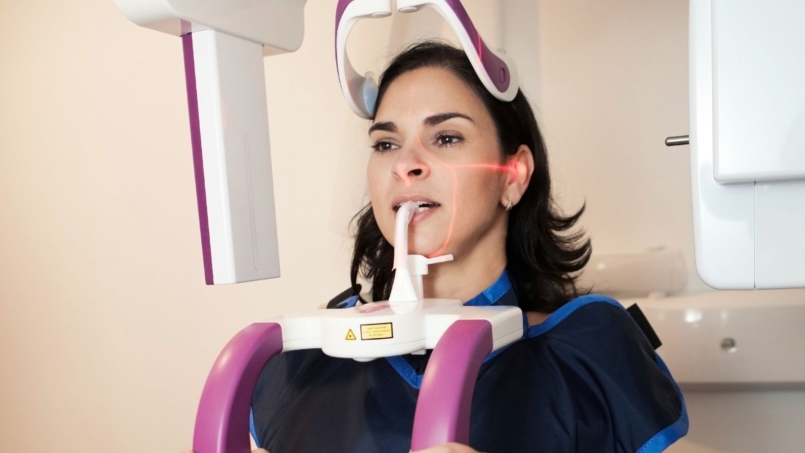
During the scan, a fixed X-ray tube is positioned opposite a digital sensor. The patient is usually scanned in a chair or standing position. The short scanning time combined with accurate positioning produces high-resolution images. At this resolution, the voxel size can be as small as 0.08 mm, allowing for the visualization of even fine details within dental structures. Some devices focus on a single jaw area, while others have a wide field of view (FOV) covering the entire head. The selectable FOV often prevents scanning of unnecessarily large areas, allowing for targeted scanning with a lower dose.
DVT devices used in dental practice are smaller than medical CT (Computed Tomography) devices and are compatible with the dental clinic environment. Many newer models are designed to perform both 2D panoramic and 3D imaging. In some models, the patient chair remains stationary, while the X-ray source and detector rotate on the same axis. It is important that the patient’s head not move during movement. Even small movements can cause image blur. Therefore, head supports or chin rests are used to minimize patient movement.
Radiation parameters are also important in technical specifications. DVT devices generally use X-ray tube voltages in the 80-120 kV range and low milliamperage values (1-10 mA). This energy level, while clearly depicting bone structure, can present difficulties in providing adequate contrast in soft tissues. However, the primary goal of Dental Volumetric Tomography is to examine bone and tooth structures with high resolution. Detector technologies developed for this purpose focus on reducing scattered and stray signals. The use of CMOS sensors or similar high-sensitivity sensors in newer-generation devices has both improved image quality and slightly reduced radiation dose.
What are the Differences Between Dental Volumetric Tomography and Other Imaging Methods?
Dental Volumetric Tomography, unlike two-dimensional X-rays, presents the entire jaw anatomy in three dimensions. While two-dimensional images carry the risk of overlapping and distortion, DVT largely overcomes these drawbacks. This allows for more accurate determination of the position and orientation of impacted teeth or pre-implant bone density assessment.
DVT generally requires a lower radiation dose compared to medical CT and has better spatial resolution for dental structures. Because medical CT scans have a larger scanning field, unnecessary tissue in dentistry is scanned, increasing the dose. Furthermore, the fan-beam technique in medical CT offers a different data collection approach than the cone-beam technique of Dental Volumetric Tomography. Fan-beam CT provides much better soft tissue contrast, while DVT excels at capturing fine details in bone.
DVT provides significantly more information than panoramic X-rays. Panoramic images are frequently used for screening because they display all teeth in a single plane. However, sensitivity is low for small cysts in the jawbones, apical lesions (around the root apex), or periodontal defects. DVT detects these smaller lesions with higher sensitivity. Studies have shown that some small foci of infection, which cannot be detected with periapical X-rays, can be seen more clearly with DVT. Similarly, the root formation pattern of impacted teeth, their relationship to neighboring tooth roots, or their proximity to the nerve canal cannot be clearly understood on panoramic X-rays, but they are easily detected on DVT scans.
Magnetic Resonance Imaging (MRI) is a leading method for detailed examination of soft tissues. While it does not provide sufficient resolution for hard tissue analysis of teeth and jawbones, it provides detailed information about joint discs and muscle structures. Therefore, MRI is preferred for soft tissue issues such as joint disc position.
Dental Volumetric Tomography, in short, addresses the need for 3D data in bone-related problems and is a more optimized method compared to medical CT in terms of dose. All these differences provide clues as to which method to choose in each situation. If you have an ordinaryFor routine checkups or the detection of minor dental caries, low-dose two-dimensional X-rays are more suitable. However, DVT becomes essential for surgical planning or complex cases.
How is Radiation Safety Achieved in Dental Volumetric Tomography?
Radiation safety in Dental Volumetric Tomography relies on achieving the highest diagnostic yield with the lowest dose. Not maintaining an unnecessarily wide field of view (FOV) is a critical safety factor for the patient. Focusing on the required area helps avoid unnecessary exposure of other anatomical regions.
While DVT involves higher radiation than traditional 2D dental X-rays, it is significantly lower than medical CT. For example, DVT scans scanning a small area can sometimes have a dose equivalent to a few periapical X-rays. In contrast, the dose of wide-field, high-resolution DVT scans can reach many times that of panoramic X-rays. Therefore, it is crucial for physicians to determine the patient’s actual needs and adjust the imaging parameters accordingly.
The use of thyroid shields and lead vests to reduce radiation-related risks is also common. The thyroid gland is particularly sensitive to radiation in children. When these accessories do not affect scan quality, wearing a thyroid shield is preferred. A headrest or chinrest during scanning also restricts the patient’s movement, thus reducing the likelihood of repeat scans. Repeat scans result in both unnecessary radiation exposure and wasted time.
The “ALARA” (As Low As Reasonably Achievable) principle is applied more strictly than ever in pediatric patients. This means that DVT scans are avoided in young children except in situations that directly impact diagnosis and treatment. In some cases, when necessary, the use of the lowest dose protocols and narrow FOV is recommended. Newer devices have developed low-dose modes, and pulse technology has significantly reduced dose by omitting X-rays when not needed.
Specialists always make decisions based on a benefit-risk assessment rather than scanning DVTs every time they are needed. In other words, choosing 3D imaging when it is crucial for diagnosis and treatment provides confidence to both healthcare professionals and patients. This avoids unnecessary radiation exposure and ensures long-term benefits with accurate treatment plans.
What are the Limitations of Dental Volumetric Tomography?
The main limitations of Dental Volumetric Tomography are primarily related to the lack of soft tissue detail. This technology is primarily optimized for imaging bones and teeth. Pathologies in muscles, joint discs, or soft tissues cannot be clearly visualized. Because there is insufficient contrast within the soft tissue, lesions such as tumors or cysts may not present as DVT unless they affect the bone margin.
Reflection and scattering artifacts, which occur in the presence of metal restorations, implants, or large fillings, pose another problem. These can cause bursts of brightness and streaks in the image, masking adjacent areas. This problem can be partially mitigated by applying some advanced software and artifact reduction algorithms. However, metal-induced distortions cannot be completely eliminated, sometimes making diagnosis difficult.
The cone-beam X-ray system used in DVT produces more scatter than the fan-beam system in medical CT. Scatter reduces image quality and triggers undesirable effects such as beam hardening. These factors become more pronounced when scanning a large area. Furthermore, pixel density values in DVT do not have a universal Hounsfield Unit (HU) equivalent, as in medical CT. Therefore, when measuring bone density, comparative assessments are necessary rather than absolute HU values.
Patient movement is a significant limitation in DVT. Although the scan is short, even a small head movement can cause loss of detail. It can be difficult to remain still, especially in children or those experiencing pain due to temporomandibular joint disorders. Therefore, repeated scans may be required, increasing the radiation dose.
Examining all the volumetric data obtained from large scans can also be time-consuming. Sometimes, unexpected lesions can be found in structures outside the physician’s primary focus. If these lesions are missed, the responsibility falls on the physician. Therefore, competent interpretation of large-volume scans requires sound radiologic knowledge or the support of a specialist oral, dental, and maxillofacial radiology specialist.
The cost of use and device installation are also limitations. DVT devices are more expensive than traditional X-rays, and certain standards for placement (radiation protection walls, adequate space, etc.) are required. Therefore, some clinics do not have DVTs, and patients may be referred to external centers. This can present logistical challenges. All these limitations make DVTs suitable for specific indications rather than for every case.It emphasizes the importance of remembering this.
What are the New Developments in Dental Volumetric Tomography?
New developments in dental volumetric tomography are focusing on low-dose protocols and artificial intelligence-supported image processing approaches. Manufacturers aim to achieve similar or better resolution with lower X-ray doses by improving sensor technology. For this purpose, pulse modes, advanced filters, and iterative reconstruction algorithms are used. Some new-generation devices claim to provide three-dimensional data at low doses approaching panoramic X-ray levels. These protocols provide significant assurance, especially for pediatric or pregnant patients.
Artificial intelligence-supported software is also a significant starting point. These software helps improve image clarity by reducing noise during and after scanning. Furthermore, functions such as automatic segmentation, lesion detection, and anatomical structure identification are being developed. For example, the location of impacted teeth can be automatically determined and a report can be provided to the physician. Some applications enable preliminary diagnosis by marking apical lesions or cyst-like formations. Such AI-based aids both save time and help detect potentially overlooked findings. Of course, the final decision always rests with the clinician.
Integration with digital dentistry is also shaping the future of Dental Volumetric Tomography. When a digital dental model obtained with intraoral scanners is combined with the bone structure obtained from the DVT, a complete 3D analysis becomes possible. This integrated model is highly advantageous for surgical guide fabrication or prosthesis design. For example, in implant planning, the implant position can be adjusted according to both bone conditions and the aesthetic requirements of the subsequent prosthesis. In orthodontic treatments, 3D facial scans can be integrated with DVT data to digitally predict jaw movements and tooth position changes.
Some researchers aim to screen for osteoporosis or osteoporosis risks by analyzing jaw bone density from Dental Volumetric Tomography data. Similarly, measurements of sinus structures or airway structures can also be helpful in cases of snoring and sleep apnea. Furthermore, the concepts of “4D DVT” or “dynamic DVT” are gaining attention. This technology aims to analyze the motion of the jaw joint using serial scans. However, due to increased radiation dose and technical challenges, it has not yet entered routine use.
Another recent development is the prototype development of micro-DVT systems. These high-resolution systems aim to provide very clear imaging of a single tooth or a small bone. The micro-DVT concept is gaining interest, particularly in the field of endodontics, for the diagnosis of complex canal anatomy or microfractures. While factors such as device size, cost, and scanning time remain limitations, this technology is expected to develop rapidly in the coming years.