Cephalometric X-ray is a significant imaging method especially in the branches of dentistry that deal with orthodontics and the jaw-face region. This method allows for detailed examination of the relationships between the face, jaw, and skull bones, identifying existing problems and providing guiding information for planning future treatments. Essentially, the X-ray images taken before, during, and after treatment enable close monitoring of changes in the dental and jaw structure. To express it in a way that an average person in daily life can easily understand, cephalometric X-rays allow for the clear measurement and analysis of the position of facial bones, the angulations of the teeth, and the compatibility of the jaws. This helps in uncovering the real cause of dental malocclusions, determining whether the jaw bones are positioned too far back or forward, and if necessary, making plans for surgical procedures.
What is Cephalometric X-ray and What is it Used For?
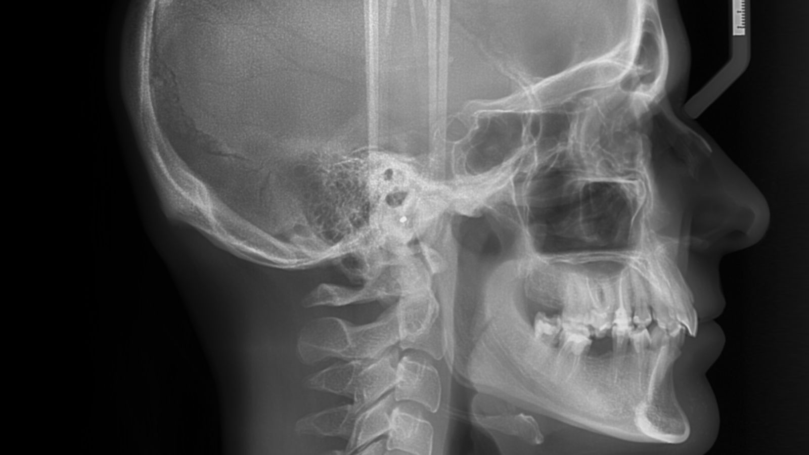
Cephalometric X-ray is a special type of X-ray image taken from the side (profile) or front of the head and face region, primarily aimed at examining the jaw-face structure, the position of the teeth, and the profile appearance in a measurable way.
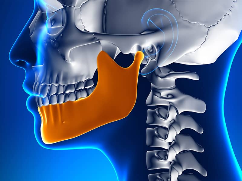
This X-ray technique serves as a very valuable guide in orthodontic treatment. The positions of the face and jaw bones, the inclinations of the teeth, and the condition of the jaw joint are all captured in a single film. These images help to show whether the jaw relationships are within normal ranges, detect potential growth issues, and determine which methods the orthodontic treatment will proceed with. Particularly, monitoring changes during the growth period of patients in adolescence becomes easier with the help of Cephalometric X-ray.
Additionally, when planning jaw surgery, this X-ray provides significant advantages. Topics such as the position of the jaws relative to each other, the potential facial profile after surgery, and the new position of the teeth can be assessed in detail during the pre-operative preparation period. This increases the likelihood of achieving the desired aesthetic and functional outcomes of the treatment.
In Which Situations is Cephalometric X-ray Preferred?
Cephalometric X-ray is frequently preferred in cases where there are discrepancies in the jaw-face region or when investigating growth disorders, particularly in situations requiring surgical planning for the jaw. It is particularly sought after by dentists for individuals considering orthodontic treatment. If teeth are not only misaligned but also the positions of the jaw bones are either retruded or protruded, if there are discrepancies in the relationships between the upper and lower jaws, general length imbalances of the face, or jaw asymmetry, Cephalometric X-ray becomes indispensable. Because in orthodontic treatments, the issue may not solely be with the teeth. In some cases, skeletal problems such as the retrusion or protrusion of the jaw can be the root cause. With this X-ray image, the doctor can clarify whether the malocclusion is skeletal or dental in origin.
Additionally, control X-rays are sometimes requested for young patients whose growth process has not been completed. This way, the speed and direction of growth in the jaw region can be determined. Whether it is the effect of orthodontic treatment or natural growth that has changed the position of the teeth can also be monitored. For individuals with more advanced jaw issues, Cephalometric X-ray is again a critical part of surgical planning.
How is Cephalometric X-ray Taken and What Equipment is Used?
Cephalometric X-ray is taken with a specially designed device where the patient’s head is fixed, and during the exposure, the patient’s head is captured from the side or front.
Sefalometrik Röntgen, özel olarak tasarlanmış bir cihazda hastanın başı sabitlenerek çekilir ve çekim sırasında hastanın başı yandan veya önden görüntülenir.
This extraction uses a head fixation device called a cephalostat. Two ear-like supports are positioned to align with the patient’s ears, and sometimes an additional support is used to stabilize the forehead. Thus, the head is held steady according to a specific reference plane. The distance between the X-ray tube and the digital sensor (or film) is predefined and is standardized for each patient. This stability ensures that measurement results can be compared.
During the extraction, the patient stands upright, and the teeth are held in normal occlusion. In the side view, the rays pass through the full profile of the head to reach the sensor. In the front view, the rays are directed from the frontal area towards the back. In both cases, the image reveals the measurable dimensions of the bones and teeth. Nowadays, since digital sensors are mostly used, the obtained images immediately reflect on the computer screen. This both ensures a lower radiation dose and allows for immediate detection if re-examination is necessary.
What Important Points Does Cephalometric X-ray Help to Identify?
Cephalometric X-ray allows us to clearly reach certain designated strategic points (landmarks) in the skull and facial area and determines the angular and distance relationships between these points.
Among these strategic points are specially named areas like Sella (S), Nasion (N), Point A (A), Point B (B), Pogonion (Pog), Menton (Me), and Gonion (Go). For instance, the Sella point is considered the center of the pituitary fossa at the base of the skull and serves as a reference point in many measurements. The Nasion point is the area where the forehead meets the nose. Point A represents the most protruded part of the upper jaw, while Point B represents the innermost part of the lower jaw.
The angles and distances calculated by determining these points clarify the position of the jawbones relative to each other. Mathematically illustrating whether the upper jaw is positioned forward or backward in relation to the base of the skull, or whether the position of the lower jaw deviates from normal, helps in making the treatment planning much more targeted. Additionally, the position of the teeth can be measured against different reference lines to see if they are excessively protruded (proclination) or retruded (retroclination).
How Are Analyses Performed on Cephalometric X-rays Interpreted?
Analyses conducted via cephalometric X-rays are interpreted by comparing the obtained angle and distance values with average (normal) ranges and contextualizing them according to the individual’s specific situation.
For instance, the SNA angle expresses the position of the upper jaw in relation to the base of the skull. The SNB indicates the position of the lower jaw relative to the base of the skull. The ratio of these two angles, ANB, clarifies the forward-backward difference between the upper and lower jaws. A very high ANB generally suggests that the upper jaw is in an advanced position or that the lower jaw is in a retracted skeletal Class II condition. Conversely, a very low or negative ANB may imply that the lower jaw is positioned forward, indicating Class III.
Regarding the teeth, measurements such as the angle of the upper incisor relative to the base of the skull (U1–SN) or the angle of the lower incisor relative to the mandibular plane (IMPA) provide important clues. It reveals whether the teeth are in the correct axis, meaning whether they are positioned excessively forward or backward. In the interpretation process, it is important for the physician to consider the patient’s facial aesthetics and the unique structure of the teeth for that individual. Sometimes deviations from average values can positively contribute to the person’s facial structure, and in such cases, decisions are made based on a comprehensive evaluation rather than solely on a “normal value” approach.
What Are the Advantages of Cephalometric X-rays?
Cephalometric X-rays offer numerous advantages, particularly in the field of orthodontics within dentistry.
- First of all, results can be compared reliably because it is taken with a standardized positioning and fixed equipment distance. Two different X-rays taken at the beginning and end of treatment clearly show actual growth or treatment-related changes. For example, it quantitatively reveals how much the upper jaw has advanced or how much change occurred in the angles of the teeth.
- Secondly, it is very successful in distinguishing between skeletal and dental issues. It becomes much easier to identify whether a person’s advanced positioning of the teeth is actually due to the retraction of the jaw or whether the teeth themselves are too far forward. The treatment plan is then shaped accordingly.
- Thirdly, thanks to the low radiation dose, it can be safely used to track growth in children or adolescents. Cephalometric X-rays taken at regular intervals from the same patient provide concrete information to the doctor about the direction, amount, and contribution of growth to treatment.
- Lastly, it plays a critical role in patients considering orthognathic surgery. Topics such as pre-surgical planning, changes in the expected facial profile after surgery, and the impact of surgical interventions on the tissue become evident through these X-ray analyses.
What Are the Limitations and Disadvantages of Cephalometric X-rays?
Although cephalometric X-rays have many benefits, they also include some limitations and disadvantages.
The most prominent limitation is that the obtained image is two-dimensional. In fact, since the face and jawbones have a three-dimensional structure, some information overlaps in a single image taken from the side or front. For example, in a side view film, the right and left jaw sections appear on top of each other, and asymmetries may not be fully displayed. This sometimes complicates the analysis.
Another disadvantage is that the determination of certain bony or soft tissue landmarks relies on observation. If the landmark points cannot be accurately identified, the margin of error in measurements increases. Therefore, the analysis of an experienced person yields more accurate results. Although computer-assisted or artificial intelligence-based automatic recognition systems have become common, the confirmation of a specialist’s eye is still required.
Additionally, a single Cephalometric X-ray does not reveal all problems. For example, issues such as detailed structures in the temporomandibular joint (TMJ) or possible cysts in the tooth roots should be investigated using different angles or 3D imaging techniques. In some complex cases, the film alone may not provide all the data the physician needs.
Why is the Integration of Cephalometric X-ray with Digital Methods Important?
The integration of cephalometric X-ray with digital methods allows for rapid and more precise measurements, providing significant convenience for both the patient and the physician.
Digital sensors enable obtaining images of the same quality with a lower dose of radiation compared to conventional film-based X-rays. Furthermore, results are visible instantly on the screen. Thus, if an error or undesirable movement is noticed during the shooting, the patient’s positioning can be corrected. In addition, analyses performed in a digital environment can be supported by automation. This means that artificial intelligence-based algorithms take over for detecting specific points, yielding faster results than a human.
Thanks to digitalization, the physician can upload these images to analysis software, allowing them to virtually move the teeth and jaw and simulate potential treatment outcomes in advance. This situation provides significant advantages, especially in surgical planning or in the digital design of orthodontic brackets. For example, it becomes easier to predict how the facial profile will appear if the upper jaw is advanced or from which area a tooth extraction will lead to the desired outcome. All of this also contributes to a transparent view of the treatment process in the hospital and strengthens patient-physician communication.
How is Radiation Safety Ensured in Cephalometric X-ray Imaging?
Although the radiation dose in cephalometric X-ray imaging is generally very low, safety measures are still taken to prevent unnecessary exposure.
To ensure this protection, the ALARA (As Low As Reasonably Achievable) principle is primarily applied. This means that an X-ray examination is performed only when it is truly necessary and with the lowest possible dose. By using a method called collimation, the X-ray beam is restricted to the relevant area of the head, thereby preventing unnecessary radiation exposure to other parts of the body.
The patient typically wears a lead apron. In some cases, a thyroid protector may also be suggested, but the projection area of the cephalometric X-ray usually does not directly affect the thyroid region. However, if necessary and if the quality of the X-ray will not be compromised, additional protective measures may be preferred. During this process, the personnel on duty perform the imaging while standing behind protective barriers such as control panels or lead walls. Maintaining a low dose with digital devices and preventing unnecessary repeat imaging is also a focus of modern clinics. This way, both the patient and the health workers’ safety are maximized.
What Innovations are Expected in the Future of Cephalometric X-rays?
In the future of cephalometric X-rays, it is expected that three-dimensional imaging technologies will become more widespread and that artificial intelligence-supported automatic analyses will develop.
Cone Beam Computed Tomography (CBCT), which is one of the three-dimensional imaging methods, can present slices of the skull, jaw, and dental structure, ensuring clear visibility of all types of asymmetry. Although this technology is becoming more common day by day, issues like radiation dose and cost are still seen as obstacles for now. However, lower dose protocols are being developed in next-generation devices.
Moreover, AI-based programs aim to mark specific bone or soft tissue points on cephalometric X-rays without the need for human intervention. Significant progress is being made in reducing error margins and accelerating analysis. Thus, it may become possible to achieve more accurate results in orthodontic treatments or jaw surgery planning in a shorter time.
With three-dimensional printing technologies, it is also possible to create patient-specific surgical guides or orthodontic appliances using the data obtained digitally. All of these innovations can be considered as complementary or transformative parts of cephalometric X-rays. Although the most frequently used basic method is still cephalometric X-ray, in the future, three-dimensional data, digital analyses, and personalized treatment planning may expand even further, partly replacing or significantly supporting this technique.