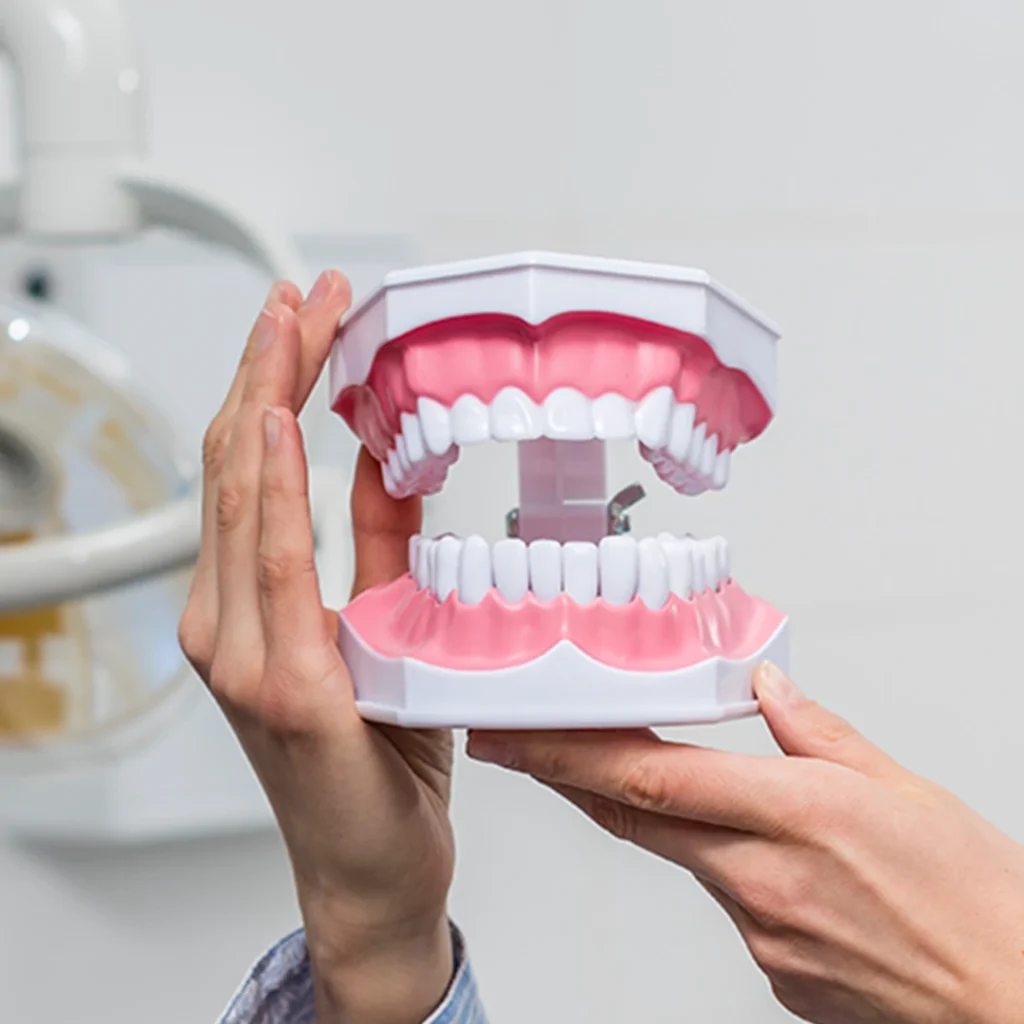
What is Gingivectomy and Gingivoplasty?
In the aesthetic perception of the smile profile, the level and contours of the gums that frame the teeth play a significant role, just as much as the size and color of the teeth themselves. In the medical literature, gingivectomy refers to the surgical removal (excision) of a portion of gum tissue, while gingivoplasty involves aesthetic adjustments made to give this tissue a more natural, thin, and tooth-surrounding form. These two procedures are often planned to complement each other within the same session. In clinical scenarios where there are inflammation-related tissue enlargements or where the gum genetically covers too much of the tooth, these surgical arrangements restore oral harmony.
The planned gum contouring procedures at the Avrupadent clinics in Izmir are meticulously carried out, taking into account the proportion of the teeth to the face and periodontal health measurements. The primary vision of these procedures is to present an individual with both visual harmony by drawing the structural form of the intraoral tissues back to their natural limits and to support long-term gum health by removing excess tissues that hinder dental hygiene (preventing brushing). Creating a proper arc for the gum contours is a critical step that directly influences the aesthetic success of the prosthetic (crown) procedures that will be planned on top of it.
In Which Situations is Gum Contouring Applied?
There are various anatomical and pathological conditions in clinical settings that require the reshaping of gum tissue. Gum swellings (hyperplasias) create “pseudopockets” between the teeth and gums, increasing food accumulation and making it difficult to clean these areas with a toothbrush. The fundamental conditions considered for evaluation are as follows:
| Clinical Case Type | Medical Purpose of the Procedure |
|---|---|
| Gingival Hyperplasia | To create space for hygiene by removing affected tissue after the use of certain systemic medications or orthodontic wire treatment. |
| Pseudopocket Formation | Facilitating plaque control by cleaning deep pocket formations caused by only gingival swelling without bone loss. |
| Gummy Smile | Lengthening the crowns (tooth heights) when the pink band between the upper lip and teeth exceeds aesthetic limits while smiling. |
| Asymmetrical Gingival Edges | Balancing the zenith points (the peak points) in case the levels of the gum tissue in symmetric areas differ from each other. |
How is Gingivectomy Performed?
The surgical process begins with a local anesthesia application, prioritizing patient comfort. After complete numbness is achieved in the area, the physician leaves small marks on the tissue to identify the true biological boundary between the tooth and the gum using special tools. These marks (bleeding points) form the guide line for the incision to be made. During the cutting of the gum tissue, while a knife can be used, modern clinical practices commonly prefer electrocautery or dental laser systems that minimize bleeding.
Laser-assisted gingivectomy allows for clearer visibility of the procedure by simultaneously coagulating the blood vessels while making cuts. Once the swollen and diseased tissue is detached from the tooth surface, the underlying healthy enamel structure becomes visible. After the cutting process is completed, the area is rinsed with special antiseptic solutions to prepare for the next stage of tissue shaping.
How is Gingivoplasty Performed?
After excessive tissue is removed, the edge of the gum can take on a thick and stepped shape. This thick edge is not suitable for periodontal health as it will create a plateau that is conducive to food debris accumulation. Gingivoplasty includes the transformation of these thick edges into a thin and smooth vestibule (fistula form) using surgical drills (special diamond tips), laser systems, or special tissue scissors.
During the procedure, the small triangular structures called papillae between the teeth are anatomically reconfigured. This not only results in an aesthetically pleasing curve but also creates an anatomical foundation that allows the patient to maintain their oral hygiene much more effectively with their toothbrush and dental floss.
Who Might Be Suitable for Gingivectomy and Gingivoplasty?
The infrastructure health of patients requesting tissue shaping for aesthetic or functional reasons is of great importance. If there is active bone loss (periodontitis) that has not been treated in the individual, simply cutting the gum does not establish tissue health; first, the deep inflammation must be cleaned. Cases where the jawbone is stable enough to allow the gum to take shape provide an ideal basis for these procedures.
Because the levels of gum tissue can vary in individuals in the adolescent period, whose growth and development process has not yet been completed, aesthetic permanent cutting procedures are often deferred until adulthood, when bone development has ceased. In patients experiencing gum enlargement related to systemic medication (such as certain blood pressure or epilepsy medications), these procedures can be performed in consultation with a physician regarding medication doses.
How is an Evaluation Conducted Before Gum Shaping?
The physician must have a clear understanding of where the jawbone begins beneath the tissue when deciding on the gum tissue removal. There is an average of a 2-3 millimeter natural protective band (biological width) between the gum boundary and the peak of the jawbone. If the gum tissue is cut to this boundary and comes very close to the bone, the tissue will chronically inflame in order to protect itself. The physical and radiological evaluations conducted provide safe working area definitions for the physician in order to avoid violating these boundaries.
On the other hand, when discussing the hospital and smiling, the position of the lips (the smile line) is examined. During the facial movements, the location of the gums on the face determines the aesthetic limits of the tissue amount to be taken. All this data is recorded to create guides that will be used during the operation.
How is the Gingivectomy and Gingivoplasty Process Planned?
Surgical procedures at İzmir Avrupadent require a meticulous preparation phase beforehand. If there is swelling (inflammation) in the gums due to plaque accumulation, the tissue will appear thick and long. In this case, it is expected that dental calculus cleaning (scaling) is performed first to achieve the natural firmness of the tissues. The boundary of the real gum will determine the actual amount to be cut once the swelling has reduced.
After controlling inflammation, the lengths of the teeth will be planned according to the principles of “Digital Smile Design” through digital photography records. Sometimes, transparent surgical guides are prepared in the laboratory using digital measurements taken from the patient. The doctor places this guide in the patient’s mouth during the procedure to create a perfect curve along the incision line.
What Steps Are Followed in Gum Contouring?
Achieving the health and aesthetic form of the tissue consists of clinically integrated stages. The steps taken by the doctor during the operation are as follows:
-
\t
- • Anesthesia and Isolation: Numbing the working area with local anesthetics and creating a clear field by clearing it of debris.
- • Clinical Marking (Bleeding Points): Marking the pocket depths and outlining the boundaries of the excess tissue to be removed.
- • Excision (Gingivectomy): The removal of hyperplastic gingival tissue that has become overgrown using either a scalpel or medical laser.
- • Contouring (Gingivoplasty): Physiologically sculpting the edges of the cut tissue to form a natural and smooth curve.
- • Post-operative Care: Placement of periodontal packing (protective paste) if necessary, to support the healing of the area and protect against external factors.
\t
What Should Be Considered Before Gingivectomy?
Surgical procedures require minimizing the amount of bacteria in the oral environment. Performing operations on bleeding and plaque-covered tissues can increase the risk of infection during the healing phase and complicate the physician’s clear assessment of aesthetic boundaries. It is clinically essential for patients to come with a clean oral flora on the day of the procedure to support the efficiency of the process.
Additionally, if anticoagulant medication that affects the clotting profile is involved, this should be discussed in detail with the physician. It is recommended to arrive at the appointment rested and slightly full to balance stress levels since local anesthesia will be applied.
What to Pay Attention to After Gingivoplasty?
After the operation, it’s essential to pause eating and drinking activities until the anesthesia effect (numbness) wears off, to prevent accidentally burning the lips or tongue. The contoured gum tissues will adapt to their new forms within a few days, while forming a thin, whitish healing (fibrin) band on them. It’s crucial not to scrape this band or disrupt it with hard foods as this is vital for wound healing.
It is strongly advised to avoid products that disrupt blood circulation (oxygenation) and hinder cell renewal during the first critical week when healing is most intense. The prescribed analgesics will help manage any mild discomfort that may arise during the tissues’ adjustment to their new positioning.
In Which Gum Problems Are Gingivectomy and Gingivoplasty Evaluated?
These periodontal surgical techniques are evaluated in a quite broad treatment spectrum aimed at correcting the structural and dimensional defects of the tissue. The primary clinical indications are as follows:
| Clinical Problem | Functional and Aesthetic Solution |
|---|---|
| False Pocket (Pseudo Pocket) Status | Making the deep plaque retention areas formed solely by the growth of the gum easily cleanable by facilitating their alignment. |
| Epulis and Fibromas | Removing hardened tissue growths (benign lesions) from the gum that are due to irritation. |
| Post-Orthodontic Treatment Condition | Restoring the gums, which have thickened due to mechanical irritation after braces are removed, to their old physiological contours. |
| Crown Margin Incompatibilities | Improving the transitions of the porcelain by thinning the gum profile that has thickened and stepped at the margins of old crowns. |
What is the Relationship Between Gum Contouring and Smile Design?
Design planning for aesthetics is like a multidisciplinary architectural setup. The peak points where the front incisors connect with the gum must be aligned according to specific aesthetic standards. If the gum of the right central incisor is a few millimeters lower than that of the left, the produced porcelain veneer will also be made shorter on one side; this disrupts facial symmetry. The shaping of the gum tissue provides the practitioner with a balanced workspace (foundation) by eliminating these inequalities before measuring the porcelains.
Moreover, in patients whose teeth are wide in form but short in length, simply extending the teeth by laminating can exceed the lip level. However, when the gums are shaped correctly in a millimetric manner, the actual length of the teeth is revealed, enabling the creation of more graceful, rectangular restorations in proportion to the face.
How Does the Healing Process Progress After Gingivectomy and Gingivoplasty?
Mucosal tissue is one of the fastest self-renewing and most vascularized structures in the body. In laser-assisted shaping procedures, this healing process occurs in a shorter time compared to the use of cautery or scalpel. The whitish layer that forms immediately after the operation is a healing band of the tissue; this appearance disappears within a few days, making way for fresh pink tissue.
While biological adaptation is completed, there may be a millimeter gap in the tissues. For this reason, if permanent zirconium or porcelain (veneer) measurements are to be taken after the procedure, it is clinically preferred to wait for an average of two weeks for the gums to achieve their final and stable position (after the remaining slight edema has completely subsided).
How Long Can Gum Shaping Take?
Eliminating a minor asymmetry in a single tooth with a small laser touch is only a few-minute procedure, while leveling a common gummy smile case involving the entire anterior aesthetic zone (6-8 teeth) will extend the time needed for more detailed measurements and shaping. The priority is to adjust the tissue contours perfectly symmetrically according to the golden ratio rules rather than speed.
In procedures performed with the prepared transparent surgical plates, since the physician’s course is digitally mapped out in advance, the procedure is conducted much more fluidly and predictably. The patient leaves the clinic having comfortably completed both the anesthetic and aesthetic adjustment phases within the same appointment.
Why is a Check-Up After Gingivectomy and Gingivoplasty Important?
After periodontal surgeries, tissues may not always fully comply with the mathematics; the rates of healing can vary according to the individual’s tissue biotype. During the control session performed approximately 10-14 days later, the physician examines whether the gums are properly positioned in the spaces between the teeth (papillae). If deemed necessary, curves can be made smoother with small laser adjustments (touch-ups).
Additionally, during these sessions, a hygiene schedule is established regarding when and how the patient will start practicing brushing on the healing tissues. Beginning aesthetic prosthetic procedures before tissue stabilization is fully confirmed may prepare the ground for the formation of clinically undesirable gaps (black triangles).
Which diagnosis and planning methods are used in gum shaping?
The physician must keep the underlying bone support in mind when removing tissue that appears excessive from the outside. When measuring the depth of the periodontal pocket, if the bone level is very close to the gum boundary, merely cutting the gum (gingivectomy) will be insufficient. In this case, the surgical plan includes slightly contouring the bone boundary to reshape it (crown lengthening). Radiological examinations play a guiding role in making this distinction.
With the integration of digital technologies (CAD/CAM systems) into the process, the patient’s intraoral scanning data is combined with studio photographs. The amount of lip mobility (movement) during smiling is measured, the amount of tissue to be obtained is determined in millimeters in a virtual environment, and the margin of error during the procedure is minimized.
Can Gingivectomy and Gingivoplasty be Planned Together with Other Dental Treatments?
In patients undergoing orthodontic treatment, if there is insufficient brushing around the brackets, the gums can become inflamed, leading to swelling that covers a large part of the tooth. Although the teeth may be aligned after the wires are removed, the aesthetic appearance may not be satisfactory due to the swollen tissue. At this stage, gingivoplasty comes into play to reveal the tooth contours.
In restorative procedures, the gums that remain beneath deep cavities must be removed slightly through a gingivectomy to allow for filling in the relevant area, providing the dentist with a dry and clear working field (isolation area). All these procedures are steps of a common treatment map where different branches of dentistry support each other.
How Should Oral Care Be Performed After Gum Contouring?
It is a medical recommendation not to change the toothbrush in the specific area of the surgical wound during the first days; as the newly formed tissue may be damaged by mechanical trauma, disrupting the healing process. Other areas of the mouth that have not undergone any procedures should continue to be brushed as usual. Mouthwash helps to eliminate microbial debris in that area.
After the doctor gives approval (usually after 1 week), delicate touches are applied to the surgical area with post-operative toothbrushes. Once the tissues reach full maturity, the use of an inter-dental brush and dental floss should become the strongest protective habit to prevent the formation of dental calculus and inflammation between the newly created physiological curves.
What Factors Affect the Treatment Plan for Gingivectomy and Gingivoplasty?
The extent of the operation and the method to be chosen are determined by the anatomical limitations and biological responses of the individual. The main clinical factors that shape the framework of the treatment planning are as follows:
| Clinical Factors | Impact on Surgical Procedure |
|---|---|
| Biological Width | If the amount of tissue desired to be obtained is very close to the bone beneath, there may be a need to shape not only the tissue but also the bone (osteotomy). |
| Gingival Biotype (Thickness) | Thick (fibrotic) tissues respond better to surgical incisions, while thin tissues carry a risk of contraction (recession) after the incision. |
| Lip Mobility Amount | When the lip muscles dramatically rise when smiling, the gingival incision may not be sufficient for aesthetics alone (botox support may be required). |
| Presence of Active Inflammation | If there is bleeding and swelling in the area, it is clinically necessary to clean and wait for the tissue to calm down before the procedure. |