What is Implantology?
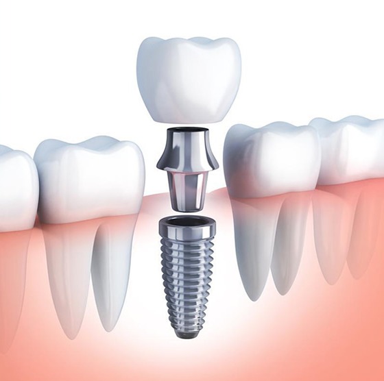
In human anatomy, teeth hold onto the jawbone through their roots, transmitting chewing forces to the bone. When a tooth is lost, not only the visible crown section but also the root support within the bone disappears. The science of implantology aims to address this biological deficiency by using artificial roots made of biocompatible materials like titanium or zirconium. Unlike traditional bridge prosthetics, which require the neighboring healthy teeth to be reduced, this method is applied directly to the area of deficiency and helps to control bone resorption due to the chewing pressure transmitted from inside the bone.
At Avrupadent clinics located in İzmir, implantology procedures are approached with a multidisciplinary perspective. In this field, where the principles of jaw surgery are combined with aesthetic prosthesis design, the patient’s facial proportion, balance of the temporomandibular joint (TMJ), and existing bone volume are evaluated in detail. The primary goal of the treatments applied is to provide the person with long-term biological compatibility to regain the missing digestive (chewing) and speech (phonetic) functions through soft tissue structures.
What Treatments Does Implantology Include?
This field of science is not just about placing a screw into the bone; constructing the biological bed where that screw will be placed in a healthy manner is also an indispensable part of the process. The fundamental implantology topics evaluated and applied in the clinic can be classified as follows:
In what situations is implantology evaluated?
There are many alternatives to replace missing tissues in dentistry. However, when traditional methods create anatomical limitations, implantology comes into play. For instance, in cases where the last molars are lost in a “free-end” situation, a fixed bridge cannot be made because there is no tooth available to serve as the posterior abutment for the bridge. In this case, the person may either be fitted with a removable partial denture or artificial roots can be placed in the gaps to obtain fixed teeth.
At the same time, patients who have been edentulous for many years and have difficulty retaining their existing dentures (removable dentures) due to the resorption of the jawbone benefit from the solutions offered by this discipline. In such clinical situations, the stability of the prosthesis is improved with retention mechanisms supported by artificial roots placed in the jaw, enhancing the patient’s chewing comfort.
How is Implant Treatment Planned?
In the management of implantology processes in İzmir Avrupadent, procedures are planned correctly from prosthetics to surgery. The doctor designs the shape of the porcelain or zirconium tooth, the biting axis, and lip support that will be in the patient’s mouth at the final stage in advance. The opening and position of the titanium body to be placed inside the jawbone are determined according to the future biomechanical needs of the tooth. This setup serves as a critical foundation for the restoration to function correctly in the future.
During the planning phase, digital systems (CAD/CAM software) are intensely utilized. The patient’s bone thickness is measured on the computer screen, and the diameter and length of the titanium root to be used are selected from a digital library and virtually placed in the bone. Safety distances to the nerve channels running under the jaw are calculated and a step-by-step route to be followed during the surgery is created.
What Diagnostic Methods Are Used in Implantology?
The reliability of surgical interventions depends on the details of the diagnostic data obtained prior to the operation. The fundamental imaging and diagnostic methods applied in the clinic are as follows:
- ● Panoramic Radiography: This is the initial examination method that shows the general condition of the upper and lower jaws, the openings of adjacent tooth roots, and the basic bone structure in two dimensions (wide view).
- ● Dental Volumetric Tomography (CBCT): This is an advanced technology system that presents bone thickness, density, and nerve canals in three-dimensional sections, creating an operational map.
- ● Intraoral Optical Scanners: These are camera systems that digitally replicate the topography of the jaws and gum tissue, allowing for the production of surgical guide plates.
- ● Clinical Casts/Digital Models: These are physical or virtual copies created to examine the occlusal contact points with the opposing jaw and to identify vertical dimension loss.
Who Can Be suitable for Implant Treatment?
To actualize this application, there is no advanced age limit; clinical procedures can be successfully applied even to healthy individuals in their nineties. What is important is the quality of the bone and the patient’s overall medical condition. In children and adolescents whose bone development is still ongoing, the titanium root to be placed for continuing jaw growth may remain buried or create asymmetry in the following years; therefore, it is expected that the growth will stop.
In individuals with chronic diseases such as diabetes (sugar disease), cardiovascular (heart) disorders, or osteoporosis, consultation with the patient’s own physician ensures that clinical values are controlled. When the medical findings reach ideal levels, implantology applications can be included in the plan.
How is an Evaluation Made Before Implant Treatment?
While placing a titanium root in a healthy jawbone can yield successful results, if there is an untreated severe gum disease (periodontitis) present in the mouth, these bacteria can also infect the newly placed artificial root area. Dentists plan preliminary treatments such as professional tooth cleaning to achieve an ideal level of oral hygiene before surgical procedures.
Additionally, the medications regularly used by the hospital are of great importance. Blood thinners, bisphosphonate group drugs used against bone loss, or treatments affecting the endocrine system should be thoroughly communicated to the doctor. Organized usage of these medications with the referral of the physician before the operation establishes the medical basis for the surgical process.
How Does the Implant Treatment Process Proceed?
The operation is generally performed under local anesthesia. Titanium bodies are positioned within millimetric sockets opened in the jawbone. At the end of the procedure, the tissues are closed with small stitches, and the patient enters the healing period. The cellular adhesion of the artificial root to the jawbone and its integration requires an average biological duration of 2-3 months for the lower jaw and 3-6 months for the upper jaw due to the more spongy structure of the bone.
Once this healing period is completed, the prosthetic phase begins. The doctor removes the gum contouring elements, places the measuring parts, and sends digital or physical impressions to the laboratory. The dental structures prepared in the laboratory (crowns or bridges) are secured by being screwed or cemented to the titanium roots after clinical trials.
What Stages Are Followed in Implant Treatment?
Implantology processes have a meticulous sequence where surgical and laboratory cycles follow each other. The basic clinical steps can be listed as follows:
What to Consider Before Implant Treatment?
Since it is a surgical operation, keeping the number of bacteria in the procedure area at a minimum reduces healing complications. If the doctor has prescribed medication (protective antibiotics) to be started a certain number of days before the operation, using them at the specified times is an important part of the clinical procedure. If there is medication that affects blood clotting, this matter should be clarified through communication between the doctors (dentist and medical doctor).
Since local anesthesia is applied, the patient’s comfort is ensured during the operation; therefore, arriving at the clinic relaxed and free from excessive anxiety and stress facilitates the process. Since there may be speech and dietary restrictions for a few days post-operation, work/social planning should be organized accordingly.
What Should Be Considered After Implant Treatment?
Mild swelling (edema) in the cheeks and minimal oozing in the wound area after surgical intervention is a physiological part of tissue repair. The prescribed analgesic medications will help you go through this process comfortably. Instead of hard, spicy, and hot foods on the first day, soft and easy-to-chew foods like warm soup, yogurt, and puree should be preferred.
Smoking slows down the healing of the wound area by reducing the oxygen level in the blood and negatively affects the integration of the artificial root with the bone. Therefore, limiting cigarette use in the first weeks after the procedure is highly recommended for the success of the treatment. Stitches in the operation area should not be brushed; only cleaning with the antiseptic solutions recommended by the doctor should be performed.
How is the Jawbone Evaluated in Implant Treatment?
For a titanium screw to remain stable within the bone, it requires a surrounding bone wall of adequate thickness. In areas that have been toothless for many years, the bone can enter a natural resorption process, becoming slender like a knife edge, or it may approach nerve canals very closely. The physician determines the geometric structure of the bone by examining three-dimensional radiological slices.
Additionally, whether the bone is spongy or hard determines the surgical technique. For example, the posterior areas of the upper jaw (D3-D4 type bones) are structurally softer and more porous, allowing for a longer waiting period required for integration (osteointegration) in these areas. If the bone volume is found to be insufficient, plans are made to prepare the area for surgery by adding an artificial bone graft (powder).
What Imaging Methods Are Used in Implant Treatment?
In order for the physician to take predictive steps during surgery, specific radiological devices that elucidate the underlying hidden anatomy are utilized. The fundamental methods used in this field are as follows:
- Panoramic X-ray: Provides a general clinical condition analysis (infection, impacted teeth, etc.) by showing the upper and lower jaws and the jaw joint in a single plane.
- Dental Tomography (CBCT – Cone Beam Computed Tomography): An advanced technology system that models the bone width not visible in two-dimensional films and the exact position of the lower jaw nerve (Inferior Alveolar Nerve) in millimeter sections.
- Periapical Imaging: Small area radiographs taken after surgery or during follow-up appointments to closely monitor the bone level around a single artificial root.
In What Cases Can Implant Treatment Be Planned for Tooth Loss?
The biomechanics of each dental deficiency differ. In the case of a single loss, the goal is to place an independent root into the empty space, avoiding the reduction of adjacent healthy teeth (such as using a crown). In multiple adjacent losses, for example, if 3 teeth are missing, instead of placing 3 screws, systems such as “implant-supported bridges” are designed by placing 2 artificial roots and connecting the missing part with the bridge structure.
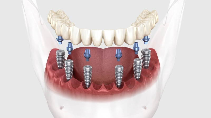
In patients who are completely toothless (having no teeth at all), for those who do not want to use removable denture prostheses, 4 or 6 titanium roots (All-on-4 / All-on-6 systems) are placed into the jawbone, and all teeth are fixed onto these roots. This way, patients achieve chewing comfort with teeth that do not cover their palate and remain fixed in the mouth.
What is the Relationship Between Implant Treatment and Implant-Supported Prostheses?
Patients often believe that the treatment is complete once the surgical procedure is finished; however, the titanium root remains in an invisible state within the bone. The primary component that will fulfill chewing and speaking functions is the prosthesis that will be built on that root months later. Therefore, regardless of how flawlessly the surgical procedure is performed, if the prosthesis placed on it is not compatible with the patient’s jaw closure, clinical success cannot be achieved.
These two disciplines are an inseparable whole. The components called “abutments” that will carry the design of the prosthesis are specifically chosen according to the thickness of the gum and the angle of the tooth. Thus, the mechanical metal root underneath is transformed into an aesthetic and natural porcelain tooth within the limits of the gum.
Why is Control Examination Important in Implant Treatment?
The healing of the wound in the hospital after the surgical procedure is usually evaluated during the control sessions in the first or second week when the stitches are removed. However, the real critical follow-up begins after the placement of the implants. Artificial roots do not have periodontal fibers that provide flexibility like natural teeth; they bond to the bone in a completely rigid manner. Therefore, a slight height formed on the implant transmits shock directly to the bone.
During six-month periodic visits, your physician examines whether there is tartar (plaque) accumulation in the gum tissue with tools. Bacterial plaques that form around the titanium body can lead to an infection called “peri-implantitis,” which can erode the bone. Routine clinical cleanings and check-ups help diagnose and prevent such complications at an early stage.
How Should Oral and Dental Care Be Performed After Implant Treatment?
Porcelain or zirconium surfaces do not wear out; however, if food residues accumulate in the neck areas where the prosthetic gums connect, bacteria can colonize in that area. Peri-implant inflammations, which progress much faster than natural gum inflammation, can result in the erosion of the titanium root and surrounding jawbone. Therefore, hygiene is clinically necessary to maintain the success of the treatment for many years.
Standard toothbrushes cannot reach between or underneath interconnected long bridges. Patients are advised to mechanically clean that area by passing dental floss under the bridges. Oral irrigators can significantly contribute to tissue health by flushing away microbial plaques in areas inaccessible to the toothbrush using pressurized water.
What Surgical Procedures Can Be Applied Within the Scope of Implantology?
In cases where the bone bed does not have enough volume to support a titanium root, advanced level procedures are performed to surgically prepare the area:
How is Personalized Planning Done in Implantology Treatments?
Each person’s oral anatomy is as unique as a fingerprint. For example, the diameter of the artificial root and porcelain material to be applied to a male patient with bruxism will be completely different from the components applied to a female patient with a high aesthetic line and thin gums. In individuals with strong chewing muscles, thicker roots and zirconium infrastructures are preferred, while in front areas where aesthetics is prioritized, ceramic (white) bases that do not show the gums as gray can be planned.
In the processes of Izmir Avrupadent, sometimes technological templates known as “Surgical Guide” are produced. The ideal width and depth determined on the computer are transferred to the patient’s bone during surgery with zero error thanks to these guides; thus, the duration of the procedure is shortened while the positioning is tailored to the individual’s anatomy.