What is Oral, Dental and Jaw Surgery?
The chewing system in human anatomy is not only composed of teeth; it is a complex whole comprising the bone structures that support these teeth, joints, and mucosal tissues. Cystic formations in any of these structures, problems with impacted teeth, or tissue losses related to trauma negatively affect the function of the system. The discipline of jaw surgery aims to bring these biological structures back to physiological norms using medical and microsurgical techniques. Procedures are based on the philosophy of protecting long-term oral health by directing the healing potential (regeneration) of tissues.
At the Avrupadent clinics located in Izmir, oral, dental and jaw surgery processes are carried out with a multidisciplinary approach that combines many parameters, from the hospital’s facial profile to the quality of bone. The focus of the procedures is to compensate for structural deficiencies using biomaterials that show cellular compatibility and current surgical principles. This wide range, extending from root apex resections to extensive bone dust grafts, carries significant medical value in preserving the integrity of the oral internal system.
Hangi Oral, Maxillofacial and Dental Surgery Hizmetine İhtiyacın Var?
What Treatments Does Oral, Dental and Jaw Surgery Include?
Oral and jaw surgery encompasses a wide range of work from soft mucosal tissues to hard bone structures. Clinical procedures vary according to the medical needs of the case, ranging from the removal of infections from inflamed roots to the replacement of lost bone. The main types of treatment that are frequently sought are:
| Type of Surgical Procedure | Clinical Purpose of the Procedure |
|---|---|
| Impacted Tooth Operations | Removal of 20-year-old and other buried teeth that could not find an outlet (surgical procedure) in the bone. |
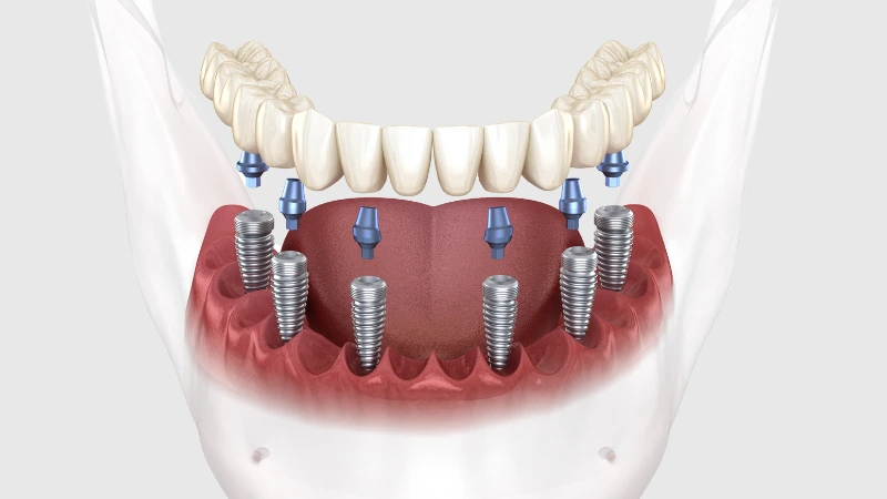
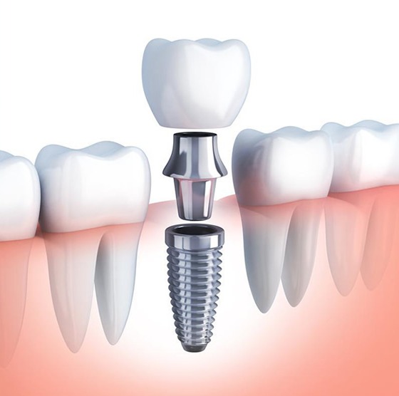
| Implantology Applications | Placement of titanium artificial roots to replace missing teeth in suitable areas of the jawbone. |
| Cyst and Lesion Resection | Removal of pathological cysts that have developed in the tooth roots or jawbone without spreading to surrounding tissues. |
| Pre-prosthetic Surgery | Shaping and contouring of bone remnants (such as tori) or tissues that are not suitable for prosthesis fabrication. |
In Which Cases is Oral, Dental, and Jaw Surgery Applied?
Structural defects in the mouth can eventually lead to pain, infection, and difficulties in chewing. Wisdom teeth that do not have enough space in the dental arch can exert pressure on adjacent teeth’s roots, disrupt alignment, or cause recurrent inflammation (pericoronitis) in the surrounding gingiva. Surgical interventions come into play to address such pressure and infection-related issues.
Similarly, when areas remain edentulous for an extended period, the bone cells become inactive and begin to resorb. Progressive bone loss limits the area for placing a porcelain bridge or artificial root. These volume deficiencies in the vertical and horizontal dimensions of the jawbone are restored to physiological standards through advanced grafting (bone powder addition) procedures.
How is an Evaluation Made Before Oral, Dental and Jaw Surgery?
Surgical interventions are directly related to the individual’s overall circulatory system and cellular repair capacity. Conditions such as cardiovascular diseases, clotting problems, or uncontrolled diabetes found in the patient’s medical history can change the clinical pathway during and after the repair phase of the operation. Therefore, the physician adjusts the process in accordance with medical protocols through consultation with the patient’s medical doctor.
In addition to physical examination, radiological findings guide the surgical operation. The size of a cyst to be removed, the distance from the lower jaw nerve (mandibular nerve) to a tooth that needs to be extracted, or the volume of bone graft to be added can be measured millimetrically with imaging tools to prevent surprises.
How Are Oral, Dental, and Jaw Surgery Treatments Planned?
In clinical practice at the Izmir Avrupadent branches, each surgical procedure is scripted step by step. For example, if a large cyst operation is planned, it is predetermined how the bone defect (cavity) created after cleaning the cyst will be filled, and the structure of the bone powder (graft) or protective membranes to be used. This planning supports time management during the operation.
If there is an acute inflammation in the area to be intervened, to prevent the reduction of anesthesia effectiveness and the spread of infection, antibacterial medication (antibiotic) is started a few days before the procedure to calm the tissue. The patient is mentally prepared for the process by being informed about dietary and rest expectations for the day of the operation.
Who Might Be Suitable for Oral, Dental, and Jaw Surgery?
A broad age range is involved in surgical interventions. While the extraction of wisdom teeth, which often presents problems in young adults, is more prevalent, volume increase related to osteoporosis and the application of artificial roots (implants) are gaining importance in older age groups. As long as the individual’s medical condition is kept stable, age alone does not pose an obstacle.
For patients using bisphosphonate-derived medications that suppress bone metabolism or those with weakened connective tissue, the approval of the physician is essential to prevent disruptions in surgical repair (wound healing). After the necessary medical arrangements are provided, steps to protect oral health are taken within the framework of safety standards.
What Diagnostic and Imaging Methods Are Used in Oral, Dental, and Jaw Surgery?
The clinical success of operations relies on specific radiological devices that elucidate the hidden anatomy beneath the tissues. The primary imaging systems guiding the process are:
What Steps Are Followed in Oral, Dental, and Jaw Surgery Procedures?
Each medical intervention is carried out with careful planning while respecting biological tissues. After anesthesia is administered, a thin flap is lifted from the gum to expose the operative area. If a buried tooth is being extracted, the resistance points around the bone are alleviated with diamond burs, and the tooth is removed in pieces without damaging the tissue.
If the procedure involves cyst removal, the pathological formation is scraped out along with its edges, and it is expected that the remaining void will be filled with a clot that supports the body’s repair, or synthetic bone powders are placed. In the final phase, the opened mucosal tissue is adapted back to its previous position and fixed with fine surgical threads; this allows the wound edges to close to the external environment, initiating the primary healing process.
What Should Be Considered Before Oral, Dental, and Jaw Surgery?
Reducing microbial load in the working area is a critical factor in preventing surgical complications. The patient should carefully brush their teeth before the day of the procedure to remove any remaining food debris. The dosage of anticoagulants or similar medications must be adjusted under the supervision of the physician, and the patient should confirm this information with their dentist on the day of the procedure.
Due to the procedure being performed under local anesthesia, it is advised not to come to the appointment on an empty stomach to maintain the patient’s blood sugar and blood pressure balance. Knowing that all tobacco products slow down cellular repair, it is beneficial to limit this habit in the days leading up to the procedure to support the healing process.
What Should Be Considered After Oral, Dental, and Jaw Surgery?
It is common to see slight swelling (edema) and small leaks in the cheeks following surgical procedures in the documents. Applying ice compresses intermittently from the outside of the cheek within the first 24 hours restricts the expansion of this swelling. Using straws that create pressure or vigorous gargling movements should be avoided in the first days as they can dislodge the protective blood clot over the wound.
When the prescribed analgesics and antibiotics are taken according to the schedule, the recovery period can be managed comfortably. Hard foods should not be chewed on the operated side; soft foods such as puree, yogurt, and broth that do not require chewing force should be preferred. To avoid irritating the healing mucosa, the consumption of acidic and spicy foods should be limited for a while.
Is Impacted Tooth Extraction Evaluated within the Scope of Oral and Maxillofacial Surgery?
Not only the 20-year molars; sometimes, canine teeth or small premolars can also be trapped beneath the bone due to bone constriction, delays in milk tooth extractions, or genetic reasons. These trapped teeth can cause damage over time by eroding the roots of adjacent healthy teeth or developing pathological cysts around them.
If a trapped tooth cannot be aligned (pulled into the mouth) with orthodontic wire treatment, it is carefully removed from its bony socket by surgical specialists. During the procedure, the anatomy of neighboring teeth and surrounding nerve pathways are preserved, adhering to the principle of minimal trauma.
How is Surgical Approach Planned for 20-Year Molar Problems?
Wisdom teeth are typically the last teeth to erupt in the mouth, so they often do not have enough space. When there is partial impaction (some part outside), the gum tissue forms a covering over the remaining 20-year molars; leftover food getting under this covering cannot be cleaned with a toothbrush and can lead to severe inflammation known as “pericoronitis.” This infection, which causes jaw tightness and pain, will result in a schedule for extraction once it subsides.
During surgical extraction, if the tooth is positioned horizontally, the surgeon uses special tools to break the tooth into smaller pieces (sections) rather than forcibly removing it in one piece to keep bone loss to a minimum. This approach is an important clinical strategy that increases the comfort of the patient’s recovery after surgery.
In What Situations Are Surgical Procedures Related to the Jaw Bone Applied?
The structural integrity of the jawbone is the main pillar that carries the health of the teeth and prosthetics in our mouth. In areas that remain vacant for many years after tooth extraction, the bone deteriorates and thins, resembling a knife’s edge. Progressive bone loss compromises the infrastructure for artificial restorations to be applied to that area. Dentists restore these areas to thickness using graft materials (synthetic or biological bone powders) with techniques known as “guided bone regeneration.”
Additionally, if there are congenital or acquired bony protrusions (torus mandibularis / palatinus) that force the use of a prosthesis, these tuberosities are flattened with pre-prosthetic surgery. The goal is always to create a porous and durable skeletal system that can withstand masticatory forces.
How is Implant Treatment Related to Oral, Dental, and Maxillofacial Surgery?
The procedure of placing an artificial root into the bone is not just a mechanical screwing; it is a biological surgery aimed at the fusion of bone cells with the titanium surface (osteointegration). Jaw surgery includes the medical knowledge of placing these screws at the most accurate angle without contacting nerve channels or sinus cavities.
If there is insufficient width in the jawbone, the jaw surgeon repairs the area by conducting bone membrane procedures simultaneously or prior to the artificial root. Therefore, implantology demonstrates its existence through the anatomy of the mouth and jaw surgery, principles of tissue healing, and sterilization management from start to finish.
In Which Situations Are Bone Grafting and Sinus Lift Procedures Evaluated?
When the back (slight) teeth of the upper jaw are lost, the air spaces (maxillary sinuses) located just above that area show a tendency for downward sagging. Over time, the bone layer becomes thin, making it impossible to place a titanium body in that area. With the “sinus lifting” procedure, the membrane of this cavity is gently pushed up and filled with bone powder (graft) in that area; thus, a new bone area in the vertical direction is gained.
In other jaw regions, horizontal resorption related to tooth loss is observed. Through directed bone regeneration, grafts (bone minerals) and barrier membranes (films) are placed in the resorbed areas to increase volume. The body converts these powders into its own living bone tissue within months, forming a healthy supporting column.
How Does the Healing Process Progress After Oral, Dental, and Maxillofacial Surgery?
The first 3-4 days after surgery is the most pronounced period of inflammation (healing response). It is common for a whitish layer (fibrin) to form on the surface of the mucosa where stitches are placed, acting as a bandage for the wound. Prescription medications support the clinical stability and smooth passage of this active phase.
The superficial (gum) closure generally occurs within 10-14 days and stitches can be removed; however, if bone grafting or cyst scraping was performed underneath, the bone’s internal healing (cellular restructuring) continues for months without being visible. It is crucial for the patient to protect the area from trauma during this hidden healing phase to ensure complete regeneration.
How Should Oral Care be Performed After Surgical Procedures?
The surgical areas where stitches are placed are quite sensitive in the early days. The hard edges of the sutures can cause tension on the stitches or separate the wound lips. Therefore, until the physician indicates otherwise, hygiene should be maintained at the wound site only with chemical cleaning (chlorhexidine-containing mouthwashes). While gargling, it is important to maneuver the liquid gently in the mouth to avoid pressure-induced turbulence.
After the wound tissue has united and the stitches are removed, light rubbing motions can begin on the area with extra soft surgical brushes as if massaging. Once healing is fully completed, standard hygiene practices and cleaning of the interferences should be resumed to maintain tissue health.
Why is Follow-Up Examination Important in Oral, Dental, and Jaw Surgery?
After surgery, the healing of the patient’s wound is generally advanced by the removal of stitches (sutures) during the control session in the first or second week. If the suture threads remain longer than the specified period, they can lead to plaque retention, causing localized infections around them. During the control appointment, the threads are removed to relieve the area.
In surgeries involving bone repair (after grafting or cyst removal), radiographic films taken months later objectively monitor the rate of osseointegration. By analyzing how the surgical area heals, customized revised hygiene training can be provided for the patient’s periodontal health in later stages.
Can Oral, Dental, and Jaw Surgery Treatments Be Planned Together with Other Dental Treatments?
In modern dentistry, no branch works independently of the others. For example, when an orthodontist aligns teeth within the bone, if they determine that the position of an impacted horizontal wisdom tooth will disrupt this arrangement, they request the extraction of that impacted tooth from the oral and maxillofacial surgery department before starting orthodontic treatment.
Similarly, in a patient planned for aesthetic design (such as aesthetic laminates), if there are bone asymmetries beneath the gum boundaries or if there is bone loss in previously extracted areas, these areas are supplemented with tissue through pre-prosthetic surgical interventions to ensure the porcelain has a symmetrical foundation. The healthy biological base provided by surgery directly supports the lifespan of all subsequent restorative and aesthetic treatments.