What is Wisdom Tooth Extraction?
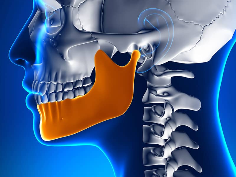
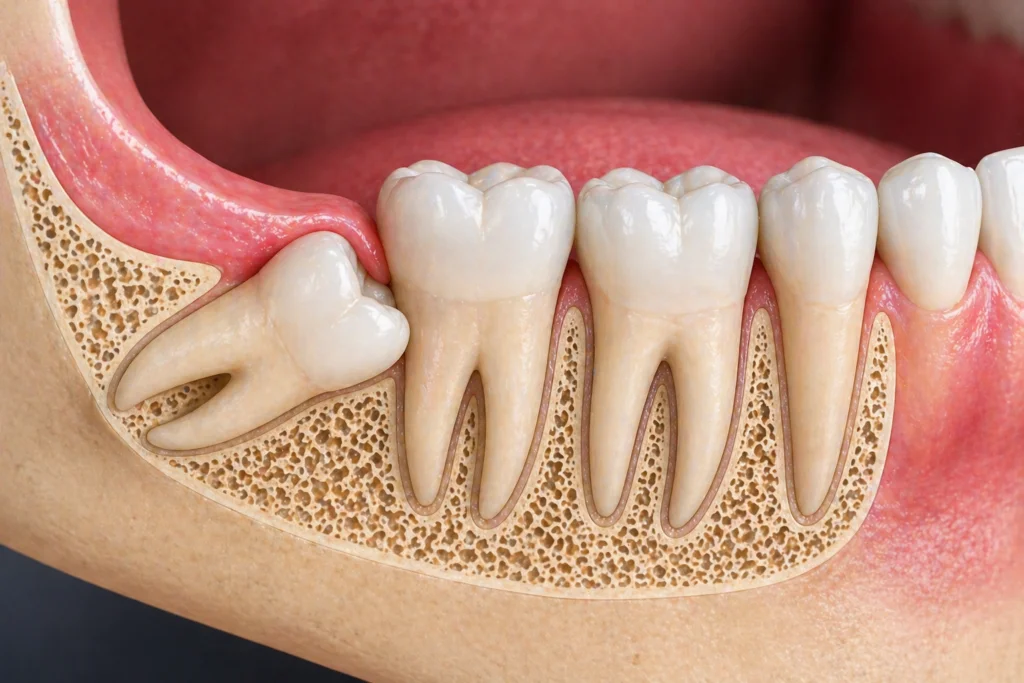
The evolution of human diet habits and the shift towards consuming softer food forms has led to a decrease in the anatomical development of jawbones. As the jaws shrink, the last wisdom teeth that erupt in the mouth struggle to find space. Although these teeth generally try to emerge between the ages of 17 and 25, they exert pressure on adjacent teeth when there is insufficient space, becoming trapped in horizontal or tilted positions within the bone. This situation not only misaligns the teeth but may also predispose surrounding tissues to cystic formations or chronic infections.
The procedures for wisdom teeth extraction at the Avrupadent clinics in Izmir are assessed within a multidisciplinary framework, considering the hospital’s jaw anatomy and chewing functions. Whether they have fully erupted into the oral cavity or remain hidden (impacted) beneath the bone, the removal of these problematic teeth carries significant clinical value to preserve oral biomechanics. The primary goal of the operations is to support the quality of chewing and to prevent more complex structural problems that may arise in the future.
Why Are Wisdom Teeth Extracted?
Cleaning these teeth, positioned at the very back of the mouth, with a toothbrush and dental floss is quite challenging. The accumulation of food debris in these dark and narrow spaces can lead to the much earlier occurrence of cavities in these teeth. The primary medical reasons for the extraction decision are as follows:
| Reason for Extraction | Explanation and Clinical Effects |
|---|---|
| Pericoronitis (Tooth Tissue Inflammation) | Pain and tissue infections resulting from food trapping against the overlying flap of partially erupted teeth. |
| Neighboring Tooth Damage | The horizontal position of the wisdom tooth causing bone resorption in the area by leaning against the root or body of the adjacent healthy tooth. |
| Risk of Malocclusion | The force of eruption (extrusion) altering the alignment of existing teeth by pushing them forward. |
| Cysts and Pathological formations | The expanding protective follicle surrounding the impacted tooth transforming into liquid-filled cysts that erode the jawbone over time. |
In Which Situations Is Wisdom Tooth Extraction Applied?
Not every wisdom tooth may be infected; however, proactive approaches are taken when a potential risk is present. In patients who have undergone or will undergo orthodontic treatment, extraction of these teeth is a frequently employed medical strategy aimed at preventing the loss of space and the distortion of the previously achieved alignment caused by the pushing force of the wisdom tooth.
Additionally, wisdom teeth that are oriented in the wrong direction (impacted) can continuously irritate the cheek mucosa while the patient is eating or talking. Since chronic cheek irritation trauma can pave the way for cellular changes over time, the removal of these misaligned teeth is a clinical necessity to keep them away from the oral environment.
How Is Wisdom Tooth Extraction Performed?
The extraction of an impacted wisdom tooth (erupted in a normal position) does not require a different procedure from that of other molars. After the administration of anesthesia to the concerned area, special instruments called elevators and levers are used to loosen the tooth from its socket and remove it. However, if the tooth is in a horizontal, level, or fully buried position within the bone, the “surgical extraction” steps are applied.
In a surgical intervention, the physician makes a small incision (flap) in the gum tissue to expose the bone surface. The layer of bone that surrounds the tooth is minimally reduced using special drills or devices operating with sound waves (piezosurgery). To minimize tissue trauma, a large buried tooth is not attempted to be removed in one piece; instead, the tooth is divided into several pieces along suitable axes and removed without damaging the bone. After the relevant cavity is cleaned, the gum tissue is stitched up with fine surgical threads, completing the procedure.
Who Can Be Evaluated for Wisdom Tooth Extraction?
Complaints about wisdom teeth most commonly manifest between the ages of 17 and 25. The healing phase of extractions performed at these younger ages tends to progress quite quickly since the roots of the teeth have not fully developed and the jawbone is more flexible. However, as age advances, a cystic wisdom tooth that is quietly waiting within the bone can become active and require intervention; therefore, there is no strict age limit for the feasibility of the procedure.
The patient’s overall systemic health status comes at the forefront of guiding factors. In individuals with diabetes (sugar disease), heart disorders, or bleeding disorders, the aim is to ensure that clinical values are aligned within ideal ranges based on consultations conducted with the treating physician. Surgical planning can be safely undertaken as long as medical values are kept stable.
How is an Evaluation Conducted Before Wisdom Tooth Extraction?
The intervention strategy is entirely based on the diagnostic data obtained. The physician analyzes the shape of the wisdom tooth roots (whether they are curved or straight), their number, and their inclination within the bone (horizontal, vertical, mesioangular, distoangular). Teeth located in the lower jaw can often be positioned very close to the nerve that provides sensation to the lower lip and chin. During the operation, safety distances are measured over X-rays to ensure there is no mechanical contact with this nerve.
If there is currently an acute (active) inflammation and swelling present in the relevant tooth area of the patient, the surgical intervention may be postponed as the effects of local anesthesia solutions may be diminished in infected tissues. Reducing the infection through medical medications (such as antibiotics) and calming the tissues is an essential step in the preparatory phase.
How is the Wisdom Tooth Extraction Process Planned?
At the İzmir Avrupadent branches, clinical procedures are designed to be the most practical and biologically least exhausting for the patient. If there are four wisdom teeth that need to be extracted from the patient’s mouth, it can be decided jointly by the physician and the patient that all these teeth can be removed (usually under sedation) in the same session; alternatively, to ensure chewing comfort, appointments can be staggered, first removing the teeth on the right side, then on the left side after recovery.
During the planning phase, the patient is provided with preliminary information about ice compresses, dietary restrictions, and medication schedules expected to be applied in the first few days after surgery. Since attention will be required for speaking and chewing habits in the days following the procedure, the appointment dates are scheduled to fall outside the busy work or exam periods of the patient.
What Steps Are Followed in Wisdom Tooth Extraction?
Clinical steps taken to support regenerative treatment and preserve anatomical boundaries are gradually implemented while adhering to surgical principles. The roadmap followed in a surgical operation is as follows:
- • Anesthesia Induction: The local nerves surrounding the operation area are blocked with local agents to ensure numbness.
- • Flap (Tissue) Elevation: A small incision is made in the gum, and the mucosa is separated to reach the underlying bone and tooth structure.
- • Bone Contouring and Partitioning: The bone obstacle over the tooth is removed using fine burrs, and it is divided into sections to enable the tooth to emerge without applying pressure to the surrounding area.
- • Extraction: Complete removal of tooth fragments and any cyst-like follicular remnants formed around it from the socket (alveolus).
- ▪ Suturing (Stitching): The surgical process of stitching the mucosa of the gum over the area that has been washed and whose bleeding has been controlled with surgical threads.
What Should Be Considered Before Wisdom Tooth Extraction?
Keeping the bacterial load in the oral flora at a minimum helps to prevent possible complications that may develop in the surgical area afterward. The patient should thoroughly brush their teeth before arriving at the clinic to remove existing food residues. The use of anticoagulants or similar systemic medications arranged under the physician’s guidance should be reconfirmed with the physician on the day of the procedure.
The patient will be conscious during the procedure under local anesthesia. Since high stress levels can cause fluctuations in blood pressure, arriving at the clinic in a slightly full and rested state positively influences clinical comfort. Getting a good night’s sleep before the day of the operation supports a calmer experience during the time spent in the doctor’s chair.
What Should Be Considered After Wisdom Tooth Extraction?
After surgical extractions, a mild to moderate swelling (edema) in the cheek area and small leaks in the mouth are a natural response of cellular repair. Applying a cold compress intermittently from the outside during the first 24 hours significantly limits this swelling. Coughing, drinking liquid through a straw, or forcefully rinsing the mouth with water can disrupt the protective blood clot formed in the extraction site and trigger bleeding.
The nutrition routine should consist of liquids and pureed foods (soup, yogurt, puree, etc.) that will not traumatize the operation area. Acidic, extremely hot, or spicy foods should be avoided in the menu for at least a few days as they may irritate the fresh mucosal surface. Consuming prescribed analgesic medications at the specified intervals will significantly help manage the process calmly.
How Are Impacted Wisdom Teeth Evaluated?
Although each impacted tooth may not cause active pain, it is a potential medical risk. Teeth silently waiting completely within the bone should be monitored periodically with X-rays against the possibility of the surrounding protective sac (follicle) filling with fluid and forming a cyst over time. If cyst formation begins, it quietly erodes the surrounding bone, weakening the jaw; at this point, it is advised that the tooth be surgically removed without delay.
If the visible tooth eruption is toward the front (compared to the neighboring larger tooth), the doctor predicts that this situation will gradually resorb the healthy tooth roots in front of it. When this structural damage risk, which cannot be observed, is confirmed by radiological findings, the doctor recommends a proactive extraction planning to the patient instead of the “wait and see” strategy.
What Diagnosis and Imaging Methods Are Used Before Wisdom Tooth Extraction?
The element that ensures the reliability of the surgical intervention is the diagnostic tools that clearly illuminate the topography of the tooth within the jawbone. The basic radiological methods applied in the clinic are as follows:
- • Panoramic Radiography: By showing the entire jaw structure and the tooth roots at a wide angle, it provides a preliminary map regarding the general position of the tooth, its inclination, and its relationship with neighboring teeth.
- • Dental Volumetric Tomography (CBCT): In cases where the root and the nerve canal overlap in two-dimensional films, it is an advanced diagnostic tool that determines whether the root passes in front of or behind the nerve with three-dimensional millimetric slices.
- • Periapical Films: Focuses on a single tooth with small film sizes, meticulously examining the curves at the root tip of the wisdom tooth or the closeness of the roots to each other.
How Does the Healing Process Progress After Wisdom Tooth Extraction?
The oral mucosa is one of the tissues with the highest blood supply and cell renewal rate in the body. This allows for rapid healing of surface wounds in the operational area, enabling the patient to return to normal eating and drinking functions within a few days. In the early days, slight restrictions when opening the mouth (during yawning) or sensitivity while swallowing can be observed; this is a temporary reaction from the muscles to the surgical intervention and decreases quickly.
However, in the deeper layers of the tissue, that is, in the bone cavity where the tooth has emerged (its socket), a deeper and longer-lasting cellular activity begins. The blood clot that fills the void gradually organizes into connective tissue and eventually turns into hard bone tissue with the work of bone-forming cells (osteoblasts). Although this inner healing (ossification) is not felt from the outside, it anatomically requires an average maturation period of between 3 to 6 months.
How Long Can the Extraction of Wisdom Teeth Take?
A wisdom tooth that is positioned straight and follows the standard extraction procedure can be removed within just a few minutes after anesthesia takes effect. However, a tooth that is horizontally positioned in the lower jaw, with roots curved like hooks and hidden beneath a deep layer of bone, will take longer to surgically extract because it will require dividing the tooth into many pieces (sections).
Another factor that determines success is the physician’s protective (minimally invasive) approach to the surrounding tissues. Procedures are carried out with extremely gentle and slow hand maneuvers to avoid excessive strain on the surrounding bone and to prevent pressure on the nerve pathways. Since the patient is under anesthesia, they go through this period without feeling any clinical discomfort.
How Should Oral Care Be Performed After Wisdom Tooth Extraction?
A sensitive blood clot that ensures bleeding stops is found in the wound area after surgical intervention. The use of hard brushing or pressured mouth washing can mechanically dislodge this clot and lead to a painful condition known as “alveolitis” (dry socket) that hinders healing. Therefore, until the physician states otherwise, hygiene should only be maintained through chemical cleaning in the operational field.
While gargling, instead of agitating the liquid in the mouth with high pressure, it is crucial to gently swirl the liquid around the wound area and gently let it out into the sink (without forcefully spitting it out). After stitches are removed and the mucosa has healed, gradually returning to the standard oral care routine can be ensured by massaging the area with extra soft (surgical) brushes.
Is It Always Necessary to Remove Wisdom Teeth?
A healthy and aligned wisdom tooth is structurally different from other molars. Although there is a widespread perception that wisdom teeth will inevitably cause pain if not removed, the potential for these teeth to create problems is entirely dependent on their anatomical positions (how easily they can be brushed). If the patient can comfortably reach the back areas to brush and maintain hygiene, the retention of that tooth in the mouth is supported.
However, even teeth that have been in a dormant state for years in the deepest layers of the jawbone (completely embedded) should be monitored by dentists with 6-month or annual radiographic follow-ups using a ‘wait and see’ strategy, even if they do not cause any discomfort to the patient. If, in the following years, the cystic formation of the follicle around the tooth is detected in the films, then the extraction decision will be implemented.
Why is Control Examination Important After Wisdom Tooth Extraction?
This control session, which is generally conducted in the first or second week following the surgical intervention, is to confirm that the healing period is progressing correctly. If the sutures used (unless they are self-dissolving) remain longer than the specified time in the oral environment, they can cause local irritations by promoting food retention. The doctor ensures that these threads are removed to allow the tissue to return to its natural form.
Moreover, if the patient indicates that their pain has increased instead of decreasing after surgery or if there is a bad odor coming from their mouth, the physician will examine the wound site to check if a clot loss (alveolitis) has occurred. If necessary, the wound will be cleaned with medical agents and dressed, and the healing process will be medically supervised.
Can Wisdom Tooth Extraction Be Planned Together with Other Oral and Dental Treatments?
Dental practices often provide infrastructure for each other. Especially in orthodontic treatments with braces or clear aligners where irregular teeth are straightened, if there is a possibility that impacted wisdom teeth may apply pressure from behind and disrupt the previously corrected alignment, it is advisable to surgically remove these teeth as a preventive (proactive) approach before orthodontic appliances are placed.
For patients who will undergo implant treatment and have an infected wisdom tooth in the same jaw, combined operation sessions are planned. While the doctor places a titanium root in one area, they also extract the problematic back tooth of the patient under anesthesia during the same surgical session, thereby protecting the patient from the stress of two separate surgeries.
How is a Personalized Treatment Plan Created for Wisdom Tooth Extraction?
Every person’s jaw structure and the shape of their tooth roots are unique. While a tooth in one patient may be in a vertical position and easily accessible, in another patient, it may be completely horizontal, leaning against the root of an adjacent tooth and buried within a labyrinth of thick bone tissue. The doctor anticipates this situation and develops an individual strategy to determine the surgical tools and which directions the tooth will be extracted from.
In the processes at Izmir Avrupadent, particularly in risky cases adjacent to nerves, the limits of surgical instruments are measured through virtual tomography sections. By blending the individual’s medical condition, bone flexibility, and psychological profile, the most ideal clinical roadmap is designed to create minimal tissue trauma (minimally invasive) and accelerate post-operative recovery.