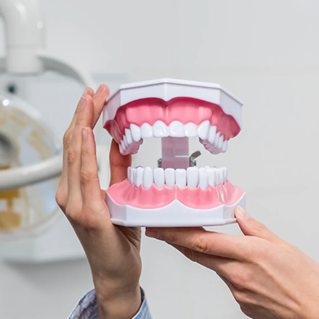
What are Diagnosis and Assessment Methods?
In order to establish an accurate treatment process in oral and dental health, it is essential to scientifically identify the source of symptoms. The methods of diagnosis and identification in dentistry encompass a wide range of medical protocols, from the anamnesis process that begins with listening to the patient’s complaints to advanced three-dimensional imaging technologies. Thanks to these methods, hard-to-see bone cysts, deep periodontal pockets, jaw joint problems, and root infections are evaluated.
The diagnosis and identification processes at the Avrupadent clinics operating in Izmir are structured with data from physician examinations, digital radiography, and intraoral (in-mouth) scanning systems. Every type of cellular or anatomical change in both hard and soft dental tissues is examined in detail through these diagnostic tools.
Hangi Diagnostic and Methodology Hizmetine İhtiyacın Var?
In dentistry, examination and imaging tools not only reveal existing pathologies; they also aim to detect asymptomatic (symptomless) bone resorptions and cyst formations in the early stages, limiting tissue losses.
Hangi Diagnostic and Methodology Hizmetine İhtiyacın Var?
How Are Diagnosis and Identification Methods Applied?
The application process begins with recording the medical history of the patient (anamnesis). The patient’s systemic discomforts, regularly used medications, and past dental treatments are also evaluated. Then, the process moves on to physical examination; the physician examines the tooth surfaces, gingival pockets, tongue, and buccal mucosa using special dental mirrors and probes.
Following the clinical examination, radiological assessments relevant to the needed areas are performed. Periapical X-rays focused on a single tooth, panoramic radiographs showing the entire jaw, or 3D Cone Beam Computed Tomography (CBCT) scans providing millimetric sections are applied. Additionally, the vitality data is completed with cold/warm vitality tests measuring the vitality of the teeth and periodontal probing procedures.
-
\t
- <span style="color: #e1660e; position: absolute; left: 0; top: 2px; font-size: 18px;▪
Visual and Physical Examination: Examination of intraoral tissues, restoration boundaries, and gingival pockets with hand tools. - <span style="color: #e1660e; position: absolute; left: 0; top: 2px; font-size: 18px;▪
Digital Radiological Examination: Displaying the jawbone and root structures on the screen using low-dose radiation sensors. - •
Digital Intraoral Scanning: Creating three-dimensional digital models of dental structures using optical cameras.
\t
Why Are Diagnostic and Evaluation Methods Important?
Intraoral structures have a complex biological network. The formation of a cyst at the root tip of a tooth that appears healthy from the outside or the presence of an embedded tooth can only be understood through biometric and radiological methods. Incorrect or incomplete diagnosis can lead to erroneous treatment applications and loss of time.
The detailed diagnostic processes implemented at Avrupadent clinics in Izmir ensure that physicians can clearly visualize all tissue boundaries before intervention. This helps prevent tissue loss and directly supports the biomechanical success of the treatment to be applied to the patient.
What Are the Advantages of Diagnosis and Assessment Methods?
The use of advanced diagnostic tools in modern dentistry makes treatment processes safer and more traceable. The benefits obtained can be summarized as follows:
| Area of Advantage | Clinical and Biological Contribution |
|---|---|
| Early Detection Opportunity | Detection of asymptomatic inflammations and initial stage lesions before tissue loss occurs. |
| Treatment Sensitivity | Performing millimetric measurements of channels and bones in procedures such as implants, surgery, and root canal treatments. |
| Open Communication | Allowing patients to be informed by viewing their own oral structures via digital data. |
| Comprehensive Assessment | Simultaneous analysis of the jaw joint, soft tissues, and tooth alignment. |
In Which Situations Are Diagnostic and Detection Methods Used?
Diagnostic tools come into play with different purposes in every branch of clinical dentistry. The main usage situations are as follows:
- ▪
Investigation of Pain and Sensitivity Source: Detection of unexplained vertical and horizontal tooth pains, root fractures, and deep caries. - ▪
Before Surgical and Implant Procedures: Assessment of the height, width of the jawbone, and distances to the mandibular nerve canal. - ▪
Classification of Periodontal (Gum) Diseases: Measurement of bone level recessions and gum pockets. - ▪
Orthodontic Planning: Examination of the jaw skeletal structure and tooth alignments using cephalometric analyses.
Who Are the Diagnosis and Detection Methods Suitable For?
Children, adolescents, adults, and elderly individuals can benefit from specially selected diagnostic protocols. In children, low-radiation periapical images and visual examinations are preferred to monitor the development of primary teeth; while in adults, digital systems that assess the entire oral structure are planned.
Those undergoing prosthetics, individuals about to begin orthodontic treatment, patients experiencing chronic gum problems, and anyone who comes for routine 6-month check-ups may be included in these examination processes.
Who Are Diagnosis and Assessment Methods Not Applicable To?
Certain special conditions are taken into account in the application of radiological diagnostic methods. It is generally a medical principle to avoid radiographic exposures involving X-rays, especially during the first three months of pregnancy. In very urgent situations, procedures can only be performed by the physician’s decision, using barriers such as lead aprons and thyroid protectors.
Additionally, for patients who have unstable general health conditions, cannot cooperate, or have significant mental/physical limitations, alternative digital scanning systems or surface examination methods may be considered instead of procedures like tomography that require remaining still.
What Should Be Considered Before Diagnostic and Screening Methods?
To improve the quality of radiological imaging, it is essential to remove earrings, necklaces, glasses, piercings, and removable dentures before extraoral (outside the mouth) exposures. These metal objects can create artifacts (reflection and shadowing) on the image, negatively affecting the accuracy of the diagnosis.
Ensuring oral hygiene before the clinical examination allows the dentist to assess the tooth surfaces and gum margins more clearly through brushing.
How Does the Diagnosis and Treatment Process Proceed?
The clinical diagnosis process consists of integrated, step-by-step actions. According to Avrupadent protocols, the tracking of the process is as follows:
| Step | Medical Procedures Applied |
|---|---|
| 1. Medical History and Registration | Patient’s complaints, medical history, allergies, and medications are recorded. |
| 2. Clinical Examination | Examination of the soft and hard tissues inside the mouth, calculus, lesions, and prostheses. |
| 3. Imaging and Tests | Panoramic, tomography, or periapical images are taken; vitality tests are applied if necessary. |
| 4. Synthesis and Information | The obtained data is consolidated by the physician, and the situation and treatment options are communicated to the patient. |
What Technologies Are Used in Diagnostic and Diagnostic Methods?
The advancing medical devices have increased the diagnostic coefficient while ensuring that radiation doses are kept to minimal levels. The main technological components are as follows:
-
\t
- 3D Cone Beam Computed Tomography (CBCT): Provides the ability to examine the volumetric map of the jawbone cross-sectionally.
- Digital Intraoral Cameras: It displays the micro cracks and initial cavities on the outer surface with high resolution on the screen.
- Digital Impression Scanners: It creates a 3D digital model of the teeth in seconds without using physical materials.
- RVG (Radiovisiography): It instantly transfers periapical images to the computer screen using low-dose X-rays.
How Does Accurate Diagnosis Affect Treatment Success?
In medicine, the principle that “an accurate diagnosis is half of the treatment” is fundamental. An accurate diagnosis allows for direct intervention in the root cause of discomfort. For instance, determining which tooth is responsible for a reflected pain prevents unnecessary intervention on the wrong tooth.
Additionally, properly delineating anatomical boundaries helps to keep complications that may arise related to nerve damage during implant procedures or sinus floor sagging during surgical interventions under control. This positively affects the clinical robustness of the treatment and tissue compatibility.
Are Diagnosis and Detection Methods Safe?
Digital radiology devices operate with significantly lower doses of radiation compared to traditional film-based X-ray devices. The amount of radiation received during a panoramic X-ray is at a level comparable to the average background radiation received in nature daily.
The use of lead aprons and thyroid protective barriers during imaging procedures supports the protection of vital organs. Oral scanners and cameras operate with completely optical light technologies that do not contain radiation.
How Long Do Diagnosis and Diagnostic Methods Take?
The duration of diagnostic procedures varies depending on the scope of the examination to be performed. A standard routine clinical examination and panoramic X-ray typically take between 15 to 30 minutes.
Three-dimensional tomography (CBCT) examinations, digital oral scans, and detailed prothetic or surgical planning sessions that include jaw joint analyses may take an average of 45 to 60 minutes. Thanks to digital technologies, the results are displayed on screens for immediate evaluation.
How is the Treatment Plan Created After Diagnosis and Identification?
After collecting diagnostic data, situations that require urgent treatment (painful teeth, active infections) are prioritized. In the second phase, periodontal (gum) treatments and cleaning procedures are planned.
Once basic oral health is established, restorative, prosthetic, surgical, or orthodontic stages begin. The comprehensive treatment plan is clarified through interdisciplinary consultation by considering the patient’s overall health status, biological structure, and expectations.
Are There Risks in Diagnosis and Identification Methods?
The diagnostic and identification methods used in dentistry are non-invasive (not damaging the tissue in a substantial manner) procedures. Therefore, they do not pose serious biological risks.
The potential cumulative radiation effects in radiological examinations are kept within medical safety limits using digital sensors and protective lead barriers. Transient discomfort that may occur in patients with sensitivity to the gag reflex during intraoral measurements is attempted to be addressed by using digital scanning cameras.
Diagnosis and Identification Methods: Is It Painful?
Radiographic images, digital scans, and visual examinations do not cause physical discomfort to the patient. Only a brief, mild sensation may be felt during probing of inflamed or sensitive gum pockets or during dental vitality tests; this is a temporary physiological response required for making a diagnosis.
Diagnosis and Identification Methods: Which Diseases Do They Help Identify?
Developmental review methods are used to detect many pathological conditions in the mouth and surrounding tissues. The main discomforts that can be identified are as follows:
-
\t
- • Interdental Spaces and Secondary Spaces: Remaining spaces at the margins or contact points of crowns.
- • Apex Lesions and Cysts: Infections developing within the jawbone and fluid-filled sacs.
- • Periodontal Bone Losses: Bone resorption occurring as a result of gum inflammation.
- • Impacted Tooth Abnormalities: The positioning of wisdom teeth or retained permanent teeth within the jaw.
- • TMJ Disorders: Joint deformities and disk displacements.
\t
\t
\t
\t
What is the Importance of Digital Technologies in Diagnosis and Assessment Methods?
While traditional diagnostic approaches may be limited to two-dimensional images, digital systems allow for the three-dimensional analysis of intra-bone structures. Computer-aided planning software enables operational simulations to be conducted before a treatment begins.
The digital diagnostic equipment used in the Avrupadent clinical infrastructure helps the patient to monitor their own treatment process through monitors and understand the procedures to be applied.
How Are the Results of Diagnosis and Assessment Methods Evaluated?
Radiographic images, digital scanning data, and clinical examination findings are evaluated (correlated) by the specialist dentist. Diagnosing based solely on radiographs can be misleading; therefore, the compatibility of physical findings and radiological findings is examined.
As a result of the evaluation, the clinical table is reported according to the standards of dental disciplines (Periodontology, Endodontics, Oral Surgery, etc.). The patient’s current oral condition, necessary interventions, and alternative methods are communicated in detail.
Our Corporate Approach and Process Standards
At Avrupadent centers in Izmir, the oral and dental health needs of our patients are clinically analyzed in conjunction with modern diagnostic and imaging technologies (CBCT, RVG, digital scanners). All radiological and physical data obtained during the diagnostic phase is evaluated in a framework of objective dialogue with our patients, free from any bias.
In all diagnostic and follow-up procedures applied, the infection control and sterilization standards specified in national health regulations are strictly followed. At the core of our service philosophy is to ensure that our patients have access to accurate information about their health status and to plan treatment processes in a safe, transparent medical environment.
Diagnostic and Methodology hakkında sıkça sorulan sorular ve cevapları
Ağız kokusu (halitosis) şikayetinin odağını bulmak için Teşhis ve Planlama kapsamında hangi kontroller yapılır?
Çürük kaviteleri, sızdıran eski dolgular ve fırçanın ulaşamadığı derin diş eti cepleri detaylı klinik taramadan geçirilerek kokunun odağı belirlenir.
Dişlerin tepe noktalarındaki aşınma yüzeylerinin incelenmesi Teşhis ve Planlama açısından diş sıkmayı kanıtlar mı?
Dişlerin sivri tepe noktalarında oluşan düzensiz sarı çukurlaşmalar ve minenin silinmesi, diş sıkmasının uzun süredir devam ettiğinin fiziksel kanıtıdır.
Diş köklerinde oluşan sinsi dikey kırıklar ileri radyolojik Teşhis ve Planlama ile saptanabilir mi?
Klasik filmlerde kök kırıkları bazen gizli kalırken, üç boyutlu tomografi kesitleri arasında gezinerek bu ince kılcal çatlaklar başarıyla teşhis edilebilir.
Yüz simetrisi ve orta hat analizleri dijital fotoğraflama ile yapılan Teşhis ve Planlama sayesinde mi kurgulanır?
Alınan resimler özel yazılımlara aktarılır ve gözbebeği hattı ile dudak çizgisi arasında çekilen dijital cetvellerle yeni dişlerin simetrisi kurgulanır.
Yapılacak işlemlere dair risklerin ve başarı oranlarının anlatılması Teşhis ve Planlama şeffaflığı açısından zorunlu mudur?
Tıpta hiçbir işlemin %100 garantisi olamayacağı için, tedavinin olası riskleri ve başarı oranları hasta hakları gereği açıkça anlatılarak şeffaf bir güven kurulur.
Kooperasyon kurulamayan (iletişim zorluğu çeken) küçük çocuklarda Teşhis ve Planlama işlemi oyunlaştırılarak travmasız atlatılabilir mi?
Kliniğe ilk adım atan çocukların iğne veya önlük korkusu yaşamaması adına, pedodonti uzmanlarımız oyun terapisi yaklaşımlarıyla muayeneyi güvenli bir masala dönüştürür.
Gece diş sıkma (bruksizm) şüphesi olanlarda Teşhis ve Planlama süreci nasıl ilerler?
Dişlerin tepe noktalarındaki düzleşmeler, yanak içindeki beyaz sürtünme çizgileri ve çene kaslarındaki sertlik muayene edilerek teşhis doğrulanır.
Çürük tespit sıvıları (boyaları) Teşhis ve Planlama arasında hastalıklı dokuyu ayırmak için güvenilir midir?
Kavite açıldığında sürülen bu özel boyalar, sadece hastalıklı bakterili dokuları renklendirerek sağlam dokudan ayırt edilmesini sağlar.
Diş taşlarının diş eti altına ne kadar indiği röntgen filmlerinin kullanıldığı Teşhis ve Planlama ile belli olur mu?
Yüzeydeki tartarlar sertleştiğinde (mineralize olduğunda), kök yüzeyinden taşan dikensi beyaz çıkıntılar olarak röntgenlerde net biçimde izlenir.
Lazer floresan cihazları gözle görünmeyen başlangıç çürüklerini saptamada modern Teşhis ve Planlama arasında yer alır mı?
Gözle görünmeyen oluk çürüklerini, lazer ışınının bakterilerden yansıması prensibiyle saptayan ve dijital bir rakamla şiddetini veren modern bir tanı yöntemidir.
Bilgilendirilmiş onam formu alınması Teşhis ve Planlama aşaması bittikten sonra tedaviye başlamanın yasal bir kuralı mıdır?
Tüm aşamalar, süreler ve medikal gerçekler hastaya anlatıldıktan sonra, hastanın tedaviye kendi rızasıyla başladığını gösteren resmi onam formunun imzalanması yasal bir şarttır.
İnatçı ağız içi lezyonlarında (iyileşmeyen yaralarda) doku örneği alınması (biyopsi) kesin Teşhis ve Planlama kapsamında mıdır?
Ağız içinde haftalarca iyileşmeyen şüpheli yaralardan ufak bir parça alınıp patoloji laboratuvarına gönderilmesi (biyopsi), hücresel karakterin kesin tanısı için şarttır.
Çene eklemi rahatsızlıklarının belirlenmesinde Teşhis ve Planlama hangi fiziksel testleri içerir?
Çenenin açılma miktarı ölçülür, eklemden gelen tıkırtı sesleri dinlenir ve parmak baskısıyla (palpasyon) kaslardaki spazmlar incelenir.
Çene yapısındaki iskeletsel uyumsuzlukların tespiti için yapılan profil analizi Teşhis ve Planlama içine girer mi?
Çene ucu, dudaklar ve burnun uyumu incelenerek çenelerin iskeletsel olarak geride veya ileride olup olmadığı analiz edilerek ortodontik planlama yapılır.
Cerrahi işlem planlanan bölgelerde kemik yoğunluğu (sertliği) dijital Teşhis ve Planlama ile radyolojik olarak ölçülebilir mi?
Tomografi yazılımları sayesinde çene kemiğinin süngerimsi veya sert yapıda olduğu tespit edilerek operasyon aletleri ve kemik tutunma tahmini buna göre yapılır.
Renk seçimi aşamasında dijital spektrofotometre cihazları kullanılarak uygulanan Teşhis ve Planlama daha mı hassastır?
Ortamın ışığından etkilenmeyen bu optik cihazlar, dişin rengini ve şeffaflığını analiz ederek laboratuvara dişin renk haritasını kusursuz bir şifreyle gönderir.
Kompleks vakalarda birden fazla uzman hekimin (konsültasyon) bir araya gelmesi Teşhis ve Planlama kalitesini artırır mı?
Cerrahi, ortodonti ve protez uzmanlarının aynı masada birleşerek hastanın radyolojik filmlerini ortak değerlendirmesi (konsültasyon) tedavi kalitesini zirveye taşır.
Avrupadent İzmir şubelerinde ilk başvuran hastalara uygulanan Teşhis ve Planlama ağız bütünlüğünü taramayı mı hedefler?
Evet, kliniğimize adım atan her misafirimizin sadece şikayetçi olduğu dişi değil; diş etleri, çene eklemi ve tüm ağız sistemi bütünsel bir yaklaşımla detaylıca taranır.
Eski ve uyumsuz protezlerin yarattığı sorunlar Teşhis ve Planlama ile nasıl saptanır?
Renkli ısırma kağıtları ve doku uyumunu gösteren özel malzemeler kullanılarak, protezin dokuya batan kısımları ve çiğneme dengesizlikleri haritalandırılır.
Dudak istirahat pozisyonunun ölçümü estetik Teşhis ve Planlama aşamalarında neden dikkate alınır?
Konuşurken veya dudak serbestken dişlerin ne kadarının göründüğü ölçülerek, yapılacak estetik kaplamaların boyu bu çerçeveye göre ayarlanır.
Contact Us for Diagnosis and Assessment Methods in Izmir
Diagnosis and Assessment Methods refer to an application area that includes the evaluation of various clinical and radiological methods for discomforts related to oral and dental health, as well as the formation of a treatment plan. At AvrupaDent, this evaluation is carried out by dentists within the framework of a treatment plan suitable for the patient's oral and dental health. You can contact us to get detailed information about the process.İletişime Geçin
- Güncelleme tarihi:25 Sep 2026