

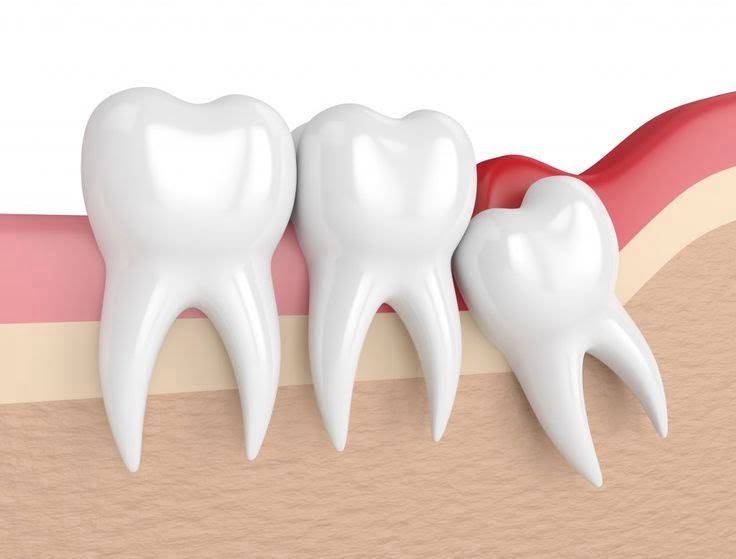
What is an Impacted Tooth?Impacted tooth; despite the time for eruption (breaking through), it is a clinical term that describes teeth that cannot reach the oral cavity due to reasons such as space limitations in the jawbone, genetic factors, or the pressure from adjacent teeth, and are wholly or partially retained beneath the gum mucosa or bone.Human jaw anatomy has shown a trend of reduction through the evolutionary process. With the shrinking of the jaws, especially the last molars in the mouth struggle to find space. A tooth that cannot find adequate room may accidentally invade the root of a neighboring tooth while searching for an exit path, may remain in a horizontal position within the bone, or may fail to penetrate thick gum tissue. This situation is commonly observed not only in the 20-year molars but also in canine teeth or small premolars. These teeth, which do not fully reach the oral environment, can over time create a foundation for medical problems such as inflammation in the surrounding tissues, cyst formation, or destruction of the roots of neighboring teeth.
In the Avrupadent clinics located in İzmir, cases of impacted teeth are evaluated in a multidisciplinary context, taking into account the patient’s overall jaw structure and chewing functions. The condition of these teeth, which are not visible in the mouth but create a subtle pressure underneath, is examined with consideration to the potential damage they may cause to the surrounding tissues. The main focus of the procedures is to preserve the biomechanics of the oral cavity and support the patient’s chewing quality while preventing more complex anatomical problems that may arise in the future.
In Which Situations Are Impacted Tooth Surgeries Applied?
Conditions Under Which the Application is Realized; while working on the eruption of wisdom teeth, the neighboring teeth can exert pressure causing misalignment, leading to recurrent inflammations in the surrounding gum tissue, creating a ground for cyst or mass formation within the jawbone, and it is medically indicated that space opening for orthodontic (braces) treatment is necessary.Not every partially erupted wisdom tooth causes issues; however, teeth that jeopardize the health of other teeth due to their anatomical position are subjects of surgical procedures. The flap of gum tissue covering partially erupted teeth creates a suitable pocket for food debris to accumulate. This situation cannot be cleaned with a toothbrush and leads to severe localized infections known as “pericoronitis.” The main conditions in which an intervention decision is made in the clinic are as follows:
How is the Impacted Tooth Surgery Performed?
The structure of the application; the anesthetic solution is applied to the surgical area to numb it, the mucosa surrounding the tooth is incised if necessary, the bone tissue above the tooth is removed with small drills, to minimize bone loss, the tooth is divided into pieces for extraction from its socket, and the area is closed with stitches.The operational process begins with the local anesthesia application, focusing on the clinical comfort of the patient. After the area is numbed, the doctor makes a small incision in the gum and exposes the bone surface. The intervention strategy is determined based on the anatomical position of the impacted tooth. If the tooth is located beneath a thick layer of bone, special surgical instruments or devices that operate with sound waves (piezosurgery) are used to carefully thin that bone and access the tooth.
To keep tissue trauma to a minimum, an impacted tooth that is large and horizontally positioned should not be attempted to be removed in one piece. The tooth is sectioned into two or three pieces using appropriate axes. This division process allows the tooth pieces to be easily extracted without damaging the surrounding bone or adjacent tooth roots. Once the remnants within the socket are cleaned, the gum tissue is adapted back to its original position and sutured with surgical threads, moving into the body’s cellular repair phase.
Who Might Be Suitable for Impacted Tooth Surgery?
This surgery; is generally indicated for individuals, usually young adults and adults, who can tolerate local surgical interventions and are experiencing pain, infection, or anatomical risks related to impacted teeth.Complaints regarding impacted teeth become pronounced mainly between the ages of 17 and 25, as these are the years when the 20-year molars are trying to erupt. However, as one ages, a cystic impacted tooth waiting silently in the bone may become active and require intervention. Therefore, there is no strict age limitation for the applicability of the procedure; what is important is the progression of the pathology in the jawbone. In older ages, extraction processes may require more intensive surgical attention due to decreased bone flexibility compared to younger individuals.
The patient’s overall systemic health status is a key determinant in this process. For individuals with diabetes, heart valve disorders, or bleeding disorders, the aim is to align clinical values with ideal ranges based on consultations with the patient’s own physician. If medical data remains within appropriate levels, surgical planning is initiated.
How is an Evaluation Made Before Impacted Tooth Surgery?Clinical evaluation phase; involves questioning the individual about their medical conditions and medications, detailed assessment of the distance of the tooth’s position to anatomical spaces (sinus or inferior alveolar nerve) using three-dimensional radiographs, and determining the degree of operational difficulty.The intervention strategy is entirely based on the diagnostic data obtained. The physician analyzes the shape and number of the roots of the impacted tooth, which are often located very close to the canal of the “mandibular nerve” (nervus alveolaris inferior) that provides sensation to the lower lip and chin. To avoid pressure on this nerve during the operation, safety distances are measured millimetrically through X-rays.
Additionally, if there is already an acute (active) inflammation in the patient’s mouth or swelling on the face, the surgical intervention may be postponed because the effect of anesthesia solutions could be diminished. Controlling the infection with medical medications (antibiotics, etc.) and calming the tissues is an important step in the evaluation phase.
How is the Impacted Tooth Surgery Process Planned?
Process Planning; involves outlining the surgical route for the tooth to be extracted based on radiological findings, selecting a local or sedation anesthesia protocol according to the psychological and physical condition of the patient, and organizing the rest period after the day of the operation.At Izmir Avrupadent, surgical procedures are designed to be the most comfortable and biologically least exhausting for the patient. If the patient has four impacted wisdom teeth in their mouth, it can be decided in mutual agreement with the doctor and the patient that all these teeth can be removed in the same session. Alternatively, to maintain chewing comfort, sequential appointments can be established where the right side is treated first, followed by the left side weeks later.
During planning, the patient will also be pre-informed about the expected application of ice compresses in the first few days after surgery, dietary restrictions, and medication schedules. Since attention will be required in the activities of speaking and swallowing in the days following the procedure, appointment dates will be scheduled during time slots that do not coincide with the hospital’s busy work or school pace.
What Steps Are Followed in Gum Surgery?
The clinical steps applied to support cellular repair and tissue integrity are carried out sequentially while adhering to surgical principles. The roadmap followed in a standard operation is as follows:
\t- • Local Anesthesia: The area surrounding the operation site is numbed by blocking the nerve endings with medical agents.
\t
- • Flap (Tissue) Lifting: A small incision is made in the gum, and the tissue is separated like a cover to reach the underlying bone and tooth structure.
\t
- • Osteotomy and Tooth Sectioning: The bone obstruction above the tooth is removed delicately using fine drills, and the tooth is separated into pieces to enable it to be extracted without applying pressure to the surrounding area.
- • Extraction (Removal): The complete removal of the teeth and any cyst-like structures formed around them (follicular remnants).
- • Suturing (Stitching): After controlling the bleeding of the cleaned cavity, the gum mucosa is sewn back to its former shape.
What Should Be Considered Before Impacted Tooth Surgery?
Elements patients should pay attention to before the procedure: Arriving with clean teeth after brushing and having a clean mouth at the appointment time, being slightly full to prevent blood sugar drops after anesthesia, and using prescribed medications on time if there are any.In surgical interventions, it is crucial to keep the microbial load in the working area as low as possible to prevent infections that may develop in the wound area after the operation. The patient should thoroughly clean the oral flora through detailed rinsing before the operation. Additionally, the use of anticoagulant medications like aspirin, which affect blood clotting, must be reported to the physician, and the doses of these medications should be adjusted according to the doctor’s instructions before the appointment.
Since the procedure will only be performed by numbing that area (locally), the patient will remain conscious. Due to the possibility of high stress and anxiety levels causing fluctuations in blood pressure, arriving at the clinic in a relaxed and rested manner will enhance clinical comfort. Having a good sleep the night before the operation and choosing comfortable clothes will help the time spent in the operating chair feel calmer.
What Should Be Considered After Impacted Tooth Surgery?
After Surgical Intervention; it is important to lightly heat the sterile tampon placed by the physician for about half an hour, not to disturb the blood clot, apply an ice compress on the cheek in the first few days, and avoid the use of substances that may hinder cell regeneration.After impacted tooth surgeries, mild to moderate swelling (edema) in the cheek area and minimal oozing inside the mouth is a natural response of cellular repair. Applying cold compress intermittently for the first 24 hours significantly limits this swelling. Chewing, drinking liquids through a straw, or vigorously rinsing the mouth can displace the protective blood clot formed at the extraction site due to vacuum effect and may lead to rebleeding.
The nutrition routine should consist of liquids and soft foods (like soup, yogurt, puree, etc.) that won’t traumatize the surgical area. Acidic, overly hot, or spicy foods should be avoided for a few days as they can irritate the fresh wound surface. Taking prescribed analgesics and antibiotics, if any, at the specified intervals greatly assists in managing the healing process calmly.
What Symptoms Can Impacted Teeth Cause?
Impacted teeth, which silently wait inside the bone, may sometimes cause no complaints, but when they try to erupt or when bacteria accumulate around them, they exhibit various irritating symptoms. Commonly encountered symptoms include:
Are 20-Year-Old Impacted Teeth Considered in Dental Operations?
Yes, 20-year-old teeth (wisdom teeth) form the largest and primary group of impacted tooth operations. The reduction of jawbone size in human evolution has led to these third molars, which are the last to emerge in the mouth, not finding sufficient space for themselves and remaining impacted within the bone.If the erupted (normally positioned in the oral cavity) wisdom teeth are functionally participating in chewing and not causing problems in surrounding tissues, they can be retained in the mouth like a standard tooth. However, in a significant portion of cases, these teeth become trapped in the bone in a horizontal, inverted, or impacted manner. These positional anomalies can cause the tooth to erode the surrounding bone and lead to the neighboring second molar’s decay.
Additionally, in patients who have received orthodontic (braces) treatment or will undergo it, the surgical removal of these teeth is often applied as a medical strategy to gain space in the jaw and prevent the disruption of the achieved alignment (occlusion) by the push of the wisdom teeth.
What Diagnosis and Imaging Methods Are Used in Impacted Teeth Treatment?
In the diagnosis and imaging phase; panoramic X-rays that show the general anatomy within the jawbone and three-dimensional dental volumetric tomography (CBCT) that allows examination of the distance to the lower jaw nerve or the upper jaw sinus cavity with millimetric sections are used.Visual examination of the oral cavity can only give an idea if just a very small tip of the tooth hiding beneath the bone has appeared. The anatomy, number, and orientation of the roots cannot be understood from the outside. To eliminate this uncertainty, the first step is to take two-dimensional panoramic films. This film provides the physician with a general map regarding the position of the tooth in the jaw and its relation to other teeth.
However, the roots of lower wisdom teeth may be very close to the main nerve canal (mandibular canal) that carries the sensation of the lip. When it appears that the root overlaps the nerve in two-dimensional films, the physician requests three-dimensional tomography to detail the risk analysis. Tomography precisely determines whether the tooth passes in front of or behind the nerve, and the operation plan is devised millimetrically based on this safety data.
How Does the Healing Process Progress After Impacted Tooth Surgery?
Healing process; the union of the incisions in the gum tissue occurs within the first 7 to 10 days (stitching phase), and the biological cycle of the area where the tooth was extracted is completed when the bone defect in that area fills with a blood clot, gradually returning to the patient’s own healthy jawbone over the course of months.The mucosa (gum) tissue is one of the body’s highest vascularized and cell renewal structures. Thus, the superficial wound healing in the operation area proceeds quite rapidly, allowing the patient to return to daily nutritional functions within a few days. During the initial days, experiencing slight restriction or sensitivity while swallowing when the mouth is open is a temporary reaction to the surgical intervention by the muscles and decreases quickly.
However, a deeper and longer-lasting activity begins in the underlying layers of the tissue, that is, in the bone socket where the tooth was extracted. The blood clot that fills the void gradually organizes into connective tissue and eventually transforms into hard bone tissue through the activity of bone-forming cells (osteoblasts). Although this internal healing (ossification) is not felt from the outside, it requires an average maturation period of 3 to 6 months anatomically.
How Long Can a Tooth Extraction Surgery Last?Duration of the surgery; depends on the depth of the tooth to be extracted within the bone, the anatomical curvature of its roots, the density of the surrounding bone tissue, and its position relative to nearby nerves, generally lasting between 20 minutes to 1 hour.In dental surgery, each case presents a unique anatomy. The bone structure of the upper jaw is softer compared to the lower jaw, making the procedures for upper impacted teeth usually quicker and easier than for the lower jaw. However, the extraction of a wisdom tooth that is lying horizontally, with roots curled like a hook and hidden beneath a dense layer of bone in the lower jaw, will take time as it may require breaking the tooth into many pieces.
Another factor that determines the duration is the surgeon’s protective (minimally invasive) approach to the surrounding tissues. The procedures are carried out with extremely gentle and slow hand movements to avoid excessive disruption to the surrounding bone and pressure on the nerve pathways. Since the patient is under anesthesia, they experience the process without any clinical discomfort or fatigue during this time.
How Should Oral Care Be Done After a Wisdom Tooth Extraction?Post-operative oral care; it involves avoiding hard brushing strokes on the wound area where the stitches are located in the first days, gently rinsing the area with only the antiseptic mouthwash recommended by the doctor, and refraining from brushing the other parts of the mouth normally.A delicate blood clot is formed to ensure the cessation of bleeding in the wound area after the surgical procedure. Using hard brushing or pressurized oral rinses can mechanically dislodge this clot and may lead to a painful infection condition known as “alveolitis,” which can hinder healing. Therefore, hygiene in the operated area should only be maintained with chemical cleaning (chlorhexidine-based mouthwashes) until the doctor says otherwise.
When gargling, it is crucial to gently circulate the liquid around the wound area and softly let it flow into the sink (without forcibly spitting), rather than vigorously agitation of the liquid in the mouth as if inflating a balloon. After the stitches are removed and the mucosa has healed, gradually returning to the standard oral care routine can be ensured by massaging the area with extra soft surgical toothbrushes.
When Should Impacted Teeth Be Monitored?
Indications for Monitoring; asymptomatic (without symptoms) scenarios where the impacted tooth is found completely isolated deep within the jawbone, with no surrounding cystic or infectious pathology observed, and where there is no mechanical pressure on the roots of adjacent teeth.There is no strict rule that every impacted tooth must be removed. Wisdom teeth that are completely buried and have not approached the gum surface at all, which have remained dormant in the deepest layers of the bone for years without causing any discomfort to the patient, and where cellular damage to surrounding tissues is not detected in the radiological examinations can be monitored with a ‘wait and see’ strategy by doctors through routine check-ups.
Efforts to remove these types of teeth through surgery may lead to structural trauma (excessive bone removal or too close proximity to nerve tissue) that outweighs the potential benefits; therefore, physicians perform a risk-benefit analysis. If in later years, panoramic images show that the follicle (sac) around the tooth has begun to enlarge and develop cystically, it will be transitioned from monitoring status to surgical intervention phase.
Why is a control examination important after a dental implant operation?
The clinical necessity of control examinations; it involves confirming that the surgical stitches placed during the operation are intact, preventing bacterial and food accumulation in the area, monitoring the closure (epithelization) performance of the wound edges, and ensuring that no secondary infection has developed in the surgical site as verified by the physician.This control session performed generally in the first or second week after the intervention is to confirm that the healing period is progressing correctly. When surgical threads remain in the oral environment longer than the designated period, they can create an ideal retention surface for food debris, leading to local irritations (inflammations). The physician ensures the relaxation of the gum tissue and its return to its natural form by removing these threads with special instruments.
Moreover, if the patient indicates that their pain is increasing after the operation or has a bad taste in their mouth, the physician will examine the wound area to check for the formation of a condition called alveolitis, which refers to the loss of the clot. If necessary, the wound can be rinsed with medical agents and dressed to support the healing process medically.
Can Impacted Tooth Surgery Be Planned Alongside Other Jaw and Dental Treatments?
Yes, it is considered as part of multidisciplinary approaches. It can be performed in an integrated manner, either by creating space in the bone before orthodontic alignment or by extracting cysts and placing artificial roots (implants) during jaw surgery sessions.Dentistry practices often form a substrate for each other. Especially in orthodontic treatments where braces are used to straighten crooked teeth, if there is a high risk that the previously corrected alignment may be disrupted by the emergence of wisdom teeth later on, dentists may request the surgical removal of these impacted teeth as a precaution before braces are installed. Additionally, instead of completely extracting impacted canine teeth remaining in the bone, they can be surgically exposed and a small bracket can be placed to guide them into the mouth (to the correct position) with orthodontics.
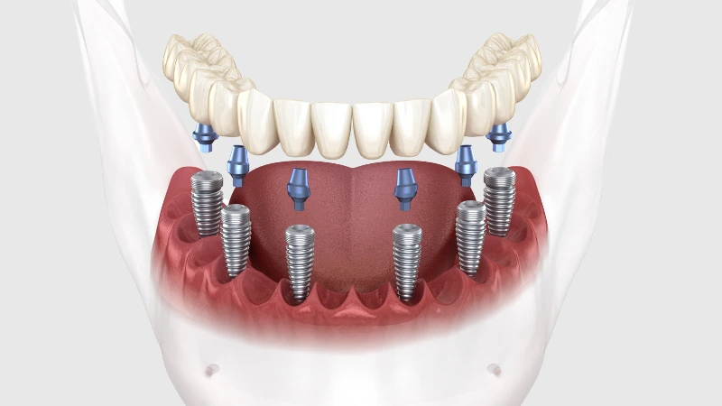
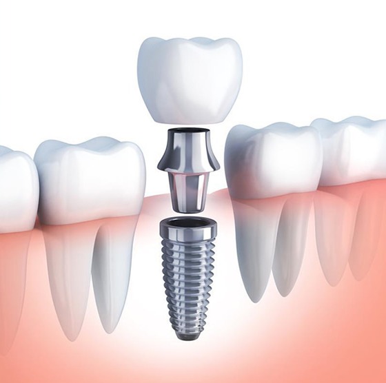
Combined sessions are also organized for patients who will undergo implant treatment and have an infected impacted tooth in the same jaw. While the doctor places a titanium root in one area, they also extract the problematic wisdom tooth under anesthesia within the same surgical session, protecting the patient from the stress of two separate surgeries.
How is a Personalized Treatment Plan Made in Impacted Tooth Surgeries?
Personalized design; the anatomical angle of the tooth in the jawbone (horizontal, mesioangular, distoangular), the shape of the root curvatures, and the millimetric distance to the inferior alveolar nerve are analyzed with three-dimensional scanning data, resulting in a cutting angle and tooth section map tailored specifically to that patient’s anatomy.The jaw structure and the shape of dental roots of every person are as unique as fingerprints. In one patient, the tooth may be in an upright position and easily accessible, while in another patient it may be completely horizontal, resting against the root of an adjacent tooth and covered by thick bone tissue. The physician anticipates this situation in advance and establishes an individualized strategy that determines the surgical instruments to be used and how the tooth will be sectioned from various angles.
In the operation planning carried out at the branches of Izmir Avrupadent, especially in cases close to nerves, the limits of where surgical instruments should not pass are measured through virtual tomography slices. By blending the individual’s medical condition, bone flexibility, and psychological profile, the most ideal clinical roadmap is designed that will create minimal tissue loss (minimally invasive) and speed up recovery after the operation.

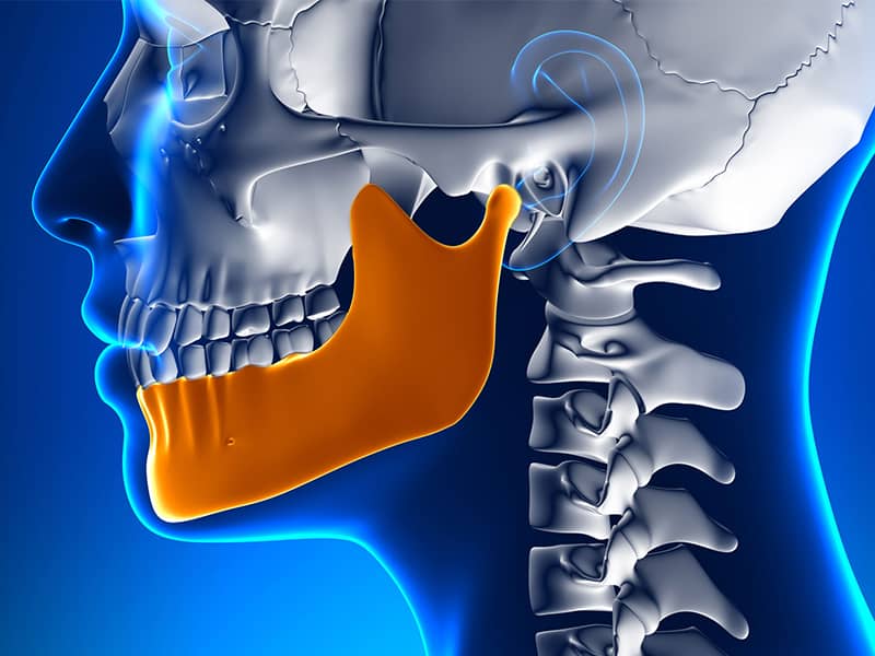
Human jaw anatomy has shown a trend of reduction through the evolutionary process. With the shrinking of the jaws, especially the last molars in the mouth struggle to find space. A tooth that cannot find adequate room may accidentally invade the root of a neighboring tooth while searching for an exit path, may remain in a horizontal position within the bone, or may fail to penetrate thick gum tissue. This situation is commonly observed not only in the 20-year molars but also in canine teeth or small premolars. These teeth, which do not fully reach the oral environment, can over time create a foundation for medical problems such as inflammation in the surrounding tissues, cyst formation, or destruction of the roots of neighboring teeth.
In the Avrupadent clinics located in İzmir, cases of impacted teeth are evaluated in a multidisciplinary context, taking into account the patient’s overall jaw structure and chewing functions. The condition of these teeth, which are not visible in the mouth but create a subtle pressure underneath, is examined with consideration to the potential damage they may cause to the surrounding tissues. The main focus of the procedures is to preserve the biomechanics of the oral cavity and support the patient’s chewing quality while preventing more complex anatomical problems that may arise in the future.
In Which Situations Are Impacted Tooth Surgeries Applied?
Not every partially erupted wisdom tooth causes issues; however, teeth that jeopardize the health of other teeth due to their anatomical position are subjects of surgical procedures. The flap of gum tissue covering partially erupted teeth creates a suitable pocket for food debris to accumulate. This situation cannot be cleaned with a toothbrush and leads to severe localized infections known as “pericoronitis.” The main conditions in which an intervention decision is made in the clinic are as follows:
How is the Impacted Tooth Surgery Performed?
The operational process begins with the local anesthesia application, focusing on the clinical comfort of the patient. After the area is numbed, the doctor makes a small incision in the gum and exposes the bone surface. The intervention strategy is determined based on the anatomical position of the impacted tooth. If the tooth is located beneath a thick layer of bone, special surgical instruments or devices that operate with sound waves (piezosurgery) are used to carefully thin that bone and access the tooth.
To keep tissue trauma to a minimum, an impacted tooth that is large and horizontally positioned should not be attempted to be removed in one piece. The tooth is sectioned into two or three pieces using appropriate axes. This division process allows the tooth pieces to be easily extracted without damaging the surrounding bone or adjacent tooth roots. Once the remnants within the socket are cleaned, the gum tissue is adapted back to its original position and sutured with surgical threads, moving into the body’s cellular repair phase.
Who Might Be Suitable for Impacted Tooth Surgery?
Complaints regarding impacted teeth become pronounced mainly between the ages of 17 and 25, as these are the years when the 20-year molars are trying to erupt. However, as one ages, a cystic impacted tooth waiting silently in the bone may become active and require intervention. Therefore, there is no strict age limitation for the applicability of the procedure; what is important is the progression of the pathology in the jawbone. In older ages, extraction processes may require more intensive surgical attention due to decreased bone flexibility compared to younger individuals.
The patient’s overall systemic health status is a key determinant in this process. For individuals with diabetes, heart valve disorders, or bleeding disorders, the aim is to align clinical values with ideal ranges based on consultations with the patient’s own physician. If medical data remains within appropriate levels, surgical planning is initiated.
How is an Evaluation Made Before Impacted Tooth Surgery?Clinical evaluation phase; involves questioning the individual about their medical conditions and medications, detailed assessment of the distance of the tooth’s position to anatomical spaces (sinus or inferior alveolar nerve) using three-dimensional radiographs, and determining the degree of operational difficulty.The intervention strategy is entirely based on the diagnostic data obtained. The physician analyzes the shape and number of the roots of the impacted tooth, which are often located very close to the canal of the “mandibular nerve” (nervus alveolaris inferior) that provides sensation to the lower lip and chin. To avoid pressure on this nerve during the operation, safety distances are measured millimetrically through X-rays.
Additionally, if there is already an acute (active) inflammation in the patient’s mouth or swelling on the face, the surgical intervention may be postponed because the effect of anesthesia solutions could be diminished. Controlling the infection with medical medications (antibiotics, etc.) and calming the tissues is an important step in the evaluation phase.
How is the Impacted Tooth Surgery Process Planned?
Process Planning; involves outlining the surgical route for the tooth to be extracted based on radiological findings, selecting a local or sedation anesthesia protocol according to the psychological and physical condition of the patient, and organizing the rest period after the day of the operation.At Izmir Avrupadent, surgical procedures are designed to be the most comfortable and biologically least exhausting for the patient. If the patient has four impacted wisdom teeth in their mouth, it can be decided in mutual agreement with the doctor and the patient that all these teeth can be removed in the same session. Alternatively, to maintain chewing comfort, sequential appointments can be established where the right side is treated first, followed by the left side weeks later.
During planning, the patient will also be pre-informed about the expected application of ice compresses in the first few days after surgery, dietary restrictions, and medication schedules. Since attention will be required in the activities of speaking and swallowing in the days following the procedure, appointment dates will be scheduled during time slots that do not coincide with the hospital’s busy work or school pace.
What Steps Are Followed in Gum Surgery?
The clinical steps applied to support cellular repair and tissue integrity are carried out sequentially while adhering to surgical principles. The roadmap followed in a standard operation is as follows:
\t- • Local Anesthesia: The area surrounding the operation site is numbed by blocking the nerve endings with medical agents.
\t
- • Flap (Tissue) Lifting: A small incision is made in the gum, and the tissue is separated like a cover to reach the underlying bone and tooth structure.
\t
- • Osteotomy and Tooth Sectioning: The bone obstruction above the tooth is removed delicately using fine drills, and the tooth is separated into pieces to enable it to be extracted without applying pressure to the surrounding area.
- • Extraction (Removal): The complete removal of the teeth and any cyst-like structures formed around them (follicular remnants).
- • Suturing (Stitching): After controlling the bleeding of the cleaned cavity, the gum mucosa is sewn back to its former shape.
What Should Be Considered Before Impacted Tooth Surgery?
Elements patients should pay attention to before the procedure: Arriving with clean teeth after brushing and having a clean mouth at the appointment time, being slightly full to prevent blood sugar drops after anesthesia, and using prescribed medications on time if there are any.In surgical interventions, it is crucial to keep the microbial load in the working area as low as possible to prevent infections that may develop in the wound area after the operation. The patient should thoroughly clean the oral flora through detailed rinsing before the operation. Additionally, the use of anticoagulant medications like aspirin, which affect blood clotting, must be reported to the physician, and the doses of these medications should be adjusted according to the doctor’s instructions before the appointment.
Since the procedure will only be performed by numbing that area (locally), the patient will remain conscious. Due to the possibility of high stress and anxiety levels causing fluctuations in blood pressure, arriving at the clinic in a relaxed and rested manner will enhance clinical comfort. Having a good sleep the night before the operation and choosing comfortable clothes will help the time spent in the operating chair feel calmer.
What Should Be Considered After Impacted Tooth Surgery?
After Surgical Intervention; it is important to lightly heat the sterile tampon placed by the physician for about half an hour, not to disturb the blood clot, apply an ice compress on the cheek in the first few days, and avoid the use of substances that may hinder cell regeneration.After impacted tooth surgeries, mild to moderate swelling (edema) in the cheek area and minimal oozing inside the mouth is a natural response of cellular repair. Applying cold compress intermittently for the first 24 hours significantly limits this swelling. Chewing, drinking liquids through a straw, or vigorously rinsing the mouth can displace the protective blood clot formed at the extraction site due to vacuum effect and may lead to rebleeding.
The nutrition routine should consist of liquids and soft foods (like soup, yogurt, puree, etc.) that won’t traumatize the surgical area. Acidic, overly hot, or spicy foods should be avoided for a few days as they can irritate the fresh wound surface. Taking prescribed analgesics and antibiotics, if any, at the specified intervals greatly assists in managing the healing process calmly.
What Symptoms Can Impacted Teeth Cause?
Impacted teeth, which silently wait inside the bone, may sometimes cause no complaints, but when they try to erupt or when bacteria accumulate around them, they exhibit various irritating symptoms. Commonly encountered symptoms include:
Are 20-Year-Old Impacted Teeth Considered in Dental Operations?
Yes, 20-year-old teeth (wisdom teeth) form the largest and primary group of impacted tooth operations. The reduction of jawbone size in human evolution has led to these third molars, which are the last to emerge in the mouth, not finding sufficient space for themselves and remaining impacted within the bone.If the erupted (normally positioned in the oral cavity) wisdom teeth are functionally participating in chewing and not causing problems in surrounding tissues, they can be retained in the mouth like a standard tooth. However, in a significant portion of cases, these teeth become trapped in the bone in a horizontal, inverted, or impacted manner. These positional anomalies can cause the tooth to erode the surrounding bone and lead to the neighboring second molar’s decay.
Additionally, in patients who have received orthodontic (braces) treatment or will undergo it, the surgical removal of these teeth is often applied as a medical strategy to gain space in the jaw and prevent the disruption of the achieved alignment (occlusion) by the push of the wisdom teeth.
What Diagnosis and Imaging Methods Are Used in Impacted Teeth Treatment?
In the diagnosis and imaging phase; panoramic X-rays that show the general anatomy within the jawbone and three-dimensional dental volumetric tomography (CBCT) that allows examination of the distance to the lower jaw nerve or the upper jaw sinus cavity with millimetric sections are used.Visual examination of the oral cavity can only give an idea if just a very small tip of the tooth hiding beneath the bone has appeared. The anatomy, number, and orientation of the roots cannot be understood from the outside. To eliminate this uncertainty, the first step is to take two-dimensional panoramic films. This film provides the physician with a general map regarding the position of the tooth in the jaw and its relation to other teeth.
However, the roots of lower wisdom teeth may be very close to the main nerve canal (mandibular canal) that carries the sensation of the lip. When it appears that the root overlaps the nerve in two-dimensional films, the physician requests three-dimensional tomography to detail the risk analysis. Tomography precisely determines whether the tooth passes in front of or behind the nerve, and the operation plan is devised millimetrically based on this safety data.
How Does the Healing Process Progress After Impacted Tooth Surgery?
Healing process; the union of the incisions in the gum tissue occurs within the first 7 to 10 days (stitching phase), and the biological cycle of the area where the tooth was extracted is completed when the bone defect in that area fills with a blood clot, gradually returning to the patient’s own healthy jawbone over the course of months.The mucosa (gum) tissue is one of the body’s highest vascularized and cell renewal structures. Thus, the superficial wound healing in the operation area proceeds quite rapidly, allowing the patient to return to daily nutritional functions within a few days. During the initial days, experiencing slight restriction or sensitivity while swallowing when the mouth is open is a temporary reaction to the surgical intervention by the muscles and decreases quickly.
However, a deeper and longer-lasting activity begins in the underlying layers of the tissue, that is, in the bone socket where the tooth was extracted. The blood clot that fills the void gradually organizes into connective tissue and eventually transforms into hard bone tissue through the activity of bone-forming cells (osteoblasts). Although this internal healing (ossification) is not felt from the outside, it requires an average maturation period of 3 to 6 months anatomically.
How Long Can a Tooth Extraction Surgery Last?Duration of the surgery; depends on the depth of the tooth to be extracted within the bone, the anatomical curvature of its roots, the density of the surrounding bone tissue, and its position relative to nearby nerves, generally lasting between 20 minutes to 1 hour.In dental surgery, each case presents a unique anatomy. The bone structure of the upper jaw is softer compared to the lower jaw, making the procedures for upper impacted teeth usually quicker and easier than for the lower jaw. However, the extraction of a wisdom tooth that is lying horizontally, with roots curled like a hook and hidden beneath a dense layer of bone in the lower jaw, will take time as it may require breaking the tooth into many pieces.
Another factor that determines the duration is the surgeon’s protective (minimally invasive) approach to the surrounding tissues. The procedures are carried out with extremely gentle and slow hand movements to avoid excessive disruption to the surrounding bone and pressure on the nerve pathways. Since the patient is under anesthesia, they experience the process without any clinical discomfort or fatigue during this time.
How Should Oral Care Be Done After a Wisdom Tooth Extraction?Post-operative oral care; it involves avoiding hard brushing strokes on the wound area where the stitches are located in the first days, gently rinsing the area with only the antiseptic mouthwash recommended by the doctor, and refraining from brushing the other parts of the mouth normally.A delicate blood clot is formed to ensure the cessation of bleeding in the wound area after the surgical procedure. Using hard brushing or pressurized oral rinses can mechanically dislodge this clot and may lead to a painful infection condition known as “alveolitis,” which can hinder healing. Therefore, hygiene in the operated area should only be maintained with chemical cleaning (chlorhexidine-based mouthwashes) until the doctor says otherwise.
When gargling, it is crucial to gently circulate the liquid around the wound area and softly let it flow into the sink (without forcibly spitting), rather than vigorously agitation of the liquid in the mouth as if inflating a balloon. After the stitches are removed and the mucosa has healed, gradually returning to the standard oral care routine can be ensured by massaging the area with extra soft surgical toothbrushes.
When Should Impacted Teeth Be Monitored?
Indications for Monitoring; asymptomatic (without symptoms) scenarios where the impacted tooth is found completely isolated deep within the jawbone, with no surrounding cystic or infectious pathology observed, and where there is no mechanical pressure on the roots of adjacent teeth.There is no strict rule that every impacted tooth must be removed. Wisdom teeth that are completely buried and have not approached the gum surface at all, which have remained dormant in the deepest layers of the bone for years without causing any discomfort to the patient, and where cellular damage to surrounding tissues is not detected in the radiological examinations can be monitored with a ‘wait and see’ strategy by doctors through routine check-ups.
Efforts to remove these types of teeth through surgery may lead to structural trauma (excessive bone removal or too close proximity to nerve tissue) that outweighs the potential benefits; therefore, physicians perform a risk-benefit analysis. If in later years, panoramic images show that the follicle (sac) around the tooth has begun to enlarge and develop cystically, it will be transitioned from monitoring status to surgical intervention phase.
Why is a control examination important after a dental implant operation?
The clinical necessity of control examinations; it involves confirming that the surgical stitches placed during the operation are intact, preventing bacterial and food accumulation in the area, monitoring the closure (epithelization) performance of the wound edges, and ensuring that no secondary infection has developed in the surgical site as verified by the physician.This control session performed generally in the first or second week after the intervention is to confirm that the healing period is progressing correctly. When surgical threads remain in the oral environment longer than the designated period, they can create an ideal retention surface for food debris, leading to local irritations (inflammations). The physician ensures the relaxation of the gum tissue and its return to its natural form by removing these threads with special instruments.
Moreover, if the patient indicates that their pain is increasing after the operation or has a bad taste in their mouth, the physician will examine the wound area to check for the formation of a condition called alveolitis, which refers to the loss of the clot. If necessary, the wound can be rinsed with medical agents and dressed to support the healing process medically.
Can Impacted Tooth Surgery Be Planned Alongside Other Jaw and Dental Treatments?
Yes, it is considered as part of multidisciplinary approaches. It can be performed in an integrated manner, either by creating space in the bone before orthodontic alignment or by extracting cysts and placing artificial roots (implants) during jaw surgery sessions.Dentistry practices often form a substrate for each other. Especially in orthodontic treatments where braces are used to straighten crooked teeth, if there is a high risk that the previously corrected alignment may be disrupted by the emergence of wisdom teeth later on, dentists may request the surgical removal of these impacted teeth as a precaution before braces are installed. Additionally, instead of completely extracting impacted canine teeth remaining in the bone, they can be surgically exposed and a small bracket can be placed to guide them into the mouth (to the correct position) with orthodontics.
Combined sessions are also organized for patients who will undergo implant treatment and have an infected impacted tooth in the same jaw. While the doctor places a titanium root in one area, they also extract the problematic wisdom tooth under anesthesia within the same surgical session, protecting the patient from the stress of two separate surgeries.
How is a Personalized Treatment Plan Made in Impacted Tooth Surgeries?
Personalized design; the anatomical angle of the tooth in the jawbone (horizontal, mesioangular, distoangular), the shape of the root curvatures, and the millimetric distance to the inferior alveolar nerve are analyzed with three-dimensional scanning data, resulting in a cutting angle and tooth section map tailored specifically to that patient’s anatomy.The jaw structure and the shape of dental roots of every person are as unique as fingerprints. In one patient, the tooth may be in an upright position and easily accessible, while in another patient it may be completely horizontal, resting against the root of an adjacent tooth and covered by thick bone tissue. The physician anticipates this situation in advance and establishes an individualized strategy that determines the surgical instruments to be used and how the tooth will be sectioned from various angles.
In the operation planning carried out at the branches of Izmir Avrupadent, especially in cases close to nerves, the limits of where surgical instruments should not pass are measured through virtual tomography slices. By blending the individual’s medical condition, bone flexibility, and psychological profile, the most ideal clinical roadmap is designed that will create minimal tissue loss (minimally invasive) and speed up recovery after the operation.
The intervention strategy is entirely based on the diagnostic data obtained. The physician analyzes the shape and number of the roots of the impacted tooth, which are often located very close to the canal of the “mandibular nerve” (nervus alveolaris inferior) that provides sensation to the lower lip and chin. To avoid pressure on this nerve during the operation, safety distances are measured millimetrically through X-rays.
Additionally, if there is already an acute (active) inflammation in the patient’s mouth or swelling on the face, the surgical intervention may be postponed because the effect of anesthesia solutions could be diminished. Controlling the infection with medical medications (antibiotics, etc.) and calming the tissues is an important step in the evaluation phase.
How is the Impacted Tooth Surgery Process Planned?
At Izmir Avrupadent, surgical procedures are designed to be the most comfortable and biologically least exhausting for the patient. If the patient has four impacted wisdom teeth in their mouth, it can be decided in mutual agreement with the doctor and the patient that all these teeth can be removed in the same session. Alternatively, to maintain chewing comfort, sequential appointments can be established where the right side is treated first, followed by the left side weeks later.
During planning, the patient will also be pre-informed about the expected application of ice compresses in the first few days after surgery, dietary restrictions, and medication schedules. Since attention will be required in the activities of speaking and swallowing in the days following the procedure, appointment dates will be scheduled during time slots that do not coincide with the hospital’s busy work or school pace.
What Steps Are Followed in Gum Surgery?
The clinical steps applied to support cellular repair and tissue integrity are carried out sequentially while adhering to surgical principles. The roadmap followed in a standard operation is as follows:
-
\t
- • Local Anesthesia: The area surrounding the operation site is numbed by blocking the nerve endings with medical agents.
- • Flap (Tissue) Lifting: A small incision is made in the gum, and the tissue is separated like a cover to reach the underlying bone and tooth structure.
- • Osteotomy and Tooth Sectioning: The bone obstruction above the tooth is removed delicately using fine drills, and the tooth is separated into pieces to enable it to be extracted without applying pressure to the surrounding area.
- • Extraction (Removal): The complete removal of the teeth and any cyst-like structures formed around them (follicular remnants).
- • Suturing (Stitching): After controlling the bleeding of the cleaned cavity, the gum mucosa is sewn back to its former shape.
\t
\t
What Should Be Considered Before Impacted Tooth Surgery?
In surgical interventions, it is crucial to keep the microbial load in the working area as low as possible to prevent infections that may develop in the wound area after the operation. The patient should thoroughly clean the oral flora through detailed rinsing before the operation. Additionally, the use of anticoagulant medications like aspirin, which affect blood clotting, must be reported to the physician, and the doses of these medications should be adjusted according to the doctor’s instructions before the appointment.
Since the procedure will only be performed by numbing that area (locally), the patient will remain conscious. Due to the possibility of high stress and anxiety levels causing fluctuations in blood pressure, arriving at the clinic in a relaxed and rested manner will enhance clinical comfort. Having a good sleep the night before the operation and choosing comfortable clothes will help the time spent in the operating chair feel calmer.
What Should Be Considered After Impacted Tooth Surgery?
After impacted tooth surgeries, mild to moderate swelling (edema) in the cheek area and minimal oozing inside the mouth is a natural response of cellular repair. Applying cold compress intermittently for the first 24 hours significantly limits this swelling. Chewing, drinking liquids through a straw, or vigorously rinsing the mouth can displace the protective blood clot formed at the extraction site due to vacuum effect and may lead to rebleeding.
The nutrition routine should consist of liquids and soft foods (like soup, yogurt, puree, etc.) that won’t traumatize the surgical area. Acidic, overly hot, or spicy foods should be avoided for a few days as they can irritate the fresh wound surface. Taking prescribed analgesics and antibiotics, if any, at the specified intervals greatly assists in managing the healing process calmly.
What Symptoms Can Impacted Teeth Cause?
Impacted teeth, which silently wait inside the bone, may sometimes cause no complaints, but when they try to erupt or when bacteria accumulate around them, they exhibit various irritating symptoms. Commonly encountered symptoms include:
Are 20-Year-Old Impacted Teeth Considered in Dental Operations?
If the erupted (normally positioned in the oral cavity) wisdom teeth are functionally participating in chewing and not causing problems in surrounding tissues, they can be retained in the mouth like a standard tooth. However, in a significant portion of cases, these teeth become trapped in the bone in a horizontal, inverted, or impacted manner. These positional anomalies can cause the tooth to erode the surrounding bone and lead to the neighboring second molar’s decay.
Additionally, in patients who have received orthodontic (braces) treatment or will undergo it, the surgical removal of these teeth is often applied as a medical strategy to gain space in the jaw and prevent the disruption of the achieved alignment (occlusion) by the push of the wisdom teeth.
What Diagnosis and Imaging Methods Are Used in Impacted Teeth Treatment?
Visual examination of the oral cavity can only give an idea if just a very small tip of the tooth hiding beneath the bone has appeared. The anatomy, number, and orientation of the roots cannot be understood from the outside. To eliminate this uncertainty, the first step is to take two-dimensional panoramic films. This film provides the physician with a general map regarding the position of the tooth in the jaw and its relation to other teeth.
However, the roots of lower wisdom teeth may be very close to the main nerve canal (mandibular canal) that carries the sensation of the lip. When it appears that the root overlaps the nerve in two-dimensional films, the physician requests three-dimensional tomography to detail the risk analysis. Tomography precisely determines whether the tooth passes in front of or behind the nerve, and the operation plan is devised millimetrically based on this safety data.
How Does the Healing Process Progress After Impacted Tooth Surgery?
The mucosa (gum) tissue is one of the body’s highest vascularized and cell renewal structures. Thus, the superficial wound healing in the operation area proceeds quite rapidly, allowing the patient to return to daily nutritional functions within a few days. During the initial days, experiencing slight restriction or sensitivity while swallowing when the mouth is open is a temporary reaction to the surgical intervention by the muscles and decreases quickly.
However, a deeper and longer-lasting activity begins in the underlying layers of the tissue, that is, in the bone socket where the tooth was extracted. The blood clot that fills the void gradually organizes into connective tissue and eventually transforms into hard bone tissue through the activity of bone-forming cells (osteoblasts). Although this internal healing (ossification) is not felt from the outside, it requires an average maturation period of 3 to 6 months anatomically.
How Long Can a Tooth Extraction Surgery Last?Duration of the surgery; depends on the depth of the tooth to be extracted within the bone, the anatomical curvature of its roots, the density of the surrounding bone tissue, and its position relative to nearby nerves, generally lasting between 20 minutes to 1 hour.In dental surgery, each case presents a unique anatomy. The bone structure of the upper jaw is softer compared to the lower jaw, making the procedures for upper impacted teeth usually quicker and easier than for the lower jaw. However, the extraction of a wisdom tooth that is lying horizontally, with roots curled like a hook and hidden beneath a dense layer of bone in the lower jaw, will take time as it may require breaking the tooth into many pieces.
Another factor that determines the duration is the surgeon’s protective (minimally invasive) approach to the surrounding tissues. The procedures are carried out with extremely gentle and slow hand movements to avoid excessive disruption to the surrounding bone and pressure on the nerve pathways. Since the patient is under anesthesia, they experience the process without any clinical discomfort or fatigue during this time.
How Should Oral Care Be Done After a Wisdom Tooth Extraction?Post-operative oral care; it involves avoiding hard brushing strokes on the wound area where the stitches are located in the first days, gently rinsing the area with only the antiseptic mouthwash recommended by the doctor, and refraining from brushing the other parts of the mouth normally.A delicate blood clot is formed to ensure the cessation of bleeding in the wound area after the surgical procedure. Using hard brushing or pressurized oral rinses can mechanically dislodge this clot and may lead to a painful infection condition known as “alveolitis,” which can hinder healing. Therefore, hygiene in the operated area should only be maintained with chemical cleaning (chlorhexidine-based mouthwashes) until the doctor says otherwise.
When gargling, it is crucial to gently circulate the liquid around the wound area and softly let it flow into the sink (without forcibly spitting), rather than vigorously agitation of the liquid in the mouth as if inflating a balloon. After the stitches are removed and the mucosa has healed, gradually returning to the standard oral care routine can be ensured by massaging the area with extra soft surgical toothbrushes.
When Should Impacted Teeth Be Monitored?
Indications for Monitoring; asymptomatic (without symptoms) scenarios where the impacted tooth is found completely isolated deep within the jawbone, with no surrounding cystic or infectious pathology observed, and where there is no mechanical pressure on the roots of adjacent teeth.There is no strict rule that every impacted tooth must be removed. Wisdom teeth that are completely buried and have not approached the gum surface at all, which have remained dormant in the deepest layers of the bone for years without causing any discomfort to the patient, and where cellular damage to surrounding tissues is not detected in the radiological examinations can be monitored with a ‘wait and see’ strategy by doctors through routine check-ups.
Efforts to remove these types of teeth through surgery may lead to structural trauma (excessive bone removal or too close proximity to nerve tissue) that outweighs the potential benefits; therefore, physicians perform a risk-benefit analysis. If in later years, panoramic images show that the follicle (sac) around the tooth has begun to enlarge and develop cystically, it will be transitioned from monitoring status to surgical intervention phase.
Why is a control examination important after a dental implant operation?
The clinical necessity of control examinations; it involves confirming that the surgical stitches placed during the operation are intact, preventing bacterial and food accumulation in the area, monitoring the closure (epithelization) performance of the wound edges, and ensuring that no secondary infection has developed in the surgical site as verified by the physician.This control session performed generally in the first or second week after the intervention is to confirm that the healing period is progressing correctly. When surgical threads remain in the oral environment longer than the designated period, they can create an ideal retention surface for food debris, leading to local irritations (inflammations). The physician ensures the relaxation of the gum tissue and its return to its natural form by removing these threads with special instruments.
Moreover, if the patient indicates that their pain is increasing after the operation or has a bad taste in their mouth, the physician will examine the wound area to check for the formation of a condition called alveolitis, which refers to the loss of the clot. If necessary, the wound can be rinsed with medical agents and dressed to support the healing process medically.
Can Impacted Tooth Surgery Be Planned Alongside Other Jaw and Dental Treatments?
Yes, it is considered as part of multidisciplinary approaches. It can be performed in an integrated manner, either by creating space in the bone before orthodontic alignment or by extracting cysts and placing artificial roots (implants) during jaw surgery sessions.Dentistry practices often form a substrate for each other. Especially in orthodontic treatments where braces are used to straighten crooked teeth, if there is a high risk that the previously corrected alignment may be disrupted by the emergence of wisdom teeth later on, dentists may request the surgical removal of these impacted teeth as a precaution before braces are installed. Additionally, instead of completely extracting impacted canine teeth remaining in the bone, they can be surgically exposed and a small bracket can be placed to guide them into the mouth (to the correct position) with orthodontics.
Combined sessions are also organized for patients who will undergo implant treatment and have an infected impacted tooth in the same jaw. While the doctor places a titanium root in one area, they also extract the problematic wisdom tooth under anesthesia within the same surgical session, protecting the patient from the stress of two separate surgeries.
How is a Personalized Treatment Plan Made in Impacted Tooth Surgeries?
Personalized design; the anatomical angle of the tooth in the jawbone (horizontal, mesioangular, distoangular), the shape of the root curvatures, and the millimetric distance to the inferior alveolar nerve are analyzed with three-dimensional scanning data, resulting in a cutting angle and tooth section map tailored specifically to that patient’s anatomy.The jaw structure and the shape of dental roots of every person are as unique as fingerprints. In one patient, the tooth may be in an upright position and easily accessible, while in another patient it may be completely horizontal, resting against the root of an adjacent tooth and covered by thick bone tissue. The physician anticipates this situation in advance and establishes an individualized strategy that determines the surgical instruments to be used and how the tooth will be sectioned from various angles.
In the operation planning carried out at the branches of Izmir Avrupadent, especially in cases close to nerves, the limits of where surgical instruments should not pass are measured through virtual tomography slices. By blending the individual’s medical condition, bone flexibility, and psychological profile, the most ideal clinical roadmap is designed that will create minimal tissue loss (minimally invasive) and speed up recovery after the operation.
In dental surgery, each case presents a unique anatomy. The bone structure of the upper jaw is softer compared to the lower jaw, making the procedures for upper impacted teeth usually quicker and easier than for the lower jaw. However, the extraction of a wisdom tooth that is lying horizontally, with roots curled like a hook and hidden beneath a dense layer of bone in the lower jaw, will take time as it may require breaking the tooth into many pieces.
Another factor that determines the duration is the surgeon’s protective (minimally invasive) approach to the surrounding tissues. The procedures are carried out with extremely gentle and slow hand movements to avoid excessive disruption to the surrounding bone and pressure on the nerve pathways. Since the patient is under anesthesia, they experience the process without any clinical discomfort or fatigue during this time.
How Should Oral Care Be Done After a Wisdom Tooth Extraction?Post-operative oral care; it involves avoiding hard brushing strokes on the wound area where the stitches are located in the first days, gently rinsing the area with only the antiseptic mouthwash recommended by the doctor, and refraining from brushing the other parts of the mouth normally.A delicate blood clot is formed to ensure the cessation of bleeding in the wound area after the surgical procedure. Using hard brushing or pressurized oral rinses can mechanically dislodge this clot and may lead to a painful infection condition known as “alveolitis,” which can hinder healing. Therefore, hygiene in the operated area should only be maintained with chemical cleaning (chlorhexidine-based mouthwashes) until the doctor says otherwise.
When gargling, it is crucial to gently circulate the liquid around the wound area and softly let it flow into the sink (without forcibly spitting), rather than vigorously agitation of the liquid in the mouth as if inflating a balloon. After the stitches are removed and the mucosa has healed, gradually returning to the standard oral care routine can be ensured by massaging the area with extra soft surgical toothbrushes.
When Should Impacted Teeth Be Monitored?
Indications for Monitoring; asymptomatic (without symptoms) scenarios where the impacted tooth is found completely isolated deep within the jawbone, with no surrounding cystic or infectious pathology observed, and where there is no mechanical pressure on the roots of adjacent teeth.There is no strict rule that every impacted tooth must be removed. Wisdom teeth that are completely buried and have not approached the gum surface at all, which have remained dormant in the deepest layers of the bone for years without causing any discomfort to the patient, and where cellular damage to surrounding tissues is not detected in the radiological examinations can be monitored with a ‘wait and see’ strategy by doctors through routine check-ups.
Efforts to remove these types of teeth through surgery may lead to structural trauma (excessive bone removal or too close proximity to nerve tissue) that outweighs the potential benefits; therefore, physicians perform a risk-benefit analysis. If in later years, panoramic images show that the follicle (sac) around the tooth has begun to enlarge and develop cystically, it will be transitioned from monitoring status to surgical intervention phase.
Why is a control examination important after a dental implant operation?
The clinical necessity of control examinations; it involves confirming that the surgical stitches placed during the operation are intact, preventing bacterial and food accumulation in the area, monitoring the closure (epithelization) performance of the wound edges, and ensuring that no secondary infection has developed in the surgical site as verified by the physician.This control session performed generally in the first or second week after the intervention is to confirm that the healing period is progressing correctly. When surgical threads remain in the oral environment longer than the designated period, they can create an ideal retention surface for food debris, leading to local irritations (inflammations). The physician ensures the relaxation of the gum tissue and its return to its natural form by removing these threads with special instruments.
Moreover, if the patient indicates that their pain is increasing after the operation or has a bad taste in their mouth, the physician will examine the wound area to check for the formation of a condition called alveolitis, which refers to the loss of the clot. If necessary, the wound can be rinsed with medical agents and dressed to support the healing process medically.
Can Impacted Tooth Surgery Be Planned Alongside Other Jaw and Dental Treatments?
Yes, it is considered as part of multidisciplinary approaches. It can be performed in an integrated manner, either by creating space in the bone before orthodontic alignment or by extracting cysts and placing artificial roots (implants) during jaw surgery sessions.Dentistry practices often form a substrate for each other. Especially in orthodontic treatments where braces are used to straighten crooked teeth, if there is a high risk that the previously corrected alignment may be disrupted by the emergence of wisdom teeth later on, dentists may request the surgical removal of these impacted teeth as a precaution before braces are installed. Additionally, instead of completely extracting impacted canine teeth remaining in the bone, they can be surgically exposed and a small bracket can be placed to guide them into the mouth (to the correct position) with orthodontics.
Combined sessions are also organized for patients who will undergo implant treatment and have an infected impacted tooth in the same jaw. While the doctor places a titanium root in one area, they also extract the problematic wisdom tooth under anesthesia within the same surgical session, protecting the patient from the stress of two separate surgeries.
How is a Personalized Treatment Plan Made in Impacted Tooth Surgeries?
Personalized design; the anatomical angle of the tooth in the jawbone (horizontal, mesioangular, distoangular), the shape of the root curvatures, and the millimetric distance to the inferior alveolar nerve are analyzed with three-dimensional scanning data, resulting in a cutting angle and tooth section map tailored specifically to that patient’s anatomy.The jaw structure and the shape of dental roots of every person are as unique as fingerprints. In one patient, the tooth may be in an upright position and easily accessible, while in another patient it may be completely horizontal, resting against the root of an adjacent tooth and covered by thick bone tissue. The physician anticipates this situation in advance and establishes an individualized strategy that determines the surgical instruments to be used and how the tooth will be sectioned from various angles.
In the operation planning carried out at the branches of Izmir Avrupadent, especially in cases close to nerves, the limits of where surgical instruments should not pass are measured through virtual tomography slices. By blending the individual’s medical condition, bone flexibility, and psychological profile, the most ideal clinical roadmap is designed that will create minimal tissue loss (minimally invasive) and speed up recovery after the operation.
A delicate blood clot is formed to ensure the cessation of bleeding in the wound area after the surgical procedure. Using hard brushing or pressurized oral rinses can mechanically dislodge this clot and may lead to a painful infection condition known as “alveolitis,” which can hinder healing. Therefore, hygiene in the operated area should only be maintained with chemical cleaning (chlorhexidine-based mouthwashes) until the doctor says otherwise.
When gargling, it is crucial to gently circulate the liquid around the wound area and softly let it flow into the sink (without forcibly spitting), rather than vigorously agitation of the liquid in the mouth as if inflating a balloon. After the stitches are removed and the mucosa has healed, gradually returning to the standard oral care routine can be ensured by massaging the area with extra soft surgical toothbrushes.
When Should Impacted Teeth Be Monitored?
There is no strict rule that every impacted tooth must be removed. Wisdom teeth that are completely buried and have not approached the gum surface at all, which have remained dormant in the deepest layers of the bone for years without causing any discomfort to the patient, and where cellular damage to surrounding tissues is not detected in the radiological examinations can be monitored with a ‘wait and see’ strategy by doctors through routine check-ups.
Efforts to remove these types of teeth through surgery may lead to structural trauma (excessive bone removal or too close proximity to nerve tissue) that outweighs the potential benefits; therefore, physicians perform a risk-benefit analysis. If in later years, panoramic images show that the follicle (sac) around the tooth has begun to enlarge and develop cystically, it will be transitioned from monitoring status to surgical intervention phase.
Why is a control examination important after a dental implant operation?
This control session performed generally in the first or second week after the intervention is to confirm that the healing period is progressing correctly. When surgical threads remain in the oral environment longer than the designated period, they can create an ideal retention surface for food debris, leading to local irritations (inflammations). The physician ensures the relaxation of the gum tissue and its return to its natural form by removing these threads with special instruments.
Moreover, if the patient indicates that their pain is increasing after the operation or has a bad taste in their mouth, the physician will examine the wound area to check for the formation of a condition called alveolitis, which refers to the loss of the clot. If necessary, the wound can be rinsed with medical agents and dressed to support the healing process medically.
Can Impacted Tooth Surgery Be Planned Alongside Other Jaw and Dental Treatments?
Dentistry practices often form a substrate for each other. Especially in orthodontic treatments where braces are used to straighten crooked teeth, if there is a high risk that the previously corrected alignment may be disrupted by the emergence of wisdom teeth later on, dentists may request the surgical removal of these impacted teeth as a precaution before braces are installed. Additionally, instead of completely extracting impacted canine teeth remaining in the bone, they can be surgically exposed and a small bracket can be placed to guide them into the mouth (to the correct position) with orthodontics.
Combined sessions are also organized for patients who will undergo implant treatment and have an infected impacted tooth in the same jaw. While the doctor places a titanium root in one area, they also extract the problematic wisdom tooth under anesthesia within the same surgical session, protecting the patient from the stress of two separate surgeries.
How is a Personalized Treatment Plan Made in Impacted Tooth Surgeries?
The jaw structure and the shape of dental roots of every person are as unique as fingerprints. In one patient, the tooth may be in an upright position and easily accessible, while in another patient it may be completely horizontal, resting against the root of an adjacent tooth and covered by thick bone tissue. The physician anticipates this situation in advance and establishes an individualized strategy that determines the surgical instruments to be used and how the tooth will be sectioned from various angles.
In the operation planning carried out at the branches of Izmir Avrupadent, especially in cases close to nerves, the limits of where surgical instruments should not pass are measured through virtual tomography slices. By blending the individual’s medical condition, bone flexibility, and psychological profile, the most ideal clinical roadmap is designed that will create minimal tissue loss (minimally invasive) and speed up recovery after the operation.