What is Apical Resection (Root Tip Resection)?
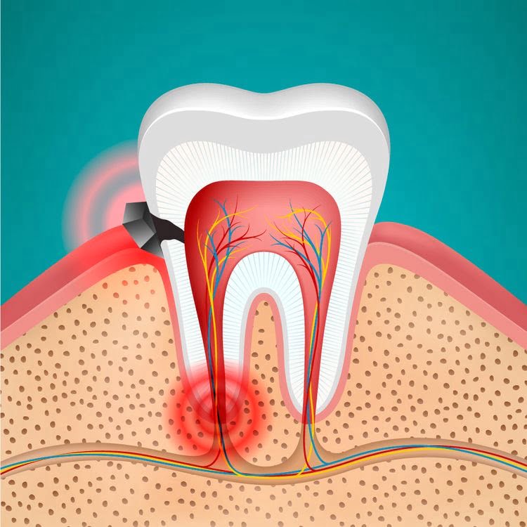
When the pulp (nerve) tissue inside the tooth becomes infected, root canal treatment is applied as a standard procedure. However, certain anatomical limitations, complex lateral canals at the root tip, or resistant stubborn bacteria may sometimes render the canal treatment insufficient to completely clean the inflammation at the root tip. The inflammation can seep from the root tip into the jawbone, creating cystic formations in that area. Root apex resection is an advanced tissue repair method designed as an alternative to tooth extraction, allowing direct external surgical access to clean this inflamed area. The infected tip of the root (approximately 3 millimeters) is cut off, and the remaining canal area is sealed with a waterproof filling material from the back (retrograde).
The planned apical resection procedures at the Avrupadent clinics located in Izmir prioritize protecting the tooth’s natural structure within the jawbone and controlling the spread of infection to neighboring tissues. Thanks to this delicate approach, which utilizes microsurgical instruments and cutting systems, chronic infections are removed and the tooth is supported in contributing to chewing function for many years. Preserving the natural tooth also creates a strong foundation for potential prosthetic (crown) restorations.
In Which Situations is Apical Resection Applied?
In clinical practice, before deciding to extract a tooth, all restorative surgical options that can retain the tooth in the mouth are considered. Root tip resection comes into play when the physical limits of endodontic (root canal) treatment have been reached. A cyst formed at the root tip can gradually erode the jawbone and begin to threaten the roots of adjacent healthy teeth. The main clinical anatomical scenarios in which the procedure is evaluated are as follows:
| Clinical Condition | Need for Surgical Intervention |
|---|---|
| Failure of Root Canal Treatment | Despite repeated canal treatments, the condition where the abscess or cystic lesion at the root tip does not decrease. |
| Anatomical Canal Curvatures | Due to the extreme curvature of the root, the canal files cannot reach the tip of the root and that area remains obstructed. |
| Presence of Fixed Restoration | When there is a long porcelain bridge or zirconium crown on the tooth, the intervention is done from the tooth’s gum to avoid removing the crown. |
| Instruments Broken Inside the Canal | Removal of broken and unretrievable rotary instrument pieces at the tip of the canal through surgical means during previous treatments. |
How is the Apical Resection Procedure Performed?
Before the procedure, the patient’s chair is prepared, and local anesthetic solutions are applied to the area to ensure clinical comfort. Once numbness is achieved, the doctor makes a delicate incision on the gum tissue and elevates it from the bone. A small window is opened on the bone surface where the root tip is located to access the chronically infected area (granuloma or cyst) directly. These tissues are extensively removed from the bone surface with special curettes.
After the cyst is cleaned, the tip of the tooth root, which is the source of the infection (approximately 3 mm segment), is cut off with a slightly angled cut (resected). After the root tip is cut, to prevent bacterial leakage inside the canal, a special material called “retrograde filling” (usually MTA or bioceramics) is used to create a backfill from the root tip. After sterilization of the surgery area is ensured, the gum is returned to its previous position and closed with fine sutures, entering the cellular healing phase.
Who Can Be Suitable for Apical Resection?
This surgical approach applied to preserve a tooth in the mouth depends on the remaining root length of the tooth being healthy enough to withstand chewing forces after the procedure. If the jawbone surrounding the tooth has significantly deteriorated due to gum disease (periodontitis) and the tooth is already loose, then removing the root tip of that tooth is not considered suitable in such cases as it would further weaken the support, and a decision may be made to extract the tooth. Teeth that have their anatomical supports preserved are ideal candidates for this surgery.
In individuals with systemic diseases, this medical intervention can be planned as long as the person’s cardiological and endocrine (diabetes, etc.) values are kept stable. Patients using bisphosphonate-type medications that affect bone structure and the healing process must consult with medical doctors before the procedure, as it is clinically necessary to confirm the biological suitability of the operation.
How is an Evaluation Made Before Apical Resection?
Success in surgical interventions depends on the details of the planning prior to the operation. The physician checks the status of the current root canal treatment. If the root filling has been poorly done from the beginning, renewing the canal treatment (retreatment) and observing afterwards may be considered before proceeding to surgery. If renewal cannot be performed (if there is a large metal post or porcelain in the tooth), direct surgical phase is planned.
Additionally, the patient’s systemic medical history and medication usage habits are thoroughly questioned. The volume of the lesion at the apex guides the physician on whether to add bone dust (graft) to the area after the operation. If the cyst is very large and has created a wide cavity in the bone, graft materials are included in the surgical design for support purposes.
How is the Apical Resection Treatment Process Planned?
The surgical protocols implemented at İzmir Avrupadent branches focus on preserving the tooth with minimal trauma. During the planning stage, it is determined where the cyst or infection is located on the tooth. The lower molars may have multiple roots; if the lesion is found in only one root, the physician designs the surgical strategy to reach just that problematic root (minimally invasive).
If the infection is very active and has caused an acute abscess on the patient’s face, antibiotic use will begin under physician supervision several days before the operation, aiming to reduce inflammation in the tissue. When the infection subsides, tissues respond better to local anesthetics, and the surgical procedure is carried out within the planned schedule inside clinical comfort.
What Steps Are Followed in Apical Resection Procedure?
The micro-surgical steps applied to support tissue repair are carried out sequentially with the principle of preserving anatomical boundaries. The basic clinical steps of the procedure are as follows:
-
\t
- • Anesthesia and Flap Removal: The anesthesia of the gum tissue surrounding the problematic tooth alignment and creating a small flap to expose the bone with the help of a scalpel.
- • Osteotomy and Cyst Excision: Creating a small window in the bone and scraping out the inflamed cystic tissues located at the root tip using curettage instruments.
- • Resection of the Root Tip: Removing an infected part of the root tip, approximately 3 mm in size, by cutting it with diamond burs at a specific angle.
- • Retrograde Filling (Closure): The cut root tip is hollowed out backward with special ultrasonic tools and sealed with biocompatible materials (such as MTA) that ensure impermeability.
- • Suturing: After ensuring the cleanliness of the opened bone cavity, the gum tissue is sutured back to its original position using fine threads, completing the procedure.
\t
\t
\t
\t
What Should Be Considered Before Apical Resection?
To perform a surgical procedure, it is essential to minimize the presence of bacteria in the working area to support wound healing. The patient should thoroughly brush their teeth before arriving at the clinic to remove any existing food residues from the area. Blood pressure medications or diabetes medications should be taken consistently at routine times.
Due to the procedure being carried out under local anesthesia, it is recommended that the patient arrives at the appointment slightly full and well-rested to keep blood sugar and blood pressure levels stable. If the use of tobacco, which slows down cellular repair, is present, limiting this habit before the day of the operation strengthens the healing process from a clinical perspective.
What Should Be Considered After Apical Resection?
Since the procedure involves intervention on the bone surface and gum, slight swelling (edema) and small leaks around the area of intervention in the cheek or lip are related to the tissue’s healing response. The cold compress applied immediately after the procedure prevents the swelling from expanding. Using a straw, which could create pressure, should be avoided in the initial days as it may disrupt the clot on the wound.
To prevent tension on the wound area during the healing stage, exaggerated facial expressions (such as stretching the lips) and consuming hard foods should be avoided. The use of tobacco should be limited until the tissue’s union is complete (especially during the crucial first week) as it disrupts the tissue’s blood supply. To avoid irritating the healing mucosa, softer, pureed foods should be chosen instead of acidic and hot foods.
In Which Dental Problems is Apical Resection Considered?
This surgical equipment serves as the last line of defense prior to tooth extraction in specific medical scenarios where the boundaries of endodontics have been exceeded. The main dental problems addressed are as follows:
- Root Tip Cysts and Granulomas: Involves cleaning pathological cavities filled with fluid or inflammation that have formed due to bacteria migrating from the root canal to the bone.
- Extruded Filling Materials: Involves the surgical removal of filling materials (such as gutta-percha) that have inadvertently migrated into the bone during root canal treatment.
- Broken Canal Instruments: The removal of rotary instruments (files) that have broken and become lodged in the final portions of the canal, depending on the anatomical structure of the root.
- Perforated Roots: If a hole has formed in the root wall due to treatment or fracture, this area can be surgically accessed and repaired with biocompatible cements.
- Extruded Filling Materials: Involves the surgical removal of filling materials (such as gutta-percha) that have inadvertently migrated into the bone during root canal treatment.
What is the Relationship Between Apical Resection and Canal Treatment?
The primary method applied when an infection is detected in a tooth is always root canal treatment (endodontics). Most of the time, a standard root canal treatment is sufficient for the body to heal the lesion at the root apex on its own. However, if the bacteria are very resistant or if there are microscopic side channels (delta channels) branching off from the main canal in the extremities of the root, normal filling materials may not fully penetrate these areas.
When these unreachable bacteria continue to proliferate in the bone, root canal treatment reaches its physical limits. At this stage, the apical resection procedure cuts and removes that blockage and the problematic apex from the outside (surgically), breaking the chain of infection. In other words, apical resection is not a replacement for root canal treatment, but rather a secondary surgical support that covers its deficiencies in inaccessible areas.
What diagnostic and imaging methods are used before apical resection?
The secure progression of the procedure is ensured by the clarity of the anatomical data obtained before entering the operation. The essential imaging tools that show the boundaries of the lesion at the root tip are as follows:
-
\t
- • Periapical Imaging (RVG): High-resolution two-dimensional small X-ray films that focus on a single tooth and its root tip. Clearly shows the extent of inflammation (radiolucent shadow).
- • Dental Volumetric Tomography (CBCT): An advanced diagnostic system used to measure the three-dimensional distance of roots to sinuses or nerves, especially in the back teeth, and analyze how much the cyst thinned the jawbone in millimeter sections.
- • Panoramic Radiography: Assessing the broad relationship of the tooth with neighboring teeth and the general structure of the mandible is also considered as supportive diagnostic data.
\t
\t
How Does the Healing Process Progress After Apical Resection?
The intervention performed externally (suture area) reaches a level where it will not affect daily functions in a short time, thanks to the high vascularization capacity of the oral mucosa. The patient can return to normal eating habits within a day. However, bone repair in the underlying layers of tissue continues for months without visible signs from the outside. After the cyst is cleaned, the remaining cavity fills with the body’s own blood clot.
Osteoblast (bone-building) cells migrate into this clot as vascularization is established. As this area gradually transforms into dense bone tissue over time, it tightly encapsulates the root tip. If the physician determines that the cyst cavity is very large, they may add bone powder (graft) to support the process. Achieving full density of the bone and the complete restoration of the lesion area to a healthy bone color in X-rays requires an average maturation cycle of 6 months.
How Long Can an Apical Resection Procedure Take?
Surgical procedures progress in a more practical and faster schedule for anterior teeth (incisors) due to the single root structure and easier access from outside to the bone. However, the presence of multiple roots in posterior molars narrows the working area due to proximity to sinus cavities or nerve canals. The dentist’s careful attention to the visual angle and anatomical boundaries in these posterior areas may cause some prolongation of the duration.
An important part of the procedure is dedicated to hollowing out the microscopic size root tip backward and sealing it with materials similar to MTA in a hermetic manner (retrograde filling). Since local anesthesia is active throughout the entire surgical procedure, the patient does not feel clinical fatigue; the process is managed in accordance with the schedule planned by the dentist.
How should oral and dental care be performed after apical resection?
Surgical sites are quite sensitive to irritation in the initial days. The stiff bristles of a toothbrush may cause tension on the stitches or lead to bleeding by pulling apart the wound edges. Therefore, until the doctor states otherwise, hygiene should be maintained in the wound area only with antiseptic mouthwashes containing chlorhexidine. While gargling, it is important to gently move the liquid in the mouth without pressure to protect the clot at the wound site.
Once the wound tissue heals and the stitches are removed, gentle dabbing motions can be applied to that area with extra soft (surgical) toothbrushes. When healing has been fully completed, standard hygiene practices using toothbrushes and dental floss can be resumed to maintain the health of the other teeth.
When is Apical Resection Considered as an Alternative Treatment Option?
No artificial material in dental practice can fully meet the advantages of the biological and chewing sensation provided by the patient’s own natural tissue. If there is a persistent infection at the root tip of a tooth and this inflammation is not resolved by conventional root canal treatments, the only fate of those teeth used to be extraction. Apical resection is conceptualized as an alternative path that breaks this extraction cycle.
Extracting the tooth and placing a titanium root (implant) is a longer, costlier process that leads to tissue loss. If the root body of that tooth is intact and the bone support around the tooth is good, the dentist can succeed in keeping the tooth in the patient’s mouth by simply cutting (resecting) that inflamed little tip. Thus, the natural life of the tooth is extended.
Why is Post-Apical Resection Control Examination Important?
After the surgical procedure, the patient’s tissue healing is usually supported by gently removing the stitches at the appointment following the first or second week. If the stitch threads remain longer than the intended duration stated above, they can lead to local infections in the wound lips by allowing food retention. During the control appointment, these threads are removed to relieve the gum tissue.
The major and diagnostic control occurs approximately 6 months after the operation. The real success criterion of the treatment is that the area around the cut root tip is not seen as black (inflammation/cavity) in X-rays, but covered with a white and dense trabecular structure (bone color), similar to the surrounding healthy bone tissues. If the doctor confirms the bone repair on the X-ray, the treatment of the tooth is considered to be completed in a definitive manner.
Can Apical Resection Be Planned Along With Other Endodontic Treatments?
The cause of inflammation at the root tip is often the bacteria hidden within the canal. If the dental canal filling is very old, porous, or fails to reach the root tip, merely cutting the root tip surgically may not permanently resolve the issue. Bacteria will continue to leak from within the canal. In such cases, the doctor will remove the old filling from inside the tooth before the surgery and disinfect the canals to place a new filling.
Immediately after this “retreatment” procedure is completed, the surgical phase begins, and the cyst is removed to unblock the root tip. This dual approach, performed simultaneously both internally and externally, maximizes the chance of the tooth remaining in the mouth and significantly reduces the risk of infection recurrence.
How is personalized planning done in Apical Resection Treatment?
The architecture of each individual’s dental roots is different. While the roots of the front teeth are usually within a flat and wide bone structure, the roots of the back teeth can be curved or can penetrate into the nasal/sinus cavities in the upper jaw. When planning, the physician examines whether the cyst is in contact with the sinus membrane in the tomographic slices; if there is a contact, the surgical path (angle) is adjusted with a special precision for the patient to ensure the membrane is not perforated during the operation.
Additionally, the size of the bone defect that will occur after the cyst is cleaned varies from patient to patient. A small defect may be sufficient for clotting to allow for bone healing, while in very large cavities, bone powder (graft) and a membrane may need to be added. In Izmir Avrupadent procedures, the selection of these materials and the repair potential of the bone are programmed with an algorithm entirely suitable for the patient’s radiological findings and medical profile.