What are Dental Traumas?
In human anatomy, although teeth and jaw bones have a resistance to withstand daily chewing forces, they can show weakness against unexpected impacts from high kinetic energy from outside. Dental traumas can range from minor cracks in the enamel layer of the tooth to deep fractures that expose the nerve tissue (pulp), root destructions, dislodgment from the socket (avulsion), or fractures of the jawbone, encompassing a wide spectrum of damage. These types of injuries are medical emergencies that significantly disrupt an individual’s chewing function, aesthetic appearance, and speaking (phonetic) ability.
In the Avrupadent clinics serving in the İzmir location, dental trauma cases are evaluated urgently and with a multidisciplinary perspective, considering the critical impact of the time on tissue repair. The biological preservation of a tooth or tissue affected by trauma depends on the time elapsed since the injury and the nature of the medical intervention to be applied. Doctors aim to restore the injured anatomical structures to their former shape and function by implementing current protocols that support tissue integrity.
In Which Situations Do Dental Traumas Occur?
The manner in which a tooth is subjected to trauma is the main factor determining the extent of the damage caused. Due to their forward angulation outside the support of the lips, the upper front incisors form the most exposed group of teeth and are the most frequently damaged. The key factors encountered in the clinic and setting the stage for trauma are as follows:
| Trauma Cause | Resulting Physical Impact and Consequences |
|---|---|
| Sport Activities | Injuries to the teeth caused by impacts to the face in activities such as boxing, basketball, or martial arts can result in fractures or displacement. |
| Daily Accidents and Falls | Especially in children, falls during walking or running can cause anterior teeth to be knocked out or impacted. |
| Traffic Accidents | In these incidents involving high kinetic energy, in addition to dental fractures, fractures of the jawbone may also be included in the process. |
| Mechanical Strains | Crown fractures resulting from opening a jar, tearing a package, or accidentally hitting a hard object like a stone/seed. |
How Are Dental Trauma Treatments Planned?
In trauma cases, time is of the essence. The physician first checks the overall health condition of the patient (neurological symptoms like dizziness, nausea) as they take the patient to the chair. Dental planning is shaped according to the type of damage. If the tooth is merely fractured and the broken piece is available, the plan is based on replacing that piece with special agents. If the tooth has been extruded (displaced upwards) or luxated (tilted) from its socket, the plan is directed towards repositioning the tooth to its original anatomical position by stabilizing it with adjacent healthy teeth (splinting).
The treatment planning for pediatric patients differs from that of adults. If the root tip of a developing permanent tooth has not yet closed, biodegradable materials (such as MTA) that will preserve the vitality of the tooth and promote continued root development are used to implement specific repair strategies (apexification or revascularization). Each case is individualized according to its dynamics, maintaining the philosophy of preserving tissue.
What Treatment Approaches Are Evaluated in Dental Trauma?
To restore the damaged tooth and surrounding tissues to their original anatomical and functional forms, various disciplines of dentistry (endodontics, surgery, restorative) are used together. The main treatment approaches evaluated in the clinic are as follows:
- Fragment Reattachment: The process of reattaching the broken piece of the tooth (fragment) with special chemical adhesives without using laboratory materials, preserving its original structure.
- Splinting (Stabilization): Stabilizing a dislodged, loose, or repositioned tooth by connecting it to adjacent healthy teeth with wires or composite materials, making it immobile within the bone.
- Endodontic Intervention (Root Canal Treatment): In cases where the nerve bundle of the tooth (pulp) is exposed due to trauma or the tooth loses vitality, cleaning and filling the tissues inside the root.
- Reimplantation: The process of placing a permanent tooth that has been knocked out (avulsed) back into its socket after it has been rinsed and cleaned, together with its intact root.
How is an Evaluation Made After Dental Trauma?
The clinical examination is conducted with great care to detect any unseen damage. The doctor attempts to feel the injury around the root by gently tapping the teeth (percussion test). Even if the tooth is not fractured, any reddish or grayish discoloration of the impacted tooth indicates a significant diagnostic finding suggesting damage or necrosis of the vascular-nervous bundle inside the tooth.
Additionally, the patient’s jaw closure is checked. If the patient cannot find the old occlusion position when they close their teeth, this is a serious indication that the tooth has shifted in its socket or that there is a fracture (fracture) in the jawbone. Soft tissues are thoroughly washed and examined to clarify the repairs of cuts or possible embedded tooth fragments during the evaluation stage.
What are the Diagnosis and Imaging Methods for Dental Trauma?
It is usually difficult to diagnose cracks in the roots, bone intra fractures, or minute displacements in the tooth socket with the naked eye. The fundamental radiographic systems determining the surgical approach are:
- ▪ Periapical Imaging: It is the most detailed small film method for detecting horizontal or vertical cracks in the tooth root due to impact, root tip fractures, and the millimetric gap with the bone socket of the tooth.
- ▪ Dental Volumetric Tomography (CBCT): It is an advanced diagnostic tool that provides depth analysis by examining structures with three-dimensional slices in cases of impacted teeth (intrusion) or suspicion of jawbone fractures.
- ▪ Soft Tissue Radiographs: These are supportive X-rays applied at low doses aimed at detecting fragments of broken teeth buried in the lips or buccal muscles.
Who Can Benefit from Dental Trauma Treatment?
Traumas do not discriminate by age. Particularly, in children aged 2-4, cases of primary teeth being knocked out or broken are common, while in the 8-12 age group, it’s quite prevalent for newly erupted permanent teeth to be broken in sports or school accidents. In young people and adults, traffic accidents or sports activities lead to the need for trauma treatment. There is no age limit for this application; age only affects the choice of material the physician will use (whether it concerns primary or permanent teeth).
Even in individuals with systemic conditions (such as bleeding disorders), the urgency of medical repair must be prioritized, with clinical necessity for rehabilitation of the damaged area to begin without delay in accordance with physician-supported bleeding control protocols.
What Treatment Options Are Considered in Case of Tooth Fracture?
Tooth fractures are classified according to the tissue layer affected by the damage. If the enamel and the underlying dentin are fractured as a result of a blow, the dentist either rebuilds the fractured part using special restorative materials or reconstructs the missing part with composite (tooth-colored) resins to fit the original form. These procedures preserve the vitality of the tooth.
However, if the fracture line exposes the nerve (pulp) chamber in the center of the tooth, intense sensitivity and pain will occur in the tooth. At this point, since the nerve tissue is exposed to bacteria in the oral environment, the dentist will take protective measures to keep the tooth in the mouth, either by removing only the infected part of the nerve (partial pulpotomy), or by cleaning all the nerves of the tooth (root canal treatment).
What Approach Should Be Followed in Case of Tooth Avulsion or Dislocation?
The fibers connecting the tooth root to the jawbone (periodontal ligaments) are elastic. In a severe impact, these fibers may tear, causing the tooth to hang outside of its socket or become embedded into the jaw. If the tooth has been displaced, the dentist will gently align the tooth back to its original position using finger pressure after anesthesia. Then, the tooth is connected to the adjacent healthy teeth with a flexible wire (splint) to facilitate the bone and fibers to wrap around the tooth again within weeks (reattachment).
If a tooth has completely erupted from the mouth along with its root, the retention of that tooth in the mouth (salvage) depends on how quickly it is handled. The tooth that is delivered under suitable storage conditions to the clinic is washed with serum by the dentist and re-implanted directly into its socket (reimplantation) and then fixed. After months, depending on the condition of the tooth, the process is supported with root canal treatment to prevent the loss of the tooth.
How Are Dental Traumas in Children Assessed?
In childhood trauma, specialists in pedodontics (pediatric dentistry) are involved. Due to bleeding and pain during the incident, the child usually experiences a very high level of anxiety. The doctor first calms the family and the child to create a safe environment. If the child’s primary tooth has been impacted, the doctor’s primary concern is not the condition of that primary tooth, but whether the underlying permanent tooth waiting to mature beneath it has been affected by the trauma.
If the primary tooth has been intruded into the depths of the jawbone due to the severity of the impact, it may damage the enamel of the underlying permanent tooth. The doctor measures this distance with X-rays. Sometimes, it is expected that these impacted primary teeth will re-erupt on their own after months (erupt downwards), while sometimes, to prevent damage to the underlying structure, extraction is designed as a clinical strategy.
What Treatments Are Applied for Traumas Occurring in Milk Teeth?
The procedure of “replacing an avulsed tooth back in its place” performed on adult permanent teeth is a definite contraindication in milk teeth (not applied). Attempting to forcibly reinsert a milk tooth that has been avulsed from its socket can cause mechanical damage to the sensitive permanent tooth bud located at the bottom of that space. If the milk tooth is completely lost, the area is washed, bleeding is controlled, and it is left to heal.
If the milk tooth is only fractured and the nerve is exposed, that tooth can remain in the mouth until the tooth falls out by performing root canal treatment or amputation. If the tooth has shifted laterally and is preventing the child’s closure (biting), the dentist can gently reposition and stabilize the tooth, or if the position cannot be repaired, can extract the tooth and design aesthetic/functioning space maintainers.
How Does the Treatment Process for Permanent Tooth Injuries Proceed?
In young individuals (between 7-12 years old), the roots of newly erupted permanent teeth may not be fully closed and possess a wide canal structure. These teeth can experience disrupted blood circulation if they sustain a blow. The dentist does not conclude the process after repairing the fractured part of the tooth or after stabilizing the tooth with a wire. Periodic vitality tests are performed to determine whether the nerves of the tooth are alive or dead.
If the tooth loses vitality, the open root apex is filled with modern tissue-repairing materials such as bioceramics or MTA (apexification procedure) to close it off and stimulate bone repair. These procedures encourage the retention of a permanent tooth, which is still undergoing development, within the bone to function throughout its lifespan.
What Should Be the First Considerations After Dental Trauma?Critical steps to consider during trauma; if the tooth or broken tooth fragment has been displaced, it should be held from its crown (the white part) without touching the root surface (the yellow part), and it should be kept in cold milk or saline and delivered to the dentist within the next hour at the latest.The awareness of first aid directly determines the chances of saving a tooth that has experienced trauma. Microscopic living cells (fibroblasts) that will reattach the tooth to the jawbone are found at the root surface of the tooth. These cells quickly die when wrapped in a dry cloth or washed with tap water, and the chance of retaining the tooth completely disappears. The ideal transport medium is cold pasteurized milk or the individual’s own saliva.
Additionally, if the person who suffered a blow shows signs of consciousness confusion, nausea, or vomiting, it is essential to visit emergency medical services with suspicion of a potential concussion prior to dental injury. After neurological danger is ruled out, the dentist will immediately start the oral emergency repair process by applying pressure to the bleeding areas with sterile compresses.
What Steps Are Followed in Dental Trauma Treatments?
Medical steps taken to limit tissue damage and restore the physiological structure are implemented sequentially within a time-sensitive framework. The clinical roadmap followed in a standard trauma intervention is as follows:
- • Emergency Assessment and Cleaning: Removal of foreign bodies from the wound area, stopping the bleeding, and determining the damage through radiological/physical means.
- • Repositioning: Gently placing a dislocated or avulsed tooth back into its alveolar socket under local anesthesia.
- • Stabilization (Splinting): Binding the tooth, which is placed in the correct position, to adjacent healthy teeth using orthodontic wires or composite materials in a flexible manner.
- • Pulp (Nerve) Surgery: Depending on the vitality of the tooth, it may be necessary to seal the broken area or to initiate the root canal treatment protocol to prevent infection.
- • Restoration: The process of reconstructing lost enamel or dentin tissues with appropriate aesthetic dental materials.
How Should Oral Care Be Provided After Dental Trauma?
Post-operative oral care; it requires avoiding hard mechanical pressure on teeth that have suffered trauma and are secured with splints, ensuring the hygiene of the area by using chlorhexidine-based chemical rinses as recommended by the dentist, and adopting a soft diet that does not require ripping.There is intense swelling (inflammation) in the root of the tooth that has been traumatized and in the surrounding connective tissue. It is essential for the tooth to rest without any impact for weeks in order for it to reattach to the jawbone. Actions like biting into an apple with front teeth or tearing a sandwich can lead to the splint (stabilizing wire) breaking or re-traumatizing the weakened root, so it is advised to consume foods by crushing them with the back teeth.
During brushing, while the healthy areas are cleaned in the usual manner, the gums and wires in the damaged area should be gently brushed with soft (surgical type) brushes. If food debris accumulates in the area, the gum can quickly become inflamed, and this inflammation disrupts bone healing. The delicate balance between maintaining impeccable oral hygiene and not applying mechanical shock to the tooth directly affects the success of the treatment.
Why Are Follow-Up Examinations Important After Dental Trauma?
The clinical necessity of follow-up examinations; is that the vascular-nervous bundle injured during the trauma may lose its vitality even months later and that radiological early detection of resorption (dissolution) foci that may insidiously begin on the root surface is possible.Dental trauma treatment does not end when the patient leaves the emergency room or clinic. A tooth may temporarily become numb due to the impact and regain circulation weeks later; or conversely, a tooth that was initially alive can lose its vitality internally (necrosis) and develop an infection months later. Often, understanding this change with the naked eye or through the patient’s pain leads to delays in treatment.
The doctor tests the vitality of the nerve by placing cold cotton on the tooth at designated periodic weeks (cold test). More importantly, the body’s immune cells may perceive the damaged tooth root as a foreign object and begin to dissolve it (root resorption). Once melting is detected in follow-up X-rays, the doctor quickly transitions to canal treatment procedures to limit the tooth’s self-dissolution process and preserve the tooth.
Can Dental Trauma Treatments Be Planned Together with Other Dental Treatments?
Yes, multidisciplinary clinical approaches are integrated as an inseparable whole. Trauma treatment is conducted simultaneously or sequentially with endodontics (root canal treatment), surgery (gum or bone repairs), and aesthetic filling and crown procedures in prosthetic/restorative dentistry.In the case of a severe fracture involving a tooth, it is not enough to just restore the fractured piece. If the nerve of the tooth is exposed during the fracture, an endodontic specialist cleans the tooth’s canals. Afterwards, an aesthetic dentist restores the fracture line using composite or porcelain (laminate) veneers to align perfectly with the patient’s natural smile line.
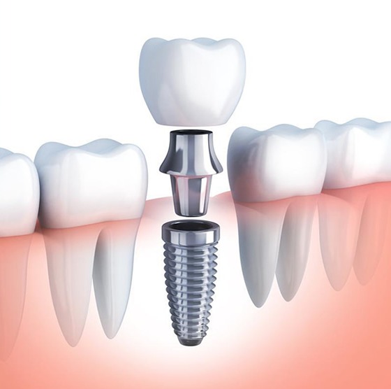
If it is determined that the tooth has sustained a deep (root-level) vertical fracture that cannot be salvaged due to trauma, the jaw surgery department will extract the tooth with minimal trauma. In the same session, to avoid narrowing of the jawbone, a titanium artificial root (implant) is placed in that space, thereby completely restoring the patient’s aesthetic and functional loss.
How is a Personalized Treatment Plan Created for Dental Traumas?
Personalized design; involves creating a specific medical intervention strategy based on the type of trauma the patient has experienced, the time elapsed since the trauma occurred, the development level of the tooth roots (apices), bone flexibility, and the individual’s overall age/growth status.Similarly, two different teeth that have been knocked out can be subjected to completely different protocols. If the patient is a child under 9 years old and the root tip of the knocked-out tooth is still in the growth phase, when the tooth is reinserted, the chances of the tooth reviving are very high due to the reattachment of the blood vessels inside; the doctor will not rush to perform root canal treatment. However, if the patient is 25 years old and the root tip is closed, there is no chance for the tooth to revive; root canal treatment must be included in the planning within 10-14 days to prevent the root of the reinserted tooth from resorbing.
The trauma management applied at the Avrupadent branches in Izmir is a platform that not only addresses the patient’s pain relief but also considers long-term biological compatibility. The flexibility of the splint (wire) to be used and how many weeks it will stay on the teeth is specifically adjusted according to whether the trauma affected just the tooth or the jawbone, preventing unnecessary fibrous tissue lockouts (ankylosis).
How Are Teeth Monitored After Dental Trauma?
Follow-up process; It is a long-term medical observation where the patient is invited to the clinic at standardized periodic intervals (IADT) such as the 1st week, 1st month, 3rd month, 6th month, and 1st year to repeat radiographic bone repair and tooth nerve vitality tests.The condition of the bone around the teeth that have been placed or fixed in their positions is monitored at each visit by comparing new films with the previous ones. The splint (wire) that holds the tooth in place usually remains in the mouth for 2 to 4 weeks, depending on the type of damage, and is removed by the doctor when its time is up, without damaging the enamel. After the wire is removed, the physiological mobility of the tooth within the bone is tested manually.
The yellowing of the tooth that may occur after months, along with defense reactions like calcification (the self-narrowing and blockage of root canals), is recorded during these follow-ups. The more closely the condition of the root cells is monitored, the more the risk of losing the tooth can be reduced by intervening in case of possible infection development in a timely manner.
The awareness of first aid directly determines the chances of saving a tooth that has experienced trauma. Microscopic living cells (fibroblasts) that will reattach the tooth to the jawbone are found at the root surface of the tooth. These cells quickly die when wrapped in a dry cloth or washed with tap water, and the chance of retaining the tooth completely disappears. The ideal transport medium is cold pasteurized milk or the individual’s own saliva.
Additionally, if the person who suffered a blow shows signs of consciousness confusion, nausea, or vomiting, it is essential to visit emergency medical services with suspicion of a potential concussion prior to dental injury. After neurological danger is ruled out, the dentist will immediately start the oral emergency repair process by applying pressure to the bleeding areas with sterile compresses.
What Steps Are Followed in Dental Trauma Treatments?
Medical steps taken to limit tissue damage and restore the physiological structure are implemented sequentially within a time-sensitive framework. The clinical roadmap followed in a standard trauma intervention is as follows:
- • Emergency Assessment and Cleaning: Removal of foreign bodies from the wound area, stopping the bleeding, and determining the damage through radiological/physical means.
- • Repositioning: Gently placing a dislocated or avulsed tooth back into its alveolar socket under local anesthesia.
- • Stabilization (Splinting): Binding the tooth, which is placed in the correct position, to adjacent healthy teeth using orthodontic wires or composite materials in a flexible manner.
- • Pulp (Nerve) Surgery: Depending on the vitality of the tooth, it may be necessary to seal the broken area or to initiate the root canal treatment protocol to prevent infection.
- • Restoration: The process of reconstructing lost enamel or dentin tissues with appropriate aesthetic dental materials.
How Should Oral Care Be Provided After Dental Trauma?
There is intense swelling (inflammation) in the root of the tooth that has been traumatized and in the surrounding connective tissue. It is essential for the tooth to rest without any impact for weeks in order for it to reattach to the jawbone. Actions like biting into an apple with front teeth or tearing a sandwich can lead to the splint (stabilizing wire) breaking or re-traumatizing the weakened root, so it is advised to consume foods by crushing them with the back teeth.
During brushing, while the healthy areas are cleaned in the usual manner, the gums and wires in the damaged area should be gently brushed with soft (surgical type) brushes. If food debris accumulates in the area, the gum can quickly become inflamed, and this inflammation disrupts bone healing. The delicate balance between maintaining impeccable oral hygiene and not applying mechanical shock to the tooth directly affects the success of the treatment.
Why Are Follow-Up Examinations Important After Dental Trauma?
Dental trauma treatment does not end when the patient leaves the emergency room or clinic. A tooth may temporarily become numb due to the impact and regain circulation weeks later; or conversely, a tooth that was initially alive can lose its vitality internally (necrosis) and develop an infection months later. Often, understanding this change with the naked eye or through the patient’s pain leads to delays in treatment.
The doctor tests the vitality of the nerve by placing cold cotton on the tooth at designated periodic weeks (cold test). More importantly, the body’s immune cells may perceive the damaged tooth root as a foreign object and begin to dissolve it (root resorption). Once melting is detected in follow-up X-rays, the doctor quickly transitions to canal treatment procedures to limit the tooth’s self-dissolution process and preserve the tooth.
Can Dental Trauma Treatments Be Planned Together with Other Dental Treatments?
In the case of a severe fracture involving a tooth, it is not enough to just restore the fractured piece. If the nerve of the tooth is exposed during the fracture, an endodontic specialist cleans the tooth’s canals. Afterwards, an aesthetic dentist restores the fracture line using composite or porcelain (laminate) veneers to align perfectly with the patient’s natural smile line.
If it is determined that the tooth has sustained a deep (root-level) vertical fracture that cannot be salvaged due to trauma, the jaw surgery department will extract the tooth with minimal trauma. In the same session, to avoid narrowing of the jawbone, a titanium artificial root (implant) is placed in that space, thereby completely restoring the patient’s aesthetic and functional loss.
How is a Personalized Treatment Plan Created for Dental Traumas?
Similarly, two different teeth that have been knocked out can be subjected to completely different protocols. If the patient is a child under 9 years old and the root tip of the knocked-out tooth is still in the growth phase, when the tooth is reinserted, the chances of the tooth reviving are very high due to the reattachment of the blood vessels inside; the doctor will not rush to perform root canal treatment. However, if the patient is 25 years old and the root tip is closed, there is no chance for the tooth to revive; root canal treatment must be included in the planning within 10-14 days to prevent the root of the reinserted tooth from resorbing.
The trauma management applied at the Avrupadent branches in Izmir is a platform that not only addresses the patient’s pain relief but also considers long-term biological compatibility. The flexibility of the splint (wire) to be used and how many weeks it will stay on the teeth is specifically adjusted according to whether the trauma affected just the tooth or the jawbone, preventing unnecessary fibrous tissue lockouts (ankylosis).
How Are Teeth Monitored After Dental Trauma?
The condition of the bone around the teeth that have been placed or fixed in their positions is monitored at each visit by comparing new films with the previous ones. The splint (wire) that holds the tooth in place usually remains in the mouth for 2 to 4 weeks, depending on the type of damage, and is removed by the doctor when its time is up, without damaging the enamel. After the wire is removed, the physiological mobility of the tooth within the bone is tested manually.
The yellowing of the tooth that may occur after months, along with defense reactions like calcification (the self-narrowing and blockage of root canals), is recorded during these follow-ups. The more closely the condition of the root cells is monitored, the more the risk of losing the tooth can be reduced by intervening in case of possible infection development in a timely manner.
Dental Traumas and Treatments hakkında sıkça sorulan sorular ve cevapları
Dişin darbe sonucu çene kemiğinin içine doğru gömülmesi Dental Travmalar ve Tedavileri ile düzeltilebilir mi?
Evet, dişin durumuna göre ya kendiliğinden geri çıkması beklenir ya da teller ve cerrahi destekle eski anatomik pozisyonuna geri çekilir.
Darbe sonrası dudakta oluşan kanamalar Dental Travmalar ve Tedavileri muayenesine gelmeden önce nasıl durdurulmalıdır?
Temiz bir gazlı bez veya peçete ile kanayan bölgeye dışarıdan hafif ve sürekli bir baskı uygulayarak kliniğe varana kadar kanama kontrol altına alınır.
Çevre yumuşak dokuların incelenmesi Dental Travmalar ve Tedavileri muayenesinin rutin bir parçası mıdır?
Düşme veya çarpma sırasında sadece dişler değil; yanak içi, dudak mukozası ve dilin de kesilip kesilmediği bütünsel olarak kontrol edilir.
Dışarı doğru uzayan veya yuvasından sarkan dişlerin Dental Travmalar ve Tedavileri ile orijinal seviyesine itilmesi mümkün müdür?
Diş yuvasından uzağa kaymışsa, hekim lokal uyuşturma altında parmak baskısıyla dişi nazikçe yuvasına geri iter ve iyileşmesi için yandaki dişlere tellerle bağlar.
Ağız gargarası kullanmak Dental Travmalar ve Tedavileri sonrası yara yerlerindeki iltihabı önlemeye yardımcı olur mu?
Hekimin önereceği klorheksidin içerikli antibakteriyel gargaralar, fırçanın ulaşamadığı dikiş veya tel bölgelerinde mikrop üremesini tamamen kimyasal olarak durdurur.
Avrupadent İzmir şubelerinde mesai saatleri sonrasına denk gelen Dental Travmalar ve Tedavileri için esnek planlamalar devreye girer mi?
Kazaların zamanı önceden bilinemeyeceği için, akşam saatlerinde gelişen travmalara yönelik mesai sonrası randevu ve nöbet opsiyonlarımızla da hizmet verilmektedir.
Süt dişlerine gelen darbeler de Dental Travmalar ve Tedavileri çerçevesinde mi incelenmektedir?
Çocuklardaki süt dişi travmaları, alttan gelen kalıcı diş tomurcuklarına zarar vermemesi için büyük bir hassasiyetle bu kapsamda değerlendirilir.
Dişin sallanması durumunda Dental Travmalar ve Tedavileri randevusuna kadar parmakla dişi yerine itmek doğru mudur?
Dişi kendi başınıza itmeye çalışmak kök ucuna ve kemiğe daha fazla zarar verebileceği için dişe dokunmadan acilen kliniğe başvurulmalıdır.
Kırık diş parçasının dudak içine saplanıp saplanmadığı Dental Travmalar ve Tedavileri röntgenlerinde görülür mü?
Bazen kırılan minik parçalar yara yerine (dudağa) gömülebilir, diş eksikse dudak bölgesine alınan özel açılı filmlerle bu parçalar rahatça bulunur.
Travma sonucu pembe veya griye dönen dişler için Dental Travmalar ve Tedavileri hangi kanal uygulamalarını içerir?
Renk değişimi sinirin öldüğünü gösterdiği için, kök kanalındaki kanamalı ve ölü doku özel solüsyonlarla yıkanarak dezenfekte edilip kök dolgusuyla kapatılır.
Darbe almış dişlerin Dental Travmalar ve Tedavileri sonrasında periyodik olarak canlılık kontrolüne çağrılması neden gereklidir?
Sinirler hasar sonrası hemen değil, aylar sonra bile yavaş yavaş canlılığını yitirip enfeksiyon üretebileceği için uzun vadeli radyolojik takip elzemdir.
İzmir içi trafik kaynaklı gecikmelerde Avrupadent İzmir acil Dental Travmalar ve Tedavileri vakalarında nasıl bir kolaylık sunar?
Gecikmeli de olsa kliniğe ulaştığınızda, hekimlerimizin zaman yönetimi esnetilerek acil hasarınız için mutlaka gerekli ilk yardım niteliğindeki işlemler aynı gün sağlanır.
Kalıcı dişlerdeki kırıklar için uygulanan Dental Travmalar ve Tedavileri prosedürleri süt dişlerinden farklı mıdır?
Kalıcı dişleri ağızda tutmak için her türlü onarım yapılırken, süt dişlerinde kök gelişimi ve alttaki kalıcı dişin durumu daha farklı yöntemler gerektirir.
Darbe alan dişi fırçalamak veya ağzı şiddetle çalkalamak Dental Travmalar ve Tedavileri öncesi sakıncalı mıdır?
Yaralı dokuları tahriş edebileceği ve kırık parçaların oynayıp kopmasına sebep olabileceği için sert çalkalama ve fırçalama yapılmamalıdır.
Diş eti cebi derinliğinin ölçülmesi Dental Travmalar ve Tedavileri sırasında kök hasarını belirlemede kullanılır mı?
Aletin dişin yanından köke doğru derinlemesine inmesi, kemik yuvasında veya kök yan duvarında dikey bir kırık olduğunun en büyük habercisidir.
İlerleyen yıllarda kararan travmalı dişlere Dental Travmalar ve Tedavileri kapsamında içten beyazlatma yapılabilir mi?
Kanal tedavisi bittikten sonra dişin içine özel beyazlatma jelleri konularak, kararmış dişin rengi diğer doğal dişlerin beyazlığına başarıyla geri döndürülebilir.
Birkaç yıl sonra bile dişte kök erimesi olup olmadığı Dental Travmalar ve Tedavileri kontrollerinde takip edilir mi?
Evet, travma gören dişlerde vücudun kendi kendine kökü içeriden eritme (rezorpsiyon) riski bulunduğu için yıllık filmlerle kök duvarlarının kalınlığı özenle izlenir.
Çocuk hastaların düştüğü acil durumlarda Avrupadent İzmir uzmanları Dental Travmalar ve Tedavileri sürecinde korkuyu nasıl yönetir?
Çocuklara işlemleri oyunlaştırarak anlatan, iğne yerine rahatlatıcı spreyler kullanan yaklaşımımız sayesinde çocuğun travma anındaki gözyaşları hızla güvene dönüştürülür.
Görünürde hiçbir kırık olmayan ancak darbe almış dişler için Dental Travmalar ve Tedavileri planlaması yapılmalı mıdır?
Dışarıdan sağlam görünse de dişin içindeki damar yapısı (sinir) kopmuş olabileceği için radyolojik ve klinik canlılık takibi yapılması şarttır.
Çocuklarda düşme sonrası ağızdan kan geliyorsa Dental Travmalar ve Tedavileri için kliniğe başvurmak acil midir?
Dokuların hızlıca dezenfekte edilmesi ve kırıkların alttaki kalıcı dişlere zarar vermeden kontrol altına alınması için oldukça acildir.
Contact Us for Dental Trauma and Treatments in Izmir
Dental Trauma and Treatments is a field of treatment that involves assessing and treating injuries to teeth and surrounding tissues resulting from accidents or impacts. At AvrupaDent, this treatment is carried out by dentists within the framework of a treatment plan suitable for the patient's oral and dental health. You can contact us to get detailed information about the treatment process.İletişime Geçin
- Güncelleme tarihi:25 Sep 2026