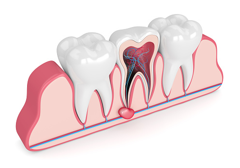
What is Tooth Cyst Removal (Cyst Enucleation)?
Cysts in the mouth and jaw area typically exhibit a slow and insidious growth characteristic. Often asymptomatic in the initial stages, these pathological formations can expand over time, leading to the erosion of healthy jawbone, shifting of neighboring tooth roots, or weakening of them. Cyst enucleation is a micro-surgical discipline designed to intervene in this destructive process. The primary goal of the procedure is to eliminate the cyst’s wall (capsule) from the area without leaving any remnants behind, minimizing the risk of lesion recurrence (nucleus).
In the Avrupadent clinics located in İzmir, the processes of cyst enucleation are carried out with a multidisciplinary perspective aimed at preserving the anatomical structure of the hospital and its chewing functions. Medical procedures are planned using the precise techniques of jaw surgery. This operation, which aims to control the spread of the cyst to surrounding tissues, prepares the biological groundwork necessary for the oral tissues to return to physiological norms. After the operation, the self-healing of the edentulous bone area is supported, or if necessary, additional materials (grafts) are applied to the area to promote cellular repair.
What Causes Dental Cysts?
The human body develops a defense mechanism to contain the situation when it detects an infection or a foreign substance. When the pulp (nerve) tissue at the center of the tooth loses its vitality due to decay or trauma (necrosis), bacteria leak from the root canal into the jawbone. To prevent these bacteria from spreading to the bone, the body’s immune system encapsulates the infected area in an epithelial membranous layer; this situation is the primary form of radicular cyst formation. The types and causes of cysts that are frequently encountered are as follows:
| Cyst Type | Necessity and Mechanism of Formation |
|---|---|
| Radicular Cysts | Lesions that occur around the root tips of infected and necrotic teeth, caused by the body’s response to trap bacteria. |
| Dentigerous Cysts | An expanding protective follicle surrounding impacted teeth (especially third molars), which eventually transforms into a pouch filled with fluid over time. |
| Residual Kistler | After a problematic tooth extraction, leaving a cyst remnant at the root tip causes it to continue to grow in that area. |
| Developmental Cysts | The activation of tissue remnants at the points of union during the embryological development phase years later, leading to the formation of a sac. |
In Which Situations is Tooth Cyst Removal Applied?
It is not medically feasible to eliminate pathological masses by dissolving them with medications (antibiotics); as cysts are independent structures surrounded by a healthy cell membrane. Surgical drainage methods come into play to prevent these formations from growing and disrupting anatomical structures. Intervention becomes a priority when it is determined that the cyst is advancing toward the lower jaw nerve (mandibular canal) or the maxillary sinuses.
In some cases, despite repeated root canal treatments, the abscess (cyst) at the root tip does not show signs of shrinking. To preserve the tooth in the oral cavity, doctors may decide to remove that stubborn tissue directly from the area. Additionally, for patients with planned orthodontic treatments (braces), it is a clinical necessity to clean the intraosseous cysts that could hinder tooth movement before starting the treatment.
How is Cyst Enucleation Performed?
The surgical process begins with a careful anesthesia phase that ensures patient comfort. Once the tissues are numb, the doctor makes a delicate incision (flap) in the gum tissue at the level of the bone where the cyst is located. If the cyst has not completely eroded the bone, a small surgical window is created using fine drills or ultrasonic (piezo) bone cutters to access the lesion. The most crucial principle of the enucleation technique is that the cyst membrane must be separated from the bone surface without tearing; even a microscopic fragment left behind can cause recurrence (residual) of the lesion in the future.
After the cyst encapsulation has been completely removed from the area, the remaining bone cavity is meticulously washed with antiseptic solutions to sterilize it. If the cyst is connected to the root tip of the tooth, the problematic root tip is also trimmed in the same session (resection). If the size of the void is large and the likelihood of it healing on its own is low, this void can be filled with bone powder (graft). Then, the gum mucosa is adapted back to its original position and the tissue is sutured with surgical threads to support the entire structure, completing the procedure.
Who May Be Suitable for Tooth Cyst Removal?
Jaw cysts can develop at any age, regardless of age group. In a child at a developmental stage, if cysts stemming from primary teeth hinder the eruption of the permanent teeth, this blockage needs to be surgically removed. In older patients, surgical decisions are made in the presence of lesions associated with untreated teeth or impacted teeth. There is no upper age limit that would create a surgical restriction, the primary metric is the patient’s systemic health.
In individuals with discomfort affecting the bleeding profile (such as the use of anticoagulants, etc.), heart failure, or uncontrolled diabetes, it is expected that through consultations with the hospital’s physician, medical values will reach ideal ranges. Once systemic values stabilize and physician approval is obtained, cyst enucleation procedures are scheduled.
How is Jaw Cyst Diagnosis Made?
Cysts at the initial stage usually do not cause any discomfort to the patient and are often accidentally noticed in routine X-rays taken for other purposes. As the cyst grows, it starts to exert pressure outward on the bone; at this stage, when the physician touches the relevant area (palpation), they may feel a sensation of flexibility (ping-pong ball sign) in the bone or detect visible asymmetry. In vitality tests performed to determine whether the dental nerve is alive, a nonresponsive tooth is also a significant finding that raises suspicion of a cyst.
The final diagnosis is made based on radiological data. Dark lesion areas observed at the tip of the tooth root or around the crown of an impacted tooth are classically radiographic appearances of a cyst. The radiographic evidence obtained is the most valuable data outlining the course of surgical planning.
How is an Evaluation Made Before Tooth Cyst Extraction?
The predictability of a surgical intervention can be established by mapping the cyst’s spread in millimeters. The physician estimates the volume of the cyst and calculates the bone gap that will form in the area post-operation. If the cyst has caused significant tissue loss, the surgical plan will include the “bone graft” (powder addition) procedure to mitigate the risk of jawbone weakening and fracture.
Additionally, if the patient currently has an acute (active and exudative) inflammatory condition in their mouth, the physician may postpone the surgical intervention as the cellular efficacy of anesthesia solutions may be reduced in these infected areas. Treating the infection with an antibiotic prescription and calming the tissues is one of the preparatory steps that support the medical comfort of the intervention phase.
How is the Cyst Enucleation Treatment Process Planned?
In the clinic operations of İzmir Avrupadent branches, it is not enough to simply remove the cyst; the aim is also to eliminate the cause of the cyst. If the cyst is originating from a tooth that is severely decayed, it is planned to perform high-quality root canal treatment on that tooth either before the surgery or in the same session as the surgery. If the tooth is too damaged to be salvaged, it is anticipated that the tooth will also be extracted during the surgical procedure, and the cyst will be cleaned by accessing it from its socket.
Another aspect of the planning involves organizing the sending of the removed pathological tissue (the cyst) to the pathology laboratory for examination of its cellular structure (biopsy). After surgery, the hospital will provide the care and nutritional guidelines that the patient needs to follow for cellular repair of the tissues, ensuring the process progresses in cooperation with the patient.
What Steps Are Followed in the Tooth Cyst Extraction Procedure?
Clinical steps designed to protect the integrity of the tissue and respect the anatomical boundaries are implemented sequentially in accordance with surgical principles. The roadmap followed in a standard procedure is as follows:
- ▪ Anesthesia Induction: The clinical numbness is achieved by blocking the nerve networks surrounding the surgery area with local anesthetics.
- ▪ Surgical Flap (Cover) Incision: A small incision is made in the gum tissue to access the underlying bone tissue by creating space in the area.
- ▪ Bone Window (Osteotomy): A thin layer of bone covering the cyst is removed using special drills to access the lesion.
- ▪ Enucleation (Removal of the Cyst): The inflamed cyst sac is removed entirely from the bone wall with delicate curettage instruments without fragmentation.
- ▪ Filling and Suturing: Cleaning the empty space, washing it, adding graft (bone powder) if necessary, and repairing the gum with surgical threads.
What Should Be Considered Before Tooth Cyst Extraction?
In surgical interventions, it is crucial to keep the microbial load of the working area as low as possible to prevent potential complications in the wound area after the operation. The patient must thoroughly brush their teeth before arriving at the clinic to remove any existing food residues from the operating area. Any systemic medications such as anticoagulants directed by the physician should be reconfirmed with the doctor on the day of the procedure.
Since the procedure will be performed under local anesthesia (or sedation), the patient’s consciousness will generally be intact. To prevent fluctuations in blood pressure and drops in blood sugar, arriving at the clinic in a slightly full and stress-free manner will enhance clinical comfort. Being rested prior to the day of the operation ensures a much calmer progression of the treatment process that will be spent in the doctor’s chair.
What to Consider After Tooth Cyst Removal?
Since an intervention was made in a large cavitation area within the jawbone, mild to moderate swelling (edema) in the cheek area and small ooze inside the mouth are expected signs of cellular repair. The application of ice, intermittently from the outside during the first 24 hours, limits this swelling to a noticeable extent. Coughing or vigorously rinsing the mouth with water can dislodge the protective clot at the wound site, disrupting healing.
The nutrition routine should be maintained until drowsiness passes, after which it should be continued with foods that will not irritate the fresh mucosal surface, such as soft and pureed foods (yogurt, soup, etc.). Acidic, overly hot, or spicy foods should be removed from the menu for the next few days. Consuming prescribed analgesics and antibiotics at the specified intervals provides significant support for managing the process calmly.
What Imaging Methods Are Used in the Diagnosis of Dental Cysts?
For the surgical intervention to proceed reliably, it is crucial to clearly delineate the anatomical borders of the lesion. The fundamental radiological methods used during the diagnosis stage in the clinic are as follows:
- Panoramic Radiography: By displaying the entire lower and upper jaw in one wide view, it provides an initial diagnostic map concerning the general location of the cyst and its relationship with adjacent teeth.
- Dental Volumetric Tomography (CBCT): An advanced diagnostic tool that detects the volume of lesions with three-dimensional millimeter slices in cases where the cyst overlaps with the mental nerve in two-dimensional films or in instances of sinus proximity.
- Periapical Films: Focusing exclusively on the problematic tooth with small film sizes, it thoroughly examines the boundaries of bone loss at the root tip with high precision.
How Does the Healing Process Progress After Cyst Enucleation?
The oral mucosa is one of the tissues with the highest repair rate in the body. Thus, the superficial wound closure in the surgical area progresses quite quickly, facilitating the patient’s return to daily nutritional functions within a few days. A slight restriction in the first days when the mouth is opened is a temporary adaptation reaction of the muscles to the surgical intervention.
In contrast, deeper cellular activity continues in the lower layers of the tissue, specifically within the bone cavity from which the cyst was removed. The blood clot that fills the void gradually transforms into firm bone tissue through the organization of connective tissue and, ultimately, bone-forming cells (osteoblasts). Depending on the size of the cyst, this internal healing requires a maturation (ossification) period that can be observed radiologically, extending on average to 6 months, even if it is not felt from the outside.
How Long Can the Tooth Cyst Removal Procedure Last?Duration of the operation; It can generally take between 30 minutes to 1.5 hours, depending on the size of the lesion within the jawbone, its anatomical relation to neighboring tooth roots, the additional bone graft procedures to be applied, and the position of the tooth.In clinical practice, each cyst case presents a unique volume. The removal of a small cyst located in the anterior region and close to the outer bone layer can generally be achieved in a relatively short time after anesthesia takes effect. However, the removal of larger cysts located in the posterior jaw area, which have unclear borders and are embedded beneath a deep layer of bone, will take more time to extract carefully without tearing the membrane.
Another factor that determines the duration is the surgeon’s microsurgical precision when working on surrounding tissues. In cases adjacent to nerve pathways or sinuses, the procedures are performed with extremely gentle and slow hand maneuvers to avoid applying pressure on anatomical tissues. Since the patient is under anesthesia, they will not feel any clinical discomfort during this duration.
How Should Oral Care Be Done After Tooth Cyst Extraction?
Post-operative oral care; it is important to avoid hard toothbrush strokes on the incision area in the first days, to gently rinse that area with antiseptic mouthwashes recommended by the doctor to ensure hygiene, and to brush other parts of the mouth normally.After the intervention, there is a blood clot that ensures the control of bleeding in the wound area and forms the basis for healing. The action of vigorous brushing or the use of strong mouthwashes can mechanically displace this clot and lead to painful conditions that obstruct wound healing. Therefore, in the operated area, hygiene should only be maintained with chemical cleaning until the doctor says otherwise.
When performing oral care with liquids, instead of swirling water in the mouth at high pressure, it is beneficial to gently circulate it in the wound area and then slowly let it flow into the sink. After the wound tissue comes together and stitches are removed, gradual return to standard oral hygiene practices is achieved by massaging the area with extra soft form surgical brushes.
What Is the Difference Between Cyst Removal and Apical Resection?
Although the two concepts are often integrated, they are based on different principles; cyst enucleation is the act of removing the diseased sac within the bone, while apical resection involves cutting a portion of the root tip of the tooth that produces the cyst in order to seal off the source of infection (mummification).If a cyst has formed at the root tip of a tooth and the tooth is to be preserved in the oral cavity, the doctor performs both procedures in the same session. The doctor first creates a window on the bone and performs cyst enucleation to remove the inflamed sac from the bone. However, simply removing that sac is not enough because bacteria inside the tooth can continue to ooze from the root tip.
To stop this seepage, the procedure of apical resection is initiated. The very tip of the root is cut back by about 3 mm, and that cut tip is sealed off with filling materials that prevent backward (retrograde) seepage. In summary, one is the cleaning of a pathological product (the cyst), while the other is cutting and sealing the source (the root tip).
Why Is Control Examination Important After Cyst Removal?
The clinical necessity of control examinations; it is monitoring the cellular closure (epithelialization) performance of the wound lips by taking surgical stitches in the first weeks and confirming whether the bone area that was released from the cyst fills healthily with new bone tissue after radiographs taken months later.A short visit usually performed in the first or second week following surgical intervention is to verify that surface healing is progressing on the right track. If the used suture threads remain longer than the specified duration in the oral environment, they can cause local irritations by allowing food retention. The doctor supports the tissue’s return to its natural form by removing these threads.
The main large and decisive control is conducted approximately 6 months after the operation. The actual clinical success of the treatment is when the area from which the cyst was removed (radiolucent) is seen to be covered with a dense trabecular structure (bone color) similar to other surrounding bone tissues in the films. If the doctor confirms bone repair in the radiograph, the risk of lesion recurrence is considered largely mitigated.
Can Cyst Removal Procedure Be Planned Along With Other Oral and Dental Treatments?
Yes, it is at the center of multidisciplinary approaches. The cyst removal procedure is often integrated with canal retreatments, the extraction of teeth that have been damaged by the cyst, or protocols such as placing bone graft and titanium root (implant) in the related area during the same session.In clinical practice, surgeries are combined to save time and reduce tissue trauma. For example, if one or two teeth need to be extracted due to a large cyst surgery, the teeth are extracted and the cyst is scraped. If the bone walls are suitable, an implant can be placed in the area made vacant by the cyst and filled with surrounding bone dust in the same session, saving the patient from undergoing a second bone surgery in the future.
If a cyst has developed around an impacted 20-year molar (dentigerous cyst), the doctor removes the impacted tooth during the same surgical session while cleaning the cyst. In orthodontic treatments, if there is any cystic lesion blocking the movement of the teeth within the bone, cleaning the area surgically before starting wire treatment provides a sterile foundation for other branches.
How is a personalized treatment plan created in the cyst removal process?
Custom design for the individual; the surgical incision size and the materials to be used for the graft (powder) are projected entirely based on the volume of the cyst in the patient’s mandible, the proximity to neighboring nerves or air (sinus) cavities, and the patient’s healing potential.Not every cyst case can be cleaned using the same biological mechanism. If the cyst has encroached upon the nerve canal in the lower jaw (nervus alveolaris), the physician may opt for a more delicate and segmental approach to avoid damaging the nerve instead of removing the cyst in one piece. Alternatively, if the cyst is very large, a special tube (drain) may be placed inside the cyst before the surgical operation (marsupialization technique) to expect the cyst to shrink over months, and it is then removed in a session designed specifically for the individual after it has shrunk.
Additionally, the size of the bone deficiency that occurs after the cyst is cleaned also individualizes the graft (bone powder) decision. While a small void can be filled with a blood clot for healing, larger weakened jaw cavities are supported with titanium plates, membranes, or bone powders compatible with the patient’s tissue. In the processes at Izmir Avrupadent, all these medical options are shaped according to the patient’s clinical radiological findings.
In clinical practice, each cyst case presents a unique volume. The removal of a small cyst located in the anterior region and close to the outer bone layer can generally be achieved in a relatively short time after anesthesia takes effect. However, the removal of larger cysts located in the posterior jaw area, which have unclear borders and are embedded beneath a deep layer of bone, will take more time to extract carefully without tearing the membrane.
Another factor that determines the duration is the surgeon’s microsurgical precision when working on surrounding tissues. In cases adjacent to nerve pathways or sinuses, the procedures are performed with extremely gentle and slow hand maneuvers to avoid applying pressure on anatomical tissues. Since the patient is under anesthesia, they will not feel any clinical discomfort during this duration.
How Should Oral Care Be Done After Tooth Cyst Extraction?
After the intervention, there is a blood clot that ensures the control of bleeding in the wound area and forms the basis for healing. The action of vigorous brushing or the use of strong mouthwashes can mechanically displace this clot and lead to painful conditions that obstruct wound healing. Therefore, in the operated area, hygiene should only be maintained with chemical cleaning until the doctor says otherwise.
When performing oral care with liquids, instead of swirling water in the mouth at high pressure, it is beneficial to gently circulate it in the wound area and then slowly let it flow into the sink. After the wound tissue comes together and stitches are removed, gradual return to standard oral hygiene practices is achieved by massaging the area with extra soft form surgical brushes.
What Is the Difference Between Cyst Removal and Apical Resection?
If a cyst has formed at the root tip of a tooth and the tooth is to be preserved in the oral cavity, the doctor performs both procedures in the same session. The doctor first creates a window on the bone and performs cyst enucleation to remove the inflamed sac from the bone. However, simply removing that sac is not enough because bacteria inside the tooth can continue to ooze from the root tip.
To stop this seepage, the procedure of apical resection is initiated. The very tip of the root is cut back by about 3 mm, and that cut tip is sealed off with filling materials that prevent backward (retrograde) seepage. In summary, one is the cleaning of a pathological product (the cyst), while the other is cutting and sealing the source (the root tip).
Why Is Control Examination Important After Cyst Removal?
A short visit usually performed in the first or second week following surgical intervention is to verify that surface healing is progressing on the right track. If the used suture threads remain longer than the specified duration in the oral environment, they can cause local irritations by allowing food retention. The doctor supports the tissue’s return to its natural form by removing these threads.
The main large and decisive control is conducted approximately 6 months after the operation. The actual clinical success of the treatment is when the area from which the cyst was removed (radiolucent) is seen to be covered with a dense trabecular structure (bone color) similar to other surrounding bone tissues in the films. If the doctor confirms bone repair in the radiograph, the risk of lesion recurrence is considered largely mitigated.
Can Cyst Removal Procedure Be Planned Along With Other Oral and Dental Treatments?
In clinical practice, surgeries are combined to save time and reduce tissue trauma. For example, if one or two teeth need to be extracted due to a large cyst surgery, the teeth are extracted and the cyst is scraped. If the bone walls are suitable, an implant can be placed in the area made vacant by the cyst and filled with surrounding bone dust in the same session, saving the patient from undergoing a second bone surgery in the future.
If a cyst has developed around an impacted 20-year molar (dentigerous cyst), the doctor removes the impacted tooth during the same surgical session while cleaning the cyst. In orthodontic treatments, if there is any cystic lesion blocking the movement of the teeth within the bone, cleaning the area surgically before starting wire treatment provides a sterile foundation for other branches.
How is a personalized treatment plan created in the cyst removal process?
Not every cyst case can be cleaned using the same biological mechanism. If the cyst has encroached upon the nerve canal in the lower jaw (nervus alveolaris), the physician may opt for a more delicate and segmental approach to avoid damaging the nerve instead of removing the cyst in one piece. Alternatively, if the cyst is very large, a special tube (drain) may be placed inside the cyst before the surgical operation (marsupialization technique) to expect the cyst to shrink over months, and it is then removed in a session designed specifically for the individual after it has shrunk.
Additionally, the size of the bone deficiency that occurs after the cyst is cleaned also individualizes the graft (bone powder) decision. While a small void can be filled with a blood clot for healing, larger weakened jaw cavities are supported with titanium plates, membranes, or bone powders compatible with the patient’s tissue. In the processes at Izmir Avrupadent, all these medical options are shaped according to the patient’s clinical radiological findings.
Tooth Cyst Removal (Cyst Enucleation) hakkında sıkça sorulan sorular ve cevapları
Kök ucunda oluşan iltihabi kistler için Diş Kisti Çıkartma (Kist Enükleasyonu) tek çözüm müdür?
Diş kanal tedavisine cevap vermediğinde, kök ucunda kronikleşen kesenin alınarak bölgenin temizlenmesi tıbben bilinen en kesin yoldur.
Hekim sadece dışarıdan elle muayene yaparak Diş Kisti Çıkartma (Kist Enükleasyonu) sınırlarını belirleyebilir mi?
Elle muayene dışarıya yansıyan şişliği ve kemiğin dış tabakasındaki incelmeyi fark ettirir, ancak kesin sınırlar daima radyoloji ile tespit edilir.
Uzun sürecek Diş Kisti Çıkartma (Kist Enükleasyonu) seanslarında çeneyi dinlendirmek için ağız takozları yerleştirilir mi?
Hastanın ağzını açık tutarak yorulmasını önlemek için yumuşak silikon dayanaklar çeneye yerleştirilip kasların zorlanmadan açık kalması sağlanır.
Kök ucu cerrahisi ile birleştirilen Diş Kisti Çıkartma (Kist Enükleasyonu) işlemi dişi kurtarmaya yönelik bir adım mıdır?
Evet, küçük apselerde sadece enfekte kök ucu kesilir ve arkasındaki kist temizlenerek dişin kemik içinde yıllarca sağlıklı kalması amaçlanır.
Atılan cerrahi dikişler Diş Kisti Çıkartma (Kist Enükleasyonu) işleminden ortalama kaç gün sonra hekim tarafından muayene ile alınır?
Genellikle doku birleşiminin güvenli bir noktaya ulaştığı 7 ile 10 gün arasındaki kısa kontrol randevusunda dikişlerin durumu değerlendirilir ve alınır.
Operasyon sonrası kemik oluşum hızı Diş Kisti Çıkartma (Kist Enükleasyonu) takiplerinde belirli aralıklarla çekilen filmlerle mi izlenir?
Boşalan kist alanının aylar içinde adım adım beyaz sert kemik dokusuyla dolması kontrol röntgenleri üzerinden İzmir şubelerimizde hekiminizce ölçülerek izlenir.
Dişi çekmeden sadece Diş Kisti Çıkartma (Kist Enükleasyonu) yapılarak diş ağızda tutulabilir mi?
Kist dişi tamamen sarmamışsa, kök ucunun bir kısmıyla birlikte sadece kist alınarak (apikal rezeksiyon) ilgili diş ağızda sağlıklı şekilde bırakılabilir.
Röntgende görülen karanlık gölgeler Diş Kisti Çıkartma (Kist Enükleasyonu) planlamasını nasıl etkiler?
Radyografide izlenen siyah (karanlık) alanlar kemiğin eridiği hacmi netleştirdiği için cerrahın müdahale edeceği pencerenin genişliğini baştan belirler.
Kist ile birlikte diş de alınacaksa Diş Kisti Çıkartma (Kist Enükleasyonu) hazırlığı aynı seansta buna göre mi planlanır?
Diş tamamen kistin içinde kalmış ve kurtarılamayacaksa, aynı operasyon anında hem kist zarı çıkarılır hem de diş çekilerek boşluk temizlenir.
İşlem sonunda aralanan dokunun kapatılması için Diş Kisti Çıkartma (Kist Enükleasyonu) bölgesine ince dikişler atılması şart mıdır?
Aralanan diş etinin kemik yüzeyine tekrar sımsıkı yapışması ve kanamanın mühürlenmesi için ince cerrahi dikişlerin atılması kesinlikle şarttır.
Dikişlerin alınması Diş Kisti Çıkartma (Kist Enükleasyonu) sonrası hiçbir sızı hissettirmeyen saniyeler süren pratik bir işlem midir?
Yara yeri tamamen birleşip duyarsızlaştığı için, hekimin ipleri nazikçe makasla kesip alması saniyeler süren ve acısız olan çok basit bir işlemdir.
Avrupadent İzmir şubelerinde Diş Kisti Çıkartma (Kist Enükleasyonu) muayene randevuları hastanın başvurusu ardından hızla planlanır mı?
İletişim hatlarımızdan bize ulaştığınızda, yaşadığınız endişeler dinlenerek vakit kaybetmeden en yakın uzman hekim takvimine randevunuz hızla oluşturulur.
İleri yaşlardaki bireylere ihtiyaç halinde Diş Kisti Çıkartma (Kist Enükleasyonu) yapılmasında sakınca var mıdır?
Hastanın genel cerrahi işlem görmesine engel tıbbi bir kısıtlaması bulunmuyorsa, kist teşhis edilen her yetişkin yaş grubuna rahatlıkla uygulanabilir.
Geçmeyen kötü ağız kokusu ve sızıntı Diş Kisti Çıkartma (Kist Enükleasyonu) gereken iltihaplı vakalarda görülür mü?
İltihaplı kist sıvısının diş eti kenarından ağza sızması durumunda, fırçalamayla geçmeyen inatçı bir koku ve kötü bir tat hissi yaşanır.
Kemik tozu kullanılacaksa Diş Kisti Çıkartma (Kist Enükleasyonu) öncesinde bu tozlar steril olarak masada nasıl hazırlanır?
Tomografiye göre hacmi belirlenen greft materyalleri, operasyondan saniyeler önce paketinden çıkarılarak tamamen steril şartlarda bekletilir.
Kendiliğinden eriyen dikiş ipleri Diş Kisti Çıkartma (Kist Enükleasyonu) operasyonlarında doku bütünlüğü için sıklıkla tercih edilir mi?
Hastanın dikiş aldırma stresini ortadan kaldıran ve dokuyu destekledikten haftalar sonra vücut sıvılarıyla eriyen medikal iplikler sıkça kullanılır.
İşlem bölgesindeki kemiğin içeriden tamamen dolması ve sertleşmesi Diş Kisti Çıkartma (Kist Enükleasyonu) sonrasında kaç ayı bulur?
Dış yüzey etle hızla kaplansa da, kemiğin içten içe kendi hücrelerini üreterek tamamen sertleşip dolması radyolojik olarak üç ile altı ay süren bir süreçtir.
Avrupadent İzmir merkezlerinde Diş Kisti Çıkartma (Kist Enükleasyonu) teşhisi için kullanılan üç boyutlu tomografiler hatasız cerrahi planlama sağlar mı?
Yüksek çözünürlüklü dijital cihazlarımız, kistin kemik içindeki hacmini ve sinirlere olan komşuluğunu ekrana yansıtarak cerrahinin kusursuzca planlanmasını destekler.
Gömülü yirmilik dişlerin etrafında gelişen kistlerde Diş Kisti Çıkartma (Kist Enükleasyonu) nasıl planlanır?
Genellikle gömülü kalan dişin kılıfı kiste dönüştüğünden, operasyon esnasında ilgili yirmilik diş ve kist kesesi aynı seansta beraberce çıkarılır.
Çiğneme anında çenede hissedilen küt baskı Diş Kisti Çıkartma (Kist Enükleasyonu) ihtiyacına işaret edebilir mi?
Kemiğin içi koflaştığı için, çiğneme kuvvetleri çeneye esnek ve tok bir baskı hissi vererek kemik dayanıklılığının azaldığını hissettirir.
Contact Us for Cyst Removal in İzmir (Cyst Enucleation)
Cyst removal (Cyst enucleation) is a surgical procedure for the removal of cysts that develop within the jawbone. At AvrupaDent, this treatment is performed by dentists as part of a treatment plan tailored to the patient’s oral and dental health. You can contact us for detailed information about the treatment process.İletişime Geçin
- Güncelleme tarihi:25 Sep 2026