



Buca Forbes Dental Clinic | Avrupadent Buca Forbes
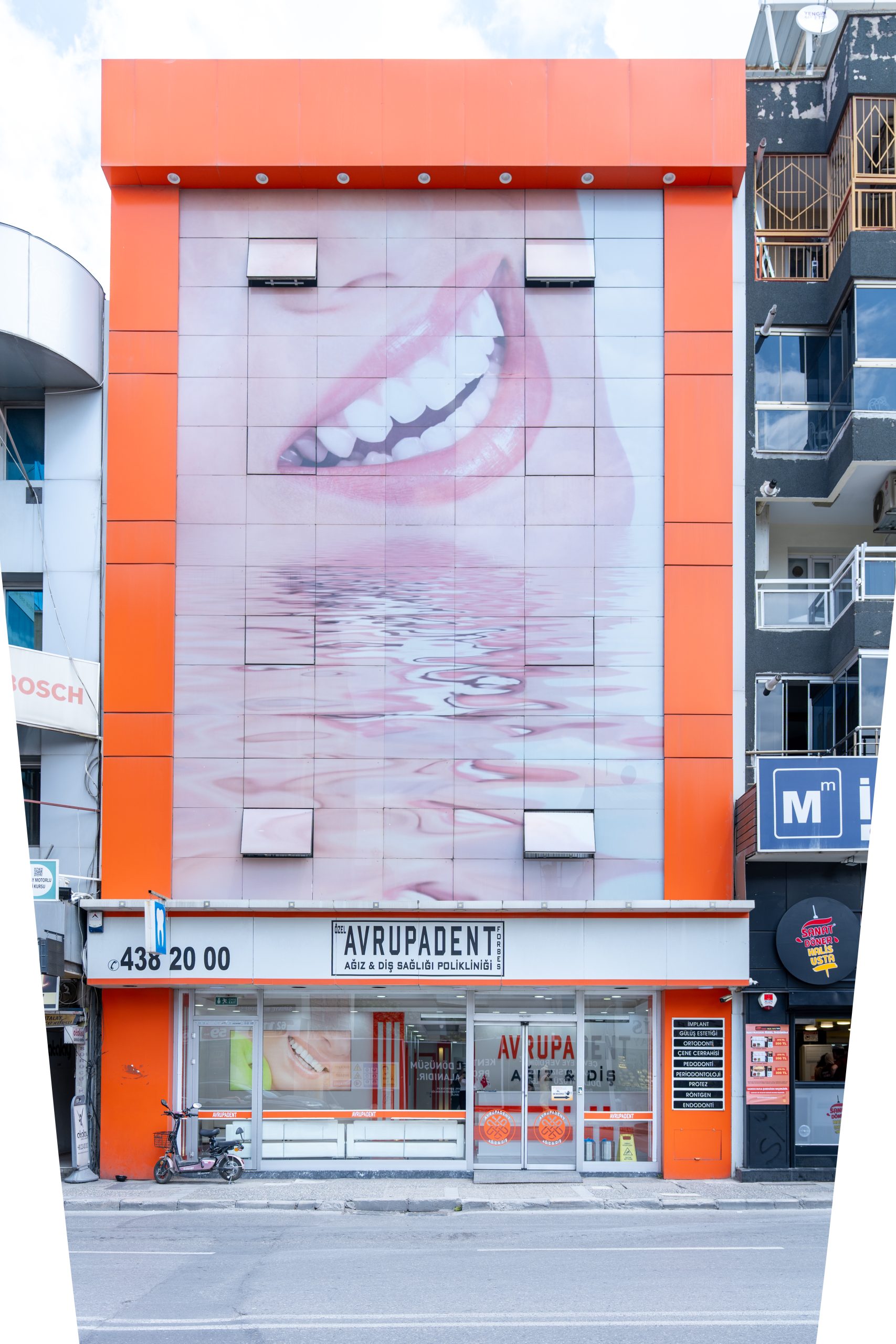
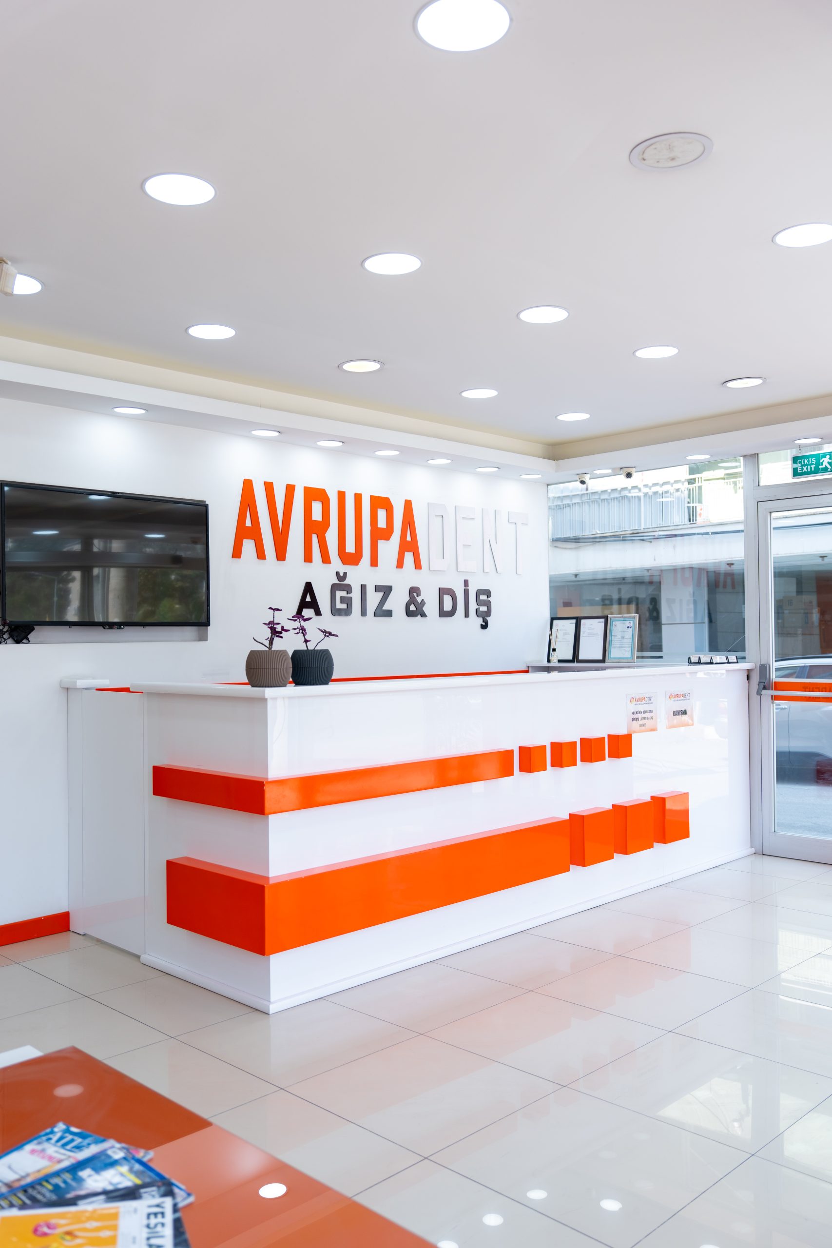
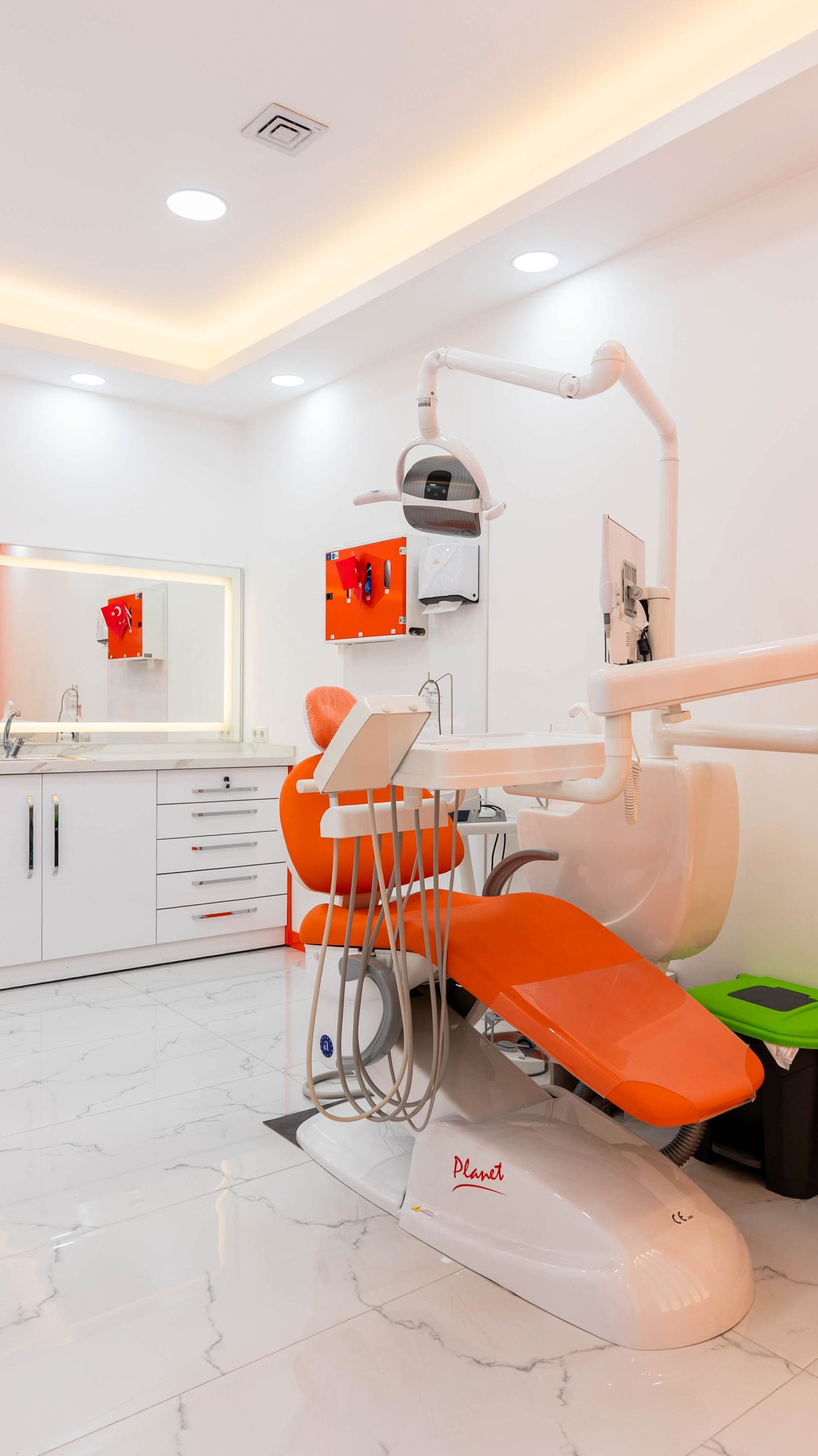
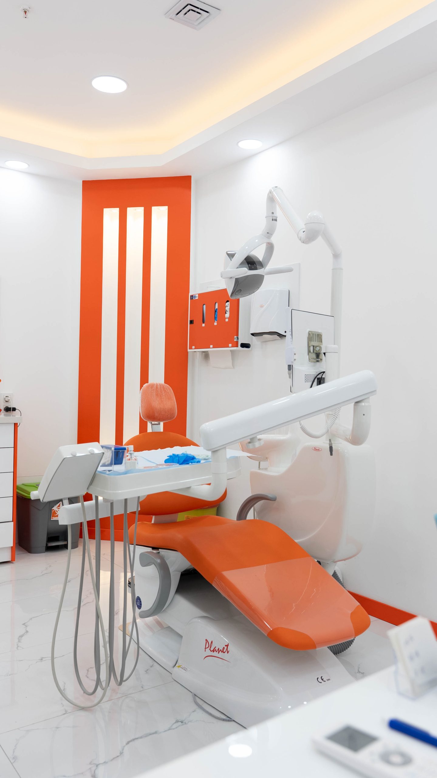
For individuals who want to get dental and oral health check-ups in the Izmir Buca Şirinyer Forbes area, the Avrupadent Buca Forbes branch offers a patient-centered health organization, where multiple specialty doctors collaborate, current clinical examination protocols are implemented, and medical hygiene standards are maintained.
Being located on major arterials is essential for individuals to attend consultations without delays. The facility located on Menderes Caddesi is within walking distance of the Forbes market area, Şirinyer İZBAN suburb station, and the neighborhood center. It is easily accessible to stops on the street served by municipal buses and minibus services from Konak, Buca Koop, and the Şirinyer transfer center.
Individuals arriving by personal vehicle can reach the clinic on Menderes Caddesi using the connecting roads of Yeşillik Caddesi or Buca Üçkuyular. The street around the branch aims to facilitate parking with vehicle parking pockets and nearby parking areas. The building’s interior arrangements are designed to support families with strollers and visitors with mobility impairments.
| Type of Transportation | Used Line / Route | Starting Point and Access |
|---|---|---|
| İZBAN Commuter Line | Aliağa – Cumaovasını / Tepeköy Main Line | Accessible by getting off at Şirinyer Station and walking distance |
| ESHOT Buses | Menderes Street and Şirinyer Transfer Routes | Stops on Menderes Street |
| Dolmuş / Minibus | Buca Statue – Şirinyer – Konak Minibus Routes | Passenger Drop-off Points on Menderes Street |
| Private Vehicle | Connection between Yeşillik Street and Menderes Street | Street-side Parking Areas and Nearby Parking Lots |
How is the First Appointment Scheduling and Clinical Registration Process Conducted?
The first application to the clinic is not just a step where the teeth are examined; it is a comprehensive registration process that analyzes the individual’s overall health profile. Factors such as blood pressure balance, blood sugar monitoring, cardiovascular history, or tissue sensitivities to certain substances are determining elements for the procedures the physician will perform and the solutions they will choose. Forms filled out at the consultation unit are simultaneously transferred to the physician’s examination screen.
The Avrupadent Buca Forbes branch accepts examinations without interruption from Monday to Saturday between 09:00 and 23:30. This working schedule, which continues until 23:30 at night, provides flexible appointment alternatives for workers and students who cannot find time after working hours or during the day.
- • The consultation staff includes the patient’s previous physician reports and examination data into the system.
- • The central staff organizes referrals to specialist physicians according to prosthetic, surgical, or orthodontic needs.
- • Thanks to the clinical database system, a comparative tracking basis is established for subsequent periodic check-ups.
What Evaluation Methods Are Applied in the First Examination of Oral and Dental Health?During the examination process, dental surfaces, gum pockets, soft tissues inside the mouth, and jaw closure relationships are examined with clinical methods. The bone level and root structures that cannot be detected visually are analyzed with radiological examinations to create a diagnosis chart by the physician.In dental practice, focusing solely on the tooth that is the source of complaints can overlook initial problems in neighboring tissues. At Avrupadent Buca Forbes clinic, the oral structure is treated as a whole. The physician meticulously evaluates the microscopic cracks in the tooth enamel, bacterial plaque at the edge of the gums, and the closure balance of the jaw joint.
The findings of the obtained radiological examination are evaluated together with the patient in the examination room. The physician clarifies the rationale and prioritization of the necessary procedures by expressing the current tissue condition in simple terms. This ensures that the individual aims to gain complete information about their treatment process.
Basic Examination Methods and Clinical Contributions Examination Method Scope of Examination Clinical Contribution Clinical Examination (Mirror and Probe) Surgical Findings, Surface Cuts, and Mobility Control Initial Detection of Physical Tissue Loss Periodontal Probing Depths of Gum Pockets and Bleeding Status Detection of Gum Recession and Bone Support Levels Radiological Analysis Root Tips, Jawbone Boundaries, and Impacted Teeth Examination of Lesions Not Visible to the Eye and Interfacial Cuts
How is Jaw Surgery and Wisdom Tooth Extraction Planned?
Due to space constraints within the jaw, wisdom teeth that cannot emerge or remain partially erupted are extracted through a planned surgical operation after clinical examination and radiological planning. The procedure is carried out considering the nerve pathways and anatomical cavities.When the third molars cannot find space in the jaw arch, they may remain horizontally positioned within the bone or may be in a partially erupted state. The gum folds around the partially erupted teeth can create a conducive environment for food debris accumulation, triggering localized tissue infections known as pericoronitis. Mechanical pressure applied to the root of the adjacent second molar can lead to root resorption.
Before the surgical intervention at the Buca Forbes branch, the distance between the nerve pathway running beneath the lower jaw and the tooth roots of the sinus floor in the upper jaw is examined. A small incision made under local anesthesia allows for the controlled extraction of the tooth, and the surgical area is disinfected and closed with stitches. The recovery period is monitored according to the care principles indicated by the physician.
• The distance to the mandibular nerve canal determines the surgical technique to be applied. • Tissues in areas with suspected cysts or lesions are removed by surgical operation. • Tissue healing is completed within an average of 7 to 10 days with the removal of stitches. What Approaches Are Adopted in the Clinical Environment for Individuals Experiencing Dental Anxiety (Dental Phobia)?
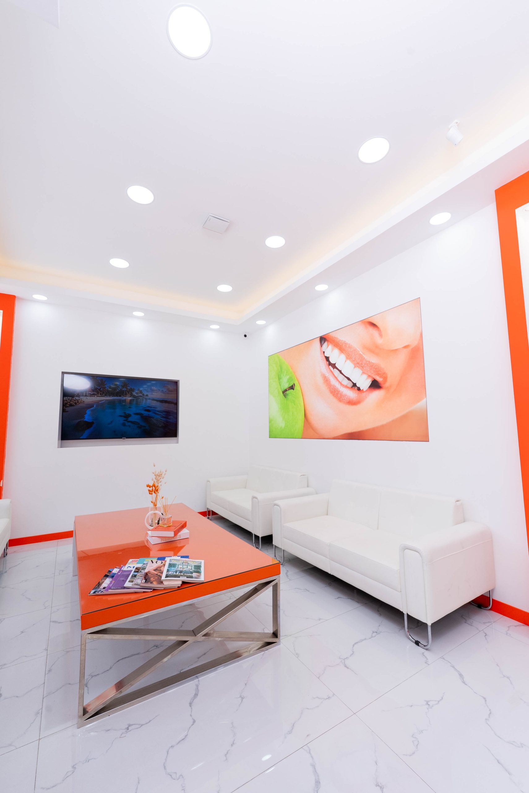
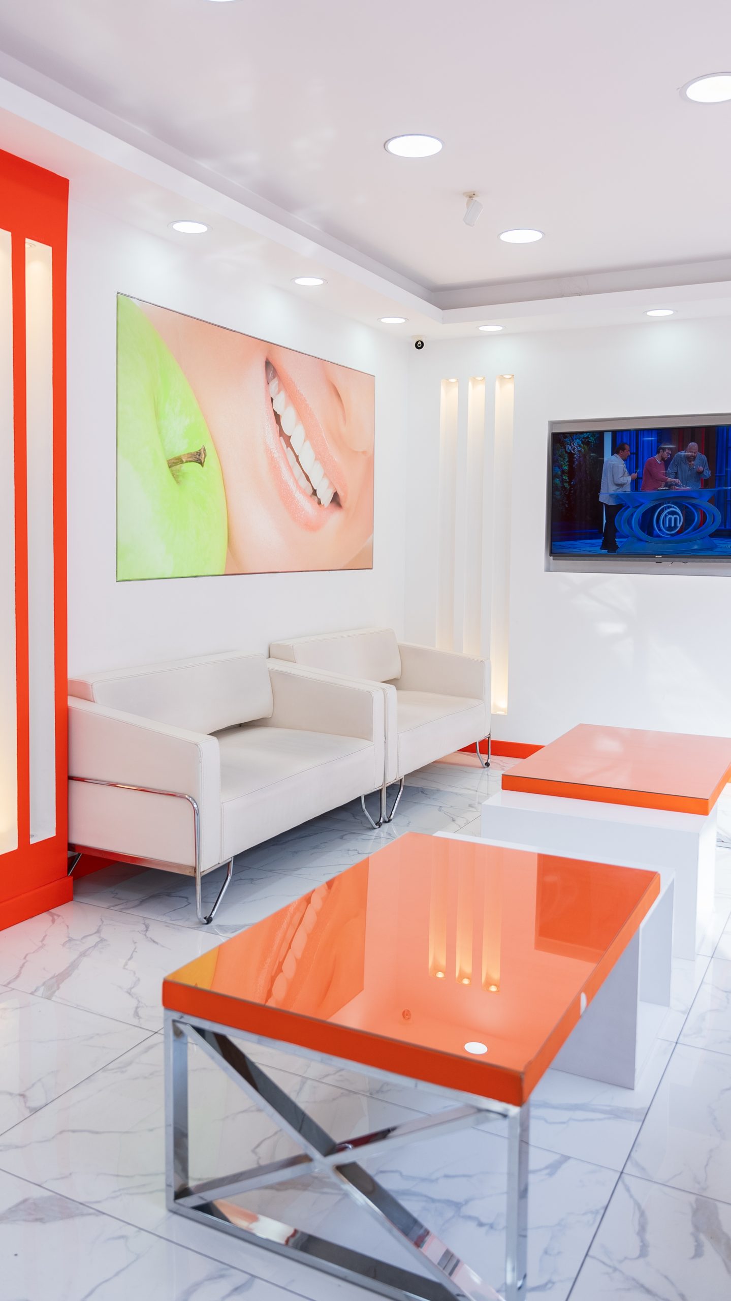
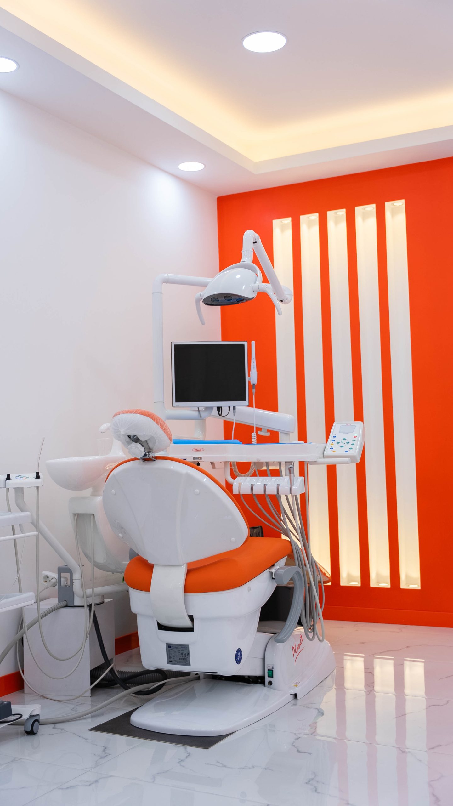
The calming interior design of the clinic helps alleviate feelings of stress; doctors inform the patient before each step of the procedure and introduce rest breaks that support a feeling of control during the session.The hesitation felt during the appointment process can stem from negative past experiences or the lack of knowledge about the steps of the procedure. In the Buca Forbes branch of Avrupadent, relaxation lounges are designed with simple colors and clean air circulation to help reduce tension factors. Acoustic insulation measures in treatment areas limit the spread of sounds to shared spaces.
A transparent dialogue is maintained in the examination chair. Aromatic gels applied to the tissue surface before local anesthesia reduce the sensation of stinging. During the session, a small hand signal from the patient to the doctor allows for a pause in the procedure, enabling progress within a collaborative working partnership.
Clinical Applications Supporting Anxiety Management Application Area Details and Explanation Topical Gel Usage Numbing the tissue surface before injection Step-by-Step Information Providing verbal information to the patient before each step Flexible Break System The session can be stopped at any desired time with the hospital’s hand gesture
In dental practice, focusing solely on the tooth that is the source of complaints can overlook initial problems in neighboring tissues. At Avrupadent Buca Forbes clinic, the oral structure is treated as a whole. The physician meticulously evaluates the microscopic cracks in the tooth enamel, bacterial plaque at the edge of the gums, and the closure balance of the jaw joint.
The findings of the obtained radiological examination are evaluated together with the patient in the examination room. The physician clarifies the rationale and prioritization of the necessary procedures by expressing the current tissue condition in simple terms. This ensures that the individual aims to gain complete information about their treatment process.
| Examination Method | Scope of Examination | Clinical Contribution |
|---|---|---|
| Clinical Examination (Mirror and Probe) | Surgical Findings, Surface Cuts, and Mobility Control | Initial Detection of Physical Tissue Loss |
| Periodontal Probing | Depths of Gum Pockets and Bleeding Status | Detection of Gum Recession and Bone Support Levels |
| Radiological Analysis | Root Tips, Jawbone Boundaries, and Impacted Teeth | Examination of Lesions Not Visible to the Eye and Interfacial Cuts |
How is Jaw Surgery and Wisdom Tooth Extraction Planned?
When the third molars cannot find space in the jaw arch, they may remain horizontally positioned within the bone or may be in a partially erupted state. The gum folds around the partially erupted teeth can create a conducive environment for food debris accumulation, triggering localized tissue infections known as pericoronitis. Mechanical pressure applied to the root of the adjacent second molar can lead to root resorption.
Before the surgical intervention at the Buca Forbes branch, the distance between the nerve pathway running beneath the lower jaw and the tooth roots of the sinus floor in the upper jaw is examined. A small incision made under local anesthesia allows for the controlled extraction of the tooth, and the surgical area is disinfected and closed with stitches. The recovery period is monitored according to the care principles indicated by the physician.
What Approaches Are Adopted in the Clinical Environment for Individuals Experiencing Dental Anxiety (Dental Phobia)?
The hesitation felt during the appointment process can stem from negative past experiences or the lack of knowledge about the steps of the procedure. In the Buca Forbes branch of Avrupadent, relaxation lounges are designed with simple colors and clean air circulation to help reduce tension factors. Acoustic insulation measures in treatment areas limit the spread of sounds to shared spaces.
A transparent dialogue is maintained in the examination chair. Aromatic gels applied to the tissue surface before local anesthesia reduce the sensation of stinging. During the session, a small hand signal from the patient to the doctor allows for a pause in the procedure, enabling progress within a collaborative working partnership.
| Application Area | Details and Explanation |
|---|---|
| Topical Gel Usage | Numbing the tissue surface before injection |
| Step-by-Step Information | Providing verbal information to the patient before each step |
| Flexible Break System | The session can be stopped at any desired time with the hospital’s hand gesture |
What Standards Are Followed for Clinical Internal Sterilization and Hygiene Procedures?
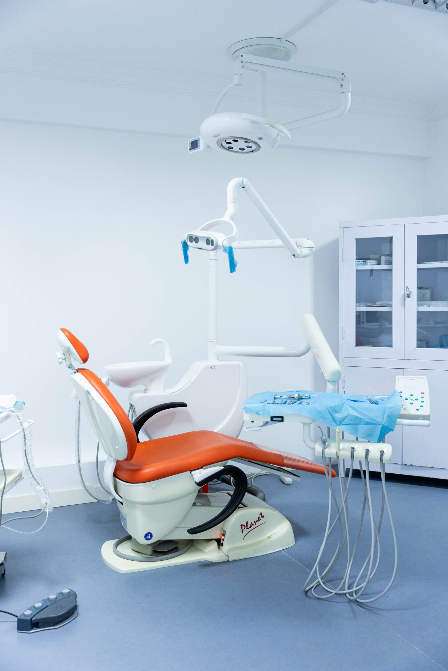
Preventing the passage of microorganisms in a clinical environment is the fundamental basis of health services. The infection control protocol is strictly applied at the Buca Forbes branch after each session. Used surgical and examination materials are transported in closed containers to the central cleaning area and soaked in enzymatic liquids for the purpose of organic residue dissolution.
The cleaned instruments are placed in airtight packages and passed through high temperature and saturated steam stages. The color-changing items on the packaging document are only opened at the patient’s head. Seating units and surfaces that come into contact with hands are cleaned with medical antiseptic solutions with each patient change.
- ■ Patient utensils, cups, syringes, and aspirator tools are designed for single use.
- ■ Steam sterilization processes are monitored through independent laboratory tests at regular intervals.
- ■ The air in clinical indoor spaces is maintained under a fresh air flow through filtration channels.
What Steps Does the Implant Procedure for Missing Teeth Involve?Implant treatment is based on placing biocompatible titanium screws into the jawbone to replace the lost teeth, followed by the attachment of a prosthesis after osseointegration occurs. It fills the gap without needing to remove neighboring healthy teeth.Missing teeth can disrupt the chewing mechanism, leading to tilting of adjacent teeth over time and bone tissue thinning. Before the surgical procedure at the Buca Forbes branch, the width and density of the jawbone are examined. In cases where the bone volume is insufficient, tissue support can be provided through bone powder (grafting) applications.
The titanium structure placed in the bone socket under local anesthesia integrates with the bone tissue at a cellular level (osseointegration). This waiting period lasts a few months depending on the individual’s metabolism and the jaw area. Once the healing phase is completed, the prosthesis phase begins.
Stages of Implant Treatment and Expected Timeline Stage Number Summary of the Procedure Estimated Duration 1. Surgical Placement Placement of the titanium screw body into the bone socket Session duration is approximately 20-40 minutes 2. Bone Integration Osteointegration (the attachment of bone cells to the screw) 2-3 months for the lower jaw, 3-6 months for the upper jaw 3. Gum Shaping Placement of healing abutment to create a natural gum profile 7-10 days of tissue adaptation period
How is the Prosthetic Phase and Superstructure Design Realized After Implant?
To attach a crown on implants with completed chemical bonding, precise intraoral measurements are taken. Zirconium or porcelain crowns, prepared considering chewing forces and aesthetic appearance, are secured to the implant with a connecting piece.The prosthetic rehabilitation phase is the stage where function and visual balance are restored. Prosthetic dentists at Buca Forbes clinic model the angle of the implant and the form of neighboring teeth based on the measurements taken. The crowns prepared in the laboratory are attached to the implant either by bonding or screwing.
After the prosthesis is placed, the balance of the occlusal contact points is meticulously checked. Measures are taken to prevent excessive loading to protect the implant body. Additionally, the contact points of dental floss and interproximal brushes are tested to plan for the prevention of food accumulation.
- ■ Zirconium surfaces are recommended for aesthetic reflection in anterior teeth.
- ■ Balanced occlusion force distribution is ensured by performing closure tests.
- ■ Daily care methods that protect the health of the gum around the implant are explained.
How is the Clear Aligner Method Applied in Orthodontic Disorders?
The transparent plate application, which is an orthodontic approach, is the gradual alignment of teeth using a series of custom-designed clear plates. It is primarily preferred by individuals seeking aesthetic expectations since it does not contain metal parts.The direction of tooth movement is planned based on precise measurements taken by the orthodontist. The prepared plate series is changed at specific intervals (usually every 7-10 days), applying light and controlled force to the teeth. This way, tooth misalignments and gaps are progressively corrected.
The plates can be removed while eating or during dental cleaning. This eliminates the food restrictions that traditional metal braces impose and allows for uninterrupted oral hygiene. To achieve the expected change, it is recommended that plates be worn for 20-22 hours a day.
Reflections of the Transparent Plate Method on Daily Life Evaluated Dimension Transparent Plate Feature External Appearance Does not show clarity during conversation with transparent material Oral Hygiene Routine tooth brushing and flossing are carried out thoroughly Eating and Drinking There are no strict food restrictions for what is taken out on that day
In Which Dental Arrangements Are Traditional Wire and Bracket Systems Preferred?In cases of advanced skeletal mismatches, alignment disorders requiring complex root movements, or situations where teeth remain within the bone’s eruption path, classic brackets and archwires are among the primary solutions.The brackets being fixed to the tooth surface and supported by archwires ensures that the roots are oriented at the targeted angle within the bone. In addition to standard metal brackets, ceramic or sapphire bracket materials can also be used for patients looking for an aesthetic appearance.
Fixed brackets are recommended to prevent the accumulation of food residues as long as they remain in the mouth. During check-up sessions, the tension of the wires is adjusted by the doctor to coordinate the alignment of the teeth to reach the targeted position step by step.
\t- ■ Provides effective mechanical control in cases of open bite and deep bite.
\t
- ■ Continuous force application is possible in younger age groups because it does not require compliance.
\t
- ■ To prevent brackets from being damaged, it is recommended to avoid consuming very hard and sticky foods.
What Factors Shape Prosthetic Applications in the Context of Aesthetic Smile Design?In the personalized smile profile; planning is done by referencing lip line, facial anatomy, skin color, and gum level. Porcelain laminates, gum level adjustments, and color adaptations are combined into a balanced composition.Smile design is a collaborative effort between prosthetic and surgical disciplines aimed at achieving harmony between an individual’s facial features and teeth. In the Buca Forbes branch, the patient’s lip movements during speaking and smiling are analyzed. In cases where the gum appears overly prominent during smiling, symmetry is achieved through pink aesthetics (gingivectomy) measures.
Before proceeding with permanent crown or laminate procedures, temporary models can be used for a mock-up trial in the mouth. The individual can see the designed form in their mouth, share their thoughts with the doctor, and the permanent porcelain production is completed based on this feedback.
Porcelain Laminate (Leaf Porcelain)
These are transparent porcelains that are applied only to the front surface of the teeth, providing color and shape changes with minimal tissue removal.
Gingivoplasty / Pink Aesthetics
It involves leveling and balancing the small tissue adjustments of asymmetric gum margins to the same height.
What Are the Structural Differences Between Zirconium and Porcelain Coating Options?
Classic metal framework porcelain provides durability but may be limited in light transmission; zirconium-backed crowns, thanks to their white cores, show strength against chewing forces while also providing light transmission close to that of natural enamel.When selecting the crown material, the position of the tooth to be restored, chewing force, and gum compatibility are taken into account. Over time, crowns with a metal framework may create a gray reflection along the gum line. Zirconium dioxide, on the other hand, does not cause discoloration of the gums due to its tissue-friendly properties and provides a natural depth.
At the Buca Forbes clinic, zirconium and porcelain materials are evaluated according to the hospital’s needs in both anterior esthetic areas and posterior molar teeth. Non-metal-containing zirconium structures also offer an alternative for individuals with metal sensitivities.
Material Selection Criteria and Qualities Feature Metal-Backed Porcelain Zirconium Crown Light Transmission Can provide a matte appearance without reflecting light from the metal support The white substructure allows for natural depth by transmitting light Gum Adaptation Grayness can be seen at the gum line over time Tissue friendly, does not cause discoloration at the gum edges Usage Area More often evaluated in the back molars Applied in both the front aesthetic area and the rear chewing group
How Are Root Canal (Endodontic) Procedures Carried Out in the Case of Dental Crisis?
Root canal treatment is the procedure of cleaning microorganisms that reach as far as the dental pulp, shaping the canals for purification, and then filling them in a leak-proof manner using tissue-friendly materials.Deep cavities penetrating the pulp cavity by extending through the enamel and dentin layers may manifest with throbbing, sensitivity to heat and cold, and nighttime pain. In the examination, canal lengths are determined using special measurement methods and radiological images.
Inflamed tissues are removed from the root canals and the canals are rinsed with disinfectant solutions. After the empty spaces are sealed with gutta-percha material, fillings or crown restorations are made depending on the tissue loss in the tooth to preserve the tooth’s presence in the jaw.
- • Comfort is ensured throughout the session because it is performed under local anesthesia.
- • In cases where a lesion is found at the root tip, drug can be placed in the canal for follow-up sessions.
- • The procedure of cleaning and re-filling old problem canals is called “canal revision” (retreatment).
How is Periodontal Care Managed in Cases of Gum Inflammation and Extraction?
Gum health is maintained by the professional removal of bacterial plaque and mineralized tartar that accumulates on the surface. In cases where inflammation progresses to the root surface and surrounding bone tissue, curettage and root planing techniques are applied.Bleeding during brushing and bad breath are signs that the accumulated plaque has turned into tartar. These hardened structures cannot be cleaned with at-home brushing. They are removed from the enamel surface through cleaning procedures performed in the clinic, resulting in a polished, smooth tissue.
In advanced cases of periodontitis, the gum pocket deepens, and bone tissue erosion can be observed. At this stage, the dentist cleans the inflamed tissues below the gums with special instruments, smoothing the root surface. This aims to allow the gum to reattach firmly to the tooth.
Gingivitis (Initial Stage)
It is an inflammatory condition characterized by redness and bleeding at the gum line, where bone loss has not yet occurred.
Periodontitis (Advanced Stage)
This stage occurs when the inflammation reaches the jawbone, leading to deeper pockets and a risk of tooth mobility.
What Does Preventive Dental Care for Children (Pedodontics) Include?
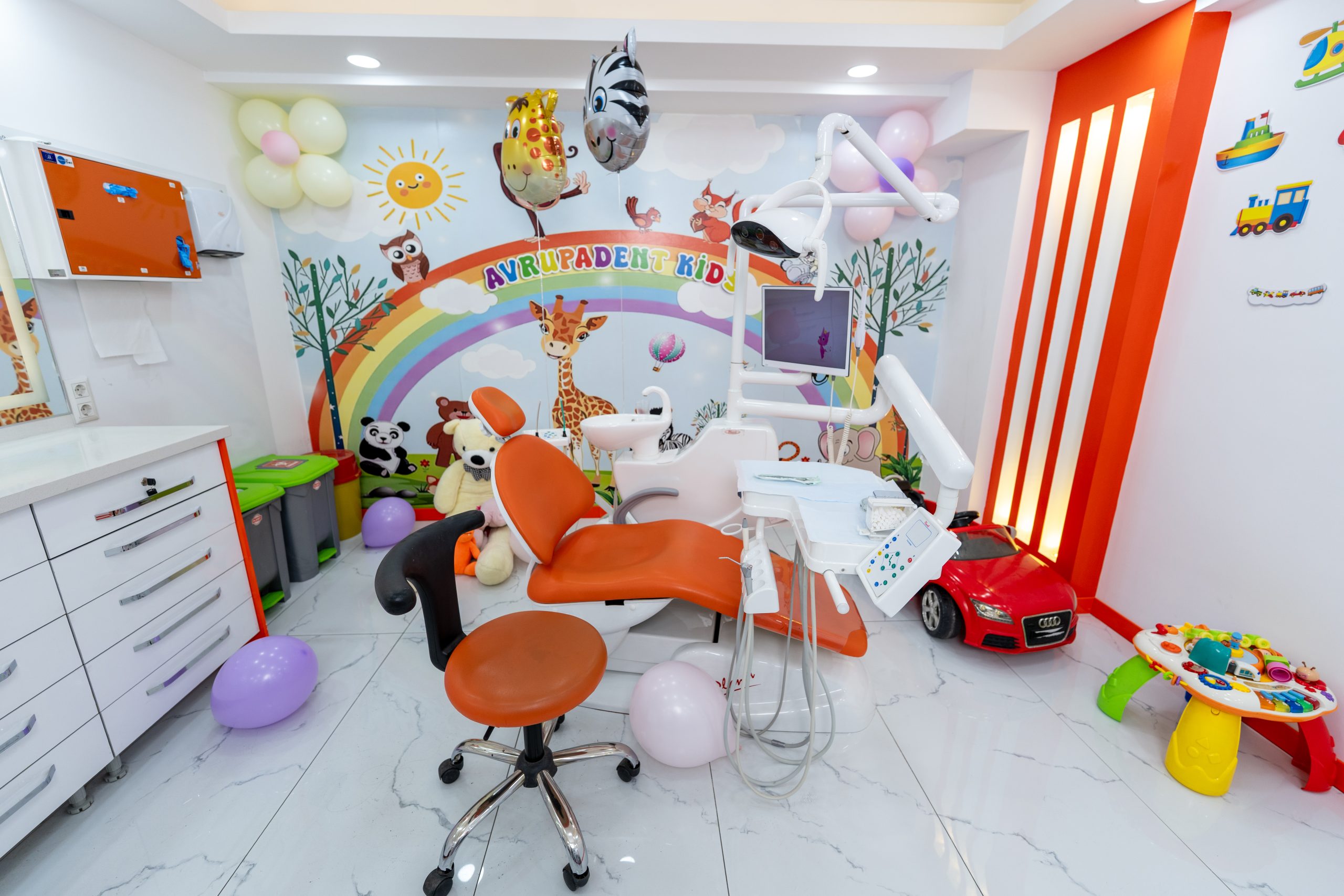
The primary goal of pediatric dentistry is to implement preventive methods that prevent the formation of cavities. The preservation of primary teeth is ensured through fissure sealants, space maintainers, and fluoride varnish that strengthens tooth enamel.Primary teeth serve as a guide for the permanent teeth that will come in. Premature loss of primary teeth can lead to the shifting of adjacent teeth into the gap and cause misalignment of the permanent teeth. Communication with pediatric patients in the clinic should be conducted in a friendly manner to prevent the development of anxiety.
The deep grooves on the chewing surfaces of molars are sealed with fissure sealants to reduce food accumulation. While fluoride applications strengthen the enamel structure against acid attacks, space maintainers placed in the gaps of early-extracted primary teeth keep the pathway for permanent teeth open.
Protective Pedodontic Approaches Method Name Summary of the Procedure Expected Benefit Fluoride Varnish Application Application of protective varnish on the enamel surface Increase the resistance of enamel to acid attacks Fissure Sealant Aligning the flow of the cavity with the lining material Preserving the chewing surface by reducing food retention Placeholder Device Placement of the shaped crown in the cavity Preventing the narrowing of the cavity and maintaining the permanent tooth space
What Principles Determine Costing and Pricing Policy in Dental Treatments?
In clinical services, pricing is determined based on the Turkish Dental Association (TDB) guideline tariffs according to the patient’s mouth structure, the number of required sessions, and the quality of materials to be used. The planned treatment steps are communicated to the patient in writing after the physician’s assessment.Due to the variations in individuals’ jaw structure, gum level, and tissue requirements in dental practices, a predetermined price list cannot be established. The clinical assessment conducted by the physician during the initial examination and the resulting radiological findings clarify the procedures to be performed. For example, if there is insufficient bone support in implant surgery, additional steps such as sinus elevation or grafting may be necessary.
At the Avrupadent Buca Forbes branch, the treatment plan prepared is explained item by item, and all stages of the process are shared transparently. The properties of the restorative materials to be used are presented to the patient in detail.
- ■ A treatment plan will be created and presented to the patient before the procedure begins, allowing for predictions.
- ■ Comparable alternative materials (for example, zirconium and porcelain) are presented comparatively.
How to Access the Full Address and Appointment Information of Avrupadent Buca Forbes Branch?
The clinic provides services at Yiğitler Neighborhood, Menderes Street No:220 Şirinyer, Buca / İzmir. For information and appointment requests, you can use the call center at +90 232 438 20 00, branch support number +90 553 832 60 35, or corporate WhatsApp channel at +90 553 832 60 35.The branch accepts uninterrupted examinations between 09:00 and 23:30 from Monday to Saturday. It is closed on Sundays. Individuals attending their first examination should bring their previous health records or any recent radiological examinations to assist the physician’s evaluation.
Summary of Clinic Communication Channels and Location Contact Information Information Description Open Address Yiğitler Mah. Menderes Cad. No:220 Şirinyer, Buca / İzmir Call Center & Switchboard Switchboard: +90 232 438 20 00 | Branch: +90 553 832 60 35 Message & Email WhatsApp: +90 553 832 60 35 | Email: info@avrupadent.com.tr Working Days and Hours Monday – Saturday: 09:00 – 23:30 (Closed on Sunday)
Missing teeth can disrupt the chewing mechanism, leading to tilting of adjacent teeth over time and bone tissue thinning. Before the surgical procedure at the Buca Forbes branch, the width and density of the jawbone are examined. In cases where the bone volume is insufficient, tissue support can be provided through bone powder (grafting) applications.
The titanium structure placed in the bone socket under local anesthesia integrates with the bone tissue at a cellular level (osseointegration). This waiting period lasts a few months depending on the individual’s metabolism and the jaw area. Once the healing phase is completed, the prosthesis phase begins.
| Stage Number | Summary of the Procedure | Estimated Duration |
|---|---|---|
| 1. Surgical Placement | Placement of the titanium screw body into the bone socket | Session duration is approximately 20-40 minutes |
| 2. Bone Integration | Osteointegration (the attachment of bone cells to the screw) | 2-3 months for the lower jaw, 3-6 months for the upper jaw |
| 3. Gum Shaping | Placement of healing abutment to create a natural gum profile | 7-10 days of tissue adaptation period |
How is the Prosthetic Phase and Superstructure Design Realized After Implant?
The prosthetic rehabilitation phase is the stage where function and visual balance are restored. Prosthetic dentists at Buca Forbes clinic model the angle of the implant and the form of neighboring teeth based on the measurements taken. The crowns prepared in the laboratory are attached to the implant either by bonding or screwing.
After the prosthesis is placed, the balance of the occlusal contact points is meticulously checked. Measures are taken to prevent excessive loading to protect the implant body. Additionally, the contact points of dental floss and interproximal brushes are tested to plan for the prevention of food accumulation.
- ■ Zirconium surfaces are recommended for aesthetic reflection in anterior teeth.
- ■ Balanced occlusion force distribution is ensured by performing closure tests.
- ■ Daily care methods that protect the health of the gum around the implant are explained.
How is the Clear Aligner Method Applied in Orthodontic Disorders?
The direction of tooth movement is planned based on precise measurements taken by the orthodontist. The prepared plate series is changed at specific intervals (usually every 7-10 days), applying light and controlled force to the teeth. This way, tooth misalignments and gaps are progressively corrected.
The plates can be removed while eating or during dental cleaning. This eliminates the food restrictions that traditional metal braces impose and allows for uninterrupted oral hygiene. To achieve the expected change, it is recommended that plates be worn for 20-22 hours a day.
| Evaluated Dimension | Transparent Plate Feature |
|---|---|
| External Appearance | Does not show clarity during conversation with transparent material |
| Oral Hygiene | Routine tooth brushing and flossing are carried out thoroughly |
| Eating and Drinking | There are no strict food restrictions for what is taken out on that day |
In Which Dental Arrangements Are Traditional Wire and Bracket Systems Preferred?In cases of advanced skeletal mismatches, alignment disorders requiring complex root movements, or situations where teeth remain within the bone’s eruption path, classic brackets and archwires are among the primary solutions.The brackets being fixed to the tooth surface and supported by archwires ensures that the roots are oriented at the targeted angle within the bone. In addition to standard metal brackets, ceramic or sapphire bracket materials can also be used for patients looking for an aesthetic appearance.
Fixed brackets are recommended to prevent the accumulation of food residues as long as they remain in the mouth. During check-up sessions, the tension of the wires is adjusted by the doctor to coordinate the alignment of the teeth to reach the targeted position step by step.
\t- ■ Provides effective mechanical control in cases of open bite and deep bite.
\t
- ■ Continuous force application is possible in younger age groups because it does not require compliance.
\t
- ■ To prevent brackets from being damaged, it is recommended to avoid consuming very hard and sticky foods.
What Factors Shape Prosthetic Applications in the Context of Aesthetic Smile Design?In the personalized smile profile; planning is done by referencing lip line, facial anatomy, skin color, and gum level. Porcelain laminates, gum level adjustments, and color adaptations are combined into a balanced composition.Smile design is a collaborative effort between prosthetic and surgical disciplines aimed at achieving harmony between an individual’s facial features and teeth. In the Buca Forbes branch, the patient’s lip movements during speaking and smiling are analyzed. In cases where the gum appears overly prominent during smiling, symmetry is achieved through pink aesthetics (gingivectomy) measures.
Before proceeding with permanent crown or laminate procedures, temporary models can be used for a mock-up trial in the mouth. The individual can see the designed form in their mouth, share their thoughts with the doctor, and the permanent porcelain production is completed based on this feedback.
Porcelain Laminate (Leaf Porcelain)
These are transparent porcelains that are applied only to the front surface of the teeth, providing color and shape changes with minimal tissue removal.
Gingivoplasty / Pink Aesthetics
It involves leveling and balancing the small tissue adjustments of asymmetric gum margins to the same height.
What Are the Structural Differences Between Zirconium and Porcelain Coating Options?
Classic metal framework porcelain provides durability but may be limited in light transmission; zirconium-backed crowns, thanks to their white cores, show strength against chewing forces while also providing light transmission close to that of natural enamel.When selecting the crown material, the position of the tooth to be restored, chewing force, and gum compatibility are taken into account. Over time, crowns with a metal framework may create a gray reflection along the gum line. Zirconium dioxide, on the other hand, does not cause discoloration of the gums due to its tissue-friendly properties and provides a natural depth.
At the Buca Forbes clinic, zirconium and porcelain materials are evaluated according to the hospital’s needs in both anterior esthetic areas and posterior molar teeth. Non-metal-containing zirconium structures also offer an alternative for individuals with metal sensitivities.
Material Selection Criteria and Qualities Feature Metal-Backed Porcelain Zirconium Crown Light Transmission Can provide a matte appearance without reflecting light from the metal support The white substructure allows for natural depth by transmitting light Gum Adaptation Grayness can be seen at the gum line over time Tissue friendly, does not cause discoloration at the gum edges Usage Area More often evaluated in the back molars Applied in both the front aesthetic area and the rear chewing group
How Are Root Canal (Endodontic) Procedures Carried Out in the Case of Dental Crisis?
Root canal treatment is the procedure of cleaning microorganisms that reach as far as the dental pulp, shaping the canals for purification, and then filling them in a leak-proof manner using tissue-friendly materials.Deep cavities penetrating the pulp cavity by extending through the enamel and dentin layers may manifest with throbbing, sensitivity to heat and cold, and nighttime pain. In the examination, canal lengths are determined using special measurement methods and radiological images.
Inflamed tissues are removed from the root canals and the canals are rinsed with disinfectant solutions. After the empty spaces are sealed with gutta-percha material, fillings or crown restorations are made depending on the tissue loss in the tooth to preserve the tooth’s presence in the jaw.
- • Comfort is ensured throughout the session because it is performed under local anesthesia.
- • In cases where a lesion is found at the root tip, drug can be placed in the canal for follow-up sessions.
- • The procedure of cleaning and re-filling old problem canals is called “canal revision” (retreatment).
How is Periodontal Care Managed in Cases of Gum Inflammation and Extraction?
Gum health is maintained by the professional removal of bacterial plaque and mineralized tartar that accumulates on the surface. In cases where inflammation progresses to the root surface and surrounding bone tissue, curettage and root planing techniques are applied.Bleeding during brushing and bad breath are signs that the accumulated plaque has turned into tartar. These hardened structures cannot be cleaned with at-home brushing. They are removed from the enamel surface through cleaning procedures performed in the clinic, resulting in a polished, smooth tissue.
In advanced cases of periodontitis, the gum pocket deepens, and bone tissue erosion can be observed. At this stage, the dentist cleans the inflamed tissues below the gums with special instruments, smoothing the root surface. This aims to allow the gum to reattach firmly to the tooth.
Gingivitis (Initial Stage)
It is an inflammatory condition characterized by redness and bleeding at the gum line, where bone loss has not yet occurred.
Periodontitis (Advanced Stage)
This stage occurs when the inflammation reaches the jawbone, leading to deeper pockets and a risk of tooth mobility.
What Does Preventive Dental Care for Children (Pedodontics) Include?
The primary goal of pediatric dentistry is to implement preventive methods that prevent the formation of cavities. The preservation of primary teeth is ensured through fissure sealants, space maintainers, and fluoride varnish that strengthens tooth enamel.Primary teeth serve as a guide for the permanent teeth that will come in. Premature loss of primary teeth can lead to the shifting of adjacent teeth into the gap and cause misalignment of the permanent teeth. Communication with pediatric patients in the clinic should be conducted in a friendly manner to prevent the development of anxiety.
The deep grooves on the chewing surfaces of molars are sealed with fissure sealants to reduce food accumulation. While fluoride applications strengthen the enamel structure against acid attacks, space maintainers placed in the gaps of early-extracted primary teeth keep the pathway for permanent teeth open.
Protective Pedodontic Approaches Method Name Summary of the Procedure Expected Benefit Fluoride Varnish Application Application of protective varnish on the enamel surface Increase the resistance of enamel to acid attacks Fissure Sealant Aligning the flow of the cavity with the lining material Preserving the chewing surface by reducing food retention Placeholder Device Placement of the shaped crown in the cavity Preventing the narrowing of the cavity and maintaining the permanent tooth space
What Principles Determine Costing and Pricing Policy in Dental Treatments?
In clinical services, pricing is determined based on the Turkish Dental Association (TDB) guideline tariffs according to the patient’s mouth structure, the number of required sessions, and the quality of materials to be used. The planned treatment steps are communicated to the patient in writing after the physician’s assessment.Due to the variations in individuals’ jaw structure, gum level, and tissue requirements in dental practices, a predetermined price list cannot be established. The clinical assessment conducted by the physician during the initial examination and the resulting radiological findings clarify the procedures to be performed. For example, if there is insufficient bone support in implant surgery, additional steps such as sinus elevation or grafting may be necessary.
At the Avrupadent Buca Forbes branch, the treatment plan prepared is explained item by item, and all stages of the process are shared transparently. The properties of the restorative materials to be used are presented to the patient in detail.
- ■ A treatment plan will be created and presented to the patient before the procedure begins, allowing for predictions.
- ■ Comparable alternative materials (for example, zirconium and porcelain) are presented comparatively.
How to Access the Full Address and Appointment Information of Avrupadent Buca Forbes Branch?
The clinic provides services at Yiğitler Neighborhood, Menderes Street No:220 Şirinyer, Buca / İzmir. For information and appointment requests, you can use the call center at +90 232 438 20 00, branch support number +90 553 832 60 35, or corporate WhatsApp channel at +90 553 832 60 35.The branch accepts uninterrupted examinations between 09:00 and 23:30 from Monday to Saturday. It is closed on Sundays. Individuals attending their first examination should bring their previous health records or any recent radiological examinations to assist the physician’s evaluation.
Summary of Clinic Communication Channels and Location Contact Information Information Description Open Address Yiğitler Mah. Menderes Cad. No:220 Şirinyer, Buca / İzmir Call Center & Switchboard Switchboard: +90 232 438 20 00 | Branch: +90 553 832 60 35 Message & Email WhatsApp: +90 553 832 60 35 | Email: info@avrupadent.com.tr Working Days and Hours Monday – Saturday: 09:00 – 23:30 (Closed on Sunday)
The brackets being fixed to the tooth surface and supported by archwires ensures that the roots are oriented at the targeted angle within the bone. In addition to standard metal brackets, ceramic or sapphire bracket materials can also be used for patients looking for an aesthetic appearance.
Fixed brackets are recommended to prevent the accumulation of food residues as long as they remain in the mouth. During check-up sessions, the tension of the wires is adjusted by the doctor to coordinate the alignment of the teeth to reach the targeted position step by step.
-
\t
- ■ Provides effective mechanical control in cases of open bite and deep bite.
- ■ Continuous force application is possible in younger age groups because it does not require compliance.
- ■ To prevent brackets from being damaged, it is recommended to avoid consuming very hard and sticky foods.
\t
\t
What Factors Shape Prosthetic Applications in the Context of Aesthetic Smile Design?In the personalized smile profile; planning is done by referencing lip line, facial anatomy, skin color, and gum level. Porcelain laminates, gum level adjustments, and color adaptations are combined into a balanced composition.Smile design is a collaborative effort between prosthetic and surgical disciplines aimed at achieving harmony between an individual’s facial features and teeth. In the Buca Forbes branch, the patient’s lip movements during speaking and smiling are analyzed. In cases where the gum appears overly prominent during smiling, symmetry is achieved through pink aesthetics (gingivectomy) measures.
Before proceeding with permanent crown or laminate procedures, temporary models can be used for a mock-up trial in the mouth. The individual can see the designed form in their mouth, share their thoughts with the doctor, and the permanent porcelain production is completed based on this feedback.
Porcelain Laminate (Leaf Porcelain)
These are transparent porcelains that are applied only to the front surface of the teeth, providing color and shape changes with minimal tissue removal.
Gingivoplasty / Pink Aesthetics
It involves leveling and balancing the small tissue adjustments of asymmetric gum margins to the same height.
What Are the Structural Differences Between Zirconium and Porcelain Coating Options?
Classic metal framework porcelain provides durability but may be limited in light transmission; zirconium-backed crowns, thanks to their white cores, show strength against chewing forces while also providing light transmission close to that of natural enamel.When selecting the crown material, the position of the tooth to be restored, chewing force, and gum compatibility are taken into account. Over time, crowns with a metal framework may create a gray reflection along the gum line. Zirconium dioxide, on the other hand, does not cause discoloration of the gums due to its tissue-friendly properties and provides a natural depth.
At the Buca Forbes clinic, zirconium and porcelain materials are evaluated according to the hospital’s needs in both anterior esthetic areas and posterior molar teeth. Non-metal-containing zirconium structures also offer an alternative for individuals with metal sensitivities.
Material Selection Criteria and Qualities Feature Metal-Backed Porcelain Zirconium Crown Light Transmission Can provide a matte appearance without reflecting light from the metal support The white substructure allows for natural depth by transmitting light Gum Adaptation Grayness can be seen at the gum line over time Tissue friendly, does not cause discoloration at the gum edges Usage Area More often evaluated in the back molars Applied in both the front aesthetic area and the rear chewing group
How Are Root Canal (Endodontic) Procedures Carried Out in the Case of Dental Crisis?
Root canal treatment is the procedure of cleaning microorganisms that reach as far as the dental pulp, shaping the canals for purification, and then filling them in a leak-proof manner using tissue-friendly materials.Deep cavities penetrating the pulp cavity by extending through the enamel and dentin layers may manifest with throbbing, sensitivity to heat and cold, and nighttime pain. In the examination, canal lengths are determined using special measurement methods and radiological images.
Inflamed tissues are removed from the root canals and the canals are rinsed with disinfectant solutions. After the empty spaces are sealed with gutta-percha material, fillings or crown restorations are made depending on the tissue loss in the tooth to preserve the tooth’s presence in the jaw.
- • Comfort is ensured throughout the session because it is performed under local anesthesia.
- • In cases where a lesion is found at the root tip, drug can be placed in the canal for follow-up sessions.
- • The procedure of cleaning and re-filling old problem canals is called “canal revision” (retreatment).
How is Periodontal Care Managed in Cases of Gum Inflammation and Extraction?
Gum health is maintained by the professional removal of bacterial plaque and mineralized tartar that accumulates on the surface. In cases where inflammation progresses to the root surface and surrounding bone tissue, curettage and root planing techniques are applied.Bleeding during brushing and bad breath are signs that the accumulated plaque has turned into tartar. These hardened structures cannot be cleaned with at-home brushing. They are removed from the enamel surface through cleaning procedures performed in the clinic, resulting in a polished, smooth tissue.
In advanced cases of periodontitis, the gum pocket deepens, and bone tissue erosion can be observed. At this stage, the dentist cleans the inflamed tissues below the gums with special instruments, smoothing the root surface. This aims to allow the gum to reattach firmly to the tooth.
Gingivitis (Initial Stage)
It is an inflammatory condition characterized by redness and bleeding at the gum line, where bone loss has not yet occurred.
Periodontitis (Advanced Stage)
This stage occurs when the inflammation reaches the jawbone, leading to deeper pockets and a risk of tooth mobility.
What Does Preventive Dental Care for Children (Pedodontics) Include?
The primary goal of pediatric dentistry is to implement preventive methods that prevent the formation of cavities. The preservation of primary teeth is ensured through fissure sealants, space maintainers, and fluoride varnish that strengthens tooth enamel.Primary teeth serve as a guide for the permanent teeth that will come in. Premature loss of primary teeth can lead to the shifting of adjacent teeth into the gap and cause misalignment of the permanent teeth. Communication with pediatric patients in the clinic should be conducted in a friendly manner to prevent the development of anxiety.
The deep grooves on the chewing surfaces of molars are sealed with fissure sealants to reduce food accumulation. While fluoride applications strengthen the enamel structure against acid attacks, space maintainers placed in the gaps of early-extracted primary teeth keep the pathway for permanent teeth open.
Protective Pedodontic Approaches Method Name Summary of the Procedure Expected Benefit Fluoride Varnish Application Application of protective varnish on the enamel surface Increase the resistance of enamel to acid attacks Fissure Sealant Aligning the flow of the cavity with the lining material Preserving the chewing surface by reducing food retention Placeholder Device Placement of the shaped crown in the cavity Preventing the narrowing of the cavity and maintaining the permanent tooth space
Smile design is a collaborative effort between prosthetic and surgical disciplines aimed at achieving harmony between an individual’s facial features and teeth. In the Buca Forbes branch, the patient’s lip movements during speaking and smiling are analyzed. In cases where the gum appears overly prominent during smiling, symmetry is achieved through pink aesthetics (gingivectomy) measures.
Before proceeding with permanent crown or laminate procedures, temporary models can be used for a mock-up trial in the mouth. The individual can see the designed form in their mouth, share their thoughts with the doctor, and the permanent porcelain production is completed based on this feedback.
Porcelain Laminate (Leaf Porcelain)
These are transparent porcelains that are applied only to the front surface of the teeth, providing color and shape changes with minimal tissue removal.
Gingivoplasty / Pink Aesthetics
It involves leveling and balancing the small tissue adjustments of asymmetric gum margins to the same height.
What Are the Structural Differences Between Zirconium and Porcelain Coating Options?
When selecting the crown material, the position of the tooth to be restored, chewing force, and gum compatibility are taken into account. Over time, crowns with a metal framework may create a gray reflection along the gum line. Zirconium dioxide, on the other hand, does not cause discoloration of the gums due to its tissue-friendly properties and provides a natural depth.
At the Buca Forbes clinic, zirconium and porcelain materials are evaluated according to the hospital’s needs in both anterior esthetic areas and posterior molar teeth. Non-metal-containing zirconium structures also offer an alternative for individuals with metal sensitivities.
| Feature | Metal-Backed Porcelain | Zirconium Crown |
|---|---|---|
| Light Transmission | Can provide a matte appearance without reflecting light from the metal support | The white substructure allows for natural depth by transmitting light |
| Gum Adaptation | Grayness can be seen at the gum line over time | Tissue friendly, does not cause discoloration at the gum edges |
| Usage Area | More often evaluated in the back molars | Applied in both the front aesthetic area and the rear chewing group |
How Are Root Canal (Endodontic) Procedures Carried Out in the Case of Dental Crisis?
Deep cavities penetrating the pulp cavity by extending through the enamel and dentin layers may manifest with throbbing, sensitivity to heat and cold, and nighttime pain. In the examination, canal lengths are determined using special measurement methods and radiological images.
Inflamed tissues are removed from the root canals and the canals are rinsed with disinfectant solutions. After the empty spaces are sealed with gutta-percha material, fillings or crown restorations are made depending on the tissue loss in the tooth to preserve the tooth’s presence in the jaw.
- • Comfort is ensured throughout the session because it is performed under local anesthesia.
- • In cases where a lesion is found at the root tip, drug can be placed in the canal for follow-up sessions.
- • The procedure of cleaning and re-filling old problem canals is called “canal revision” (retreatment).
How is Periodontal Care Managed in Cases of Gum Inflammation and Extraction?
Bleeding during brushing and bad breath are signs that the accumulated plaque has turned into tartar. These hardened structures cannot be cleaned with at-home brushing. They are removed from the enamel surface through cleaning procedures performed in the clinic, resulting in a polished, smooth tissue.
In advanced cases of periodontitis, the gum pocket deepens, and bone tissue erosion can be observed. At this stage, the dentist cleans the inflamed tissues below the gums with special instruments, smoothing the root surface. This aims to allow the gum to reattach firmly to the tooth.
Gingivitis (Initial Stage)
It is an inflammatory condition characterized by redness and bleeding at the gum line, where bone loss has not yet occurred.
Periodontitis (Advanced Stage)
This stage occurs when the inflammation reaches the jawbone, leading to deeper pockets and a risk of tooth mobility.
What Does Preventive Dental Care for Children (Pedodontics) Include?
Primary teeth serve as a guide for the permanent teeth that will come in. Premature loss of primary teeth can lead to the shifting of adjacent teeth into the gap and cause misalignment of the permanent teeth. Communication with pediatric patients in the clinic should be conducted in a friendly manner to prevent the development of anxiety.
The deep grooves on the chewing surfaces of molars are sealed with fissure sealants to reduce food accumulation. While fluoride applications strengthen the enamel structure against acid attacks, space maintainers placed in the gaps of early-extracted primary teeth keep the pathway for permanent teeth open.
| Method Name | Summary of the Procedure | Expected Benefit |
|---|---|---|
| Fluoride Varnish Application | Application of protective varnish on the enamel surface | Increase the resistance of enamel to acid attacks |
| Fissure Sealant | Aligning the flow of the cavity with the lining material | Preserving the chewing surface by reducing food retention |
| Placeholder Device | Placement of the shaped crown in the cavity | Preventing the narrowing of the cavity and maintaining the permanent tooth space |
What Principles Determine Costing and Pricing Policy in Dental Treatments?
Due to the variations in individuals’ jaw structure, gum level, and tissue requirements in dental practices, a predetermined price list cannot be established. The clinical assessment conducted by the physician during the initial examination and the resulting radiological findings clarify the procedures to be performed. For example, if there is insufficient bone support in implant surgery, additional steps such as sinus elevation or grafting may be necessary.
At the Avrupadent Buca Forbes branch, the treatment plan prepared is explained item by item, and all stages of the process are shared transparently. The properties of the restorative materials to be used are presented to the patient in detail.
- ■ A treatment plan will be created and presented to the patient before the procedure begins, allowing for predictions.
- ■ Comparable alternative materials (for example, zirconium and porcelain) are presented comparatively.
How to Access the Full Address and Appointment Information of Avrupadent Buca Forbes Branch?
The branch accepts uninterrupted examinations between 09:00 and 23:30 from Monday to Saturday. It is closed on Sundays. Individuals attending their first examination should bring their previous health records or any recent radiological examinations to assist the physician’s evaluation.
| Contact Information | Information Description |
|---|---|
| Open Address | Yiğitler Mah. Menderes Cad. No:220 Şirinyer, Buca / İzmir |
| Call Center & Switchboard | Switchboard: +90 232 438 20 00 | Branch: +90 553 832 60 35 |
| Message & Email | WhatsApp: +90 553 832 60 35 | Email: info@avrupadent.com.tr |
| Working Days and Hours | Monday – Saturday: 09:00 – 23:30 (Closed on Sunday) |