What is Tooth Extraction?
In human anatomy, teeth play a significant role in both the initial stage of the digestion process known as chewing and in phonetics (speech) and aesthetics overall. The primary goal of dentistry is to always maintain and restore natural tissue in the mouth. However, the existence of teeth that have undergone severe degradation, lost their root support, or continuously produce infections in the oral flora can turn into a risk factor for the health of neighboring teeth and overall body health. In instances where such conditions arise, the removal of the problematic tooth becomes an essential medical intervention. The extraction procedures applied in modern dentistry aim to preserve the surrounding jawbone (alveolar ridge) to the greatest extent possible.
The tooth extraction processes at Avrupadent clinics, located in Izmir, are carried out with a tissue-friendly approach, taking into account the dynamics of the patient’s face and jaw. This procedure aims to leave a healthy bone structure for potential future prosthetic or titanium root (implant) repairs rather than merely being a simple extraction. Careful attention to preserving the supporting bony walls around the tooth is an important factor that increases the healing speed post-extraction and helps control bone loss (resorption).
In Which Situations is Tooth Extraction Applied?
Before deciding to extract a tooth from the mouth in clinical practice, detailed repair analyses are conducted. However, in cases where the repair boundaries are exceeded, leaving the tooth in the mouth may lead to cystic lesions or systemic infections. The main clinical situations that warrant the extraction decision are as follows:
| Clinical Condition | Explanation and Medical Justification |
|---|---|
| Advanced Level Decay | Complete breakdown of the dental crown area, with damage so severe that restoration is not possible. |
| Periodontal Damages | The inflammation causing the surrounding bone tissue of the tooth to erode, resulting in the tooth becoming severely loose. |
| Orthodontic Space Problems | When there isn’t enough bone area (space) in the dental arch to align existing teeth, plans made to create space for movement. |
| Vertical/Horizontal Root Fractures | Fracture of the dental root to a level that cannot be repaired due to trauma, remaining below the bone alignment. |
How is Tooth Extraction Performed?
The procedure begins with an anesthesia phase that ensures the clinical comfort of the patient in the hospital. After blocking the nerve transmission in the area, the physician proceeds to a stage called “syndesmotomy”; in this stage, the surrounding soft tissue and gum fibers are delicately separated from the tooth. Once the neck portion of the tooth is exposed, tools called “elevators” are inserted between the tooth and the bone wall to gently loosen the tooth.
This loosening process allows for the expansion of the microfibers (periodontal ligaments) that anchor the tooth root to the bone. When the tooth reaches sufficient mobility, it is grasped with a holding instrument called a “forceps” and removed with maneuvers appropriate to its anatomical root structure. The area vacated by the roots is carefully inspected by the physician; any cystic formations or inflammatory granulation tissues are removed from the area. Afterward, a sterile gauze is placed over the wound, and the patient is instructed to apply pressure to this area.
Who Can Be Evaluated for Tooth Extraction?
In dental practices, age alone does not constitute a barrier criterion. In developing children, removal of primary teeth that disrupt the eruption path of the underlying permanent teeth is of great importance, while in adults and older age groups, cases arising from more decay or bone loss are considered. The primary determinant for the procedure to be performed is the patient’s current medical condition.
For individuals with diseases affecting the bleeding profile (e.g., those using anticoagulant medications), it is ensured that a consultation is established with a doctor before performing the procedure to adjust medication levels. Every individual whose systemic values are brought to ideal levels can be included in these medical processes under physician supervision.
How is an Evaluation Made Before Tooth Extraction?
The intervention strategy is built upon the diagnostic data obtained. The physician analyzes the shape of the tooth roots (whether they are curved or straight), their number, and the retention area within the bone. The lower molars in the back region may be positioned close to the mandibular nerve canal, which carries sensation to the lip, while the roots of the upper molars can be very close to the sinus cavities. The positions of these anatomical formations guide the manual maneuvers during the procedure.
If there is an acute (active and pus-producing) inflammation and facial swelling in the area related to the tooth at the hospital, the physician may postpone the intervention for a day due to the potential decrease in the cellular efficacy of anesthetic agents in these infected tissues. Reducing the inflammation with medical support (antibiotic prescription) and calming the tissues is an essential part of the preparation phase.
How is the Tooth Extraction Process Planned?
In the clinical procedures at the İzmir Avrupadent branches, each procedure is tailored according to the patient’s physical suitability. If multiple teeth are to be treated in the patient’s mouth, appointments can be scheduled in sequence, first treating the right side and then the left side, to maintain chewing comfort. This way, the patient can continue eating with the area that has not undergone the procedure.
During the planning phase, preliminary information is provided to the patient regarding the restorative treatment (implant or bridge) to be applied to that space in the months following the extraction. How the space will be evaluated is a planning factor that directly impacts the protective interventions to be performed on the bone tissue during the extraction.
What Steps Are Followed in Tooth Extraction?
The clinical steps applied to support cellular integrity and protect anatomical boundaries are carried out sequentially following medical principles. The roadmap followed in a standard procedure is as follows:
- Regional Anesthesia: Achieving clinical numbness by blocking the nerve fibers surrounding the surgical area with local agents.
- Gingival Separation: The delicate tissue surrounding the neck of the tooth is separated from the tooth using fine handheld instruments.
- Luxation (Mobility): Moving the tooth within its socket (without damaging the bone walls) using gentle maneuvers.
- Extraction: Grasping the sufficiently loosened tooth with special forceps and fully separating it from the bone socket.
- Curettage and Compression: Placing a sterile gauze to clean the inflamed tissues remaining in the cavity and control bleeding.
What Should Be Considered Before Tooth Extraction?
Keeping the bacterial load in the oral flora at a minimum level helps prevent complications that may develop at the wound site after the procedure. The patient should thoroughly brush their teeth before arriving at the clinic to eliminate food residues from the treatment area. Any systemic medications (especially blood thinners) as directed by the physician should be re-confirmed with the doctor on the day of the procedure.
Since the procedure will only be performed by numbing that specific area (locally), the patient will be conscious. To minimize fluctuations in blood pressure, arriving at the clinic in a well-rested and full state increases clinical comfort. Considering that there will be no food intake for a few hours after the procedure, arriving at the clinic with a full stomach allows the process to proceed more comfortably.
What Should Be Considered After Tooth Extraction?
It is a physiological condition to observe small leaks as a response to cellular repair in the area. However, sucking or drinking liquid through a straw can create negative pressure in the mouth, which may dislodge the valuable clot in the wound and lead to renewed bleeding. Therefore, swallowing liquid on the first day is recommended.
The dietary routine should be maintained until numbness wears off, after which soft foods that won’t cause trauma (such as soup, yogurt, etc.) should be consumed. Acidic, extremely hot, or spicy foods should be kept off the menu for a few days as they may irritate fresh wound tissue. Regular use of prescribed pain-relief medications will significantly contribute to managing the healing process calmly.
In Which Dental Problems is Tooth Extraction Applicable?
Although the fundamental principle of modern dentistry is to preserve the tissue, in some advanced pathologies, tooth extraction may become the only way to maintain overall oral health. The main problems that may warrant intervention are as follows:
- Deep Dentin Caries: Cavities that have progressed to the root canals and completely eroded the crown structure, eliminating the possibility of restoration.
- Radicular Cysts: Persistent cystic formations that cannot be healed with root canal treatment or surgical repairs and severely erode the surrounding jawbone.
- Retained Deciduous Teeth: Temporary teeth that do not resorb due to preservation of the root despite the emergence of the permanent tooth below, disrupting the trajectory of the permanent tooth.
- Vertical Root Fractures: Traumatic fracture cases where the root is split along its long axis, unable to be repaired with restorative materials.
What Diagnosis and Imaging Methods are Used Before Tooth Extraction?
A visual physical examination only shows the part of the tooth that is outside the bone; X-ray films are required to examine the underlying root morphology. Panoramic X-rays reflect the rate of bone resorption around the tooth and the health status of adjacent teeth from a broad perspective by showing the entire jaw in a single plane.
When a more detailed examination is needed, for example, when there is suspicion of a twisted structure at the root tip or a small cyst, the physician examines the region in detail by taking periapical (small) films focused solely on that tooth. These radiological diagnostics determine the direction of the force to be applied when extracting the tooth and significantly reduce the likelihood of fracture inside the root.
What is the Difference Between Tooth Extraction and Surgical Tooth Extraction?
The shape of the procedure is determined by the anatomical presence of the tooth. Clinical differences can be summarized as follows:
| Comparison Criteria | Normal Tooth Extraction | Surgical Tooth Extraction |
|---|---|---|
| Position of the Tooth | It is visible above the gum. | It is hidden/broken below the bone or mucosa. |
| Medical Intervention | Incisions are not made in the gum tissue, and the bone is not exposed. | The gum tissue is raised in a flap shape and the bone is arranged. |
| Closure Method | Typically heals with a blood clot without stitches. | Fine stitches are placed for the repositioning of the separated tissue. |
How Does the Healing Process Progress After Tooth Extraction?
The oral mucosa is one of the highly vascularized tissues in the body, so healing of wounds on the outer surface occurs quite rapidly. This condition prepares the ground for the patient to confidently return to their daily nutritional functions within a day. It is normal to experience mild sensitivity in the area where the procedure was performed during the first few days.
On the other hand, deeper cellular activity begins in the lower layers of the tissue. The blood clot formed in the hollow left by the root gradually organizes, turning into a hard bone tissue through the work of the bone-forming cells (osteoblasts). Although this internal repair cannot be felt by the naked eye, it is a process that can be monitored radiologically and requires a maturation period of between 3 to 6 months.
How Long Can Tooth Extraction Take?
Each tooth exhibits a unique anatomy in dental practice. The extraction of a single-rooted anterior tooth can be performed within just a few minutes after the anesthesia takes effect. However, the extraction of molars located in the posterior region, which have three distinct roots positioned separately and broadly, requires slower and more careful maneuvers to avoid damaging the bone walls.
Another factor that determines the duration is the loss of substance (crown) of the tooth. If the upper part of the tooth is very crumbly, the dentist may need to place the instruments that grasp the tooth more delicately. Since the patient is under local anesthesia, they can complete the process during this time period without feeling any discomfort, thus ensuring clinical comfort.
How Should Oral Care Be Performed After Tooth Extraction?
A clot is found in the wound area that occurs after intervention, which ensures the control of bleeding and is a fundamental building block of healing. The act of vigorous rinsing or using a pressurized mouthwash can mechanically dislodge this clot and lead to a painful bone situation (alveolitis) that disrupts wound repair. Therefore, until the doctor says otherwise, only chemical cleaning should be continued in the relevant area.
When performing oral care with liquids, instead of agitating water inside the mouth with high pressure, gently circulating it around the wound area and allowing it to drain into the sink supports the process positively. Once the wound tissue has healed and closed, gradually returning to the standard oral care routine can be achieved by massaging the area with extra soft-form brushes.
Can Alternative Treatments be Considered for Tooth Extraction?
If a tooth experiences severe pain or is significantly broken, it can still be restored if its roots are securely anchored to the jawbone. An inflamed tooth can function for many years with zirconium or porcelain crowns placed after the nerves are removed through root canal treatment. If there is a stubborn cyst at the root tip, instead of extracting the tooth, surgical methods that cut away only the cyst and root tip (apical resection) help preserve the tooth.
However, if there is no support left to hold the tooth due to osteoporosis (periodontitis), or if it has completely destroyed the integrity of the tooth by extending to the bifurcation points of the roots, in this case, keeping the tooth in the mouth could harm other healthy teeth, and alternatives will be considered as exhausted, leading to extraction protocols being implemented.
Why is Control Examination Important After Tooth Extraction?
After a routine intervention, tissue often heals itself without the need for a second checkup. However, if the patient experiences increasing discomfort, a bad odor in the mouth, or discomfort radiating to the ear/neck area a few days after the procedure, a follow-up visit is very important. The doctor examines the wound site during this session.
When a possible “dry socket” (loss of clot) situation is detected, the wound area is washed with special antiseptic solutions, and dressing is applied with therapeutic materials to support the healing process. Additionally, the planning of restorative measures such as implants or bridges to be applied to replace the lost tooth is clarified during these healing checkups.
Can Tooth Extraction Be Planned Together with Other Oral and Dental Treatments?
Dental practices often form the medical foundation for each other. In orthodontic treatments where crooked teeth are corrected, if there is not enough bone space to align all the teeth next to each other in the arch, the dentist may request the extraction of some small molars (premolars) to ensure symmetry. This way, the braces align the teeth by properly recording them into these newly created gaps.
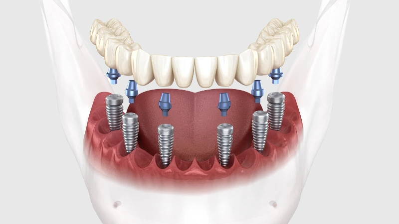
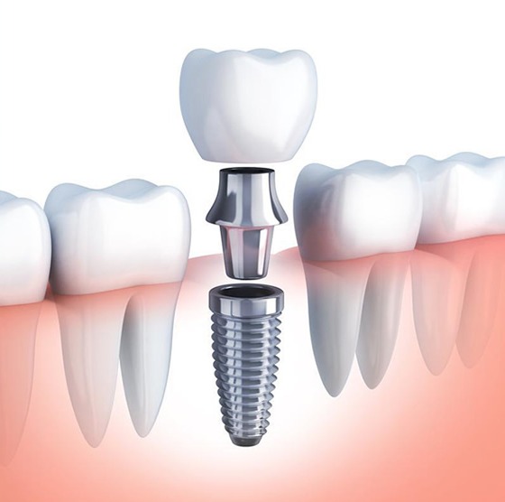
Additionally, techniques referred to as “immediate implant placement” can be designed for patients who will undergo implant treatment. If there is no acute inflammation in the area and the bone is thick enough, the titanium root can be placed immediately into the extraction socket during the same session when the tooth is removed. This integrated approach protects the patient from two separate anesthesia processes and prevents the bone from collapsing internally.
How is a Customized Treatment Plan Created for Tooth Extraction?
The morphology of each individual’s tooth roots is different. In one patient, the tooth root may be shaped like a blunt cone and easily extensible; while in another patient, it may be bent like a hook, resting on the neighboring tooth’s root or structurally fused to the surrounding bone (ankylosis). The physician creates an individual strategy by predicting this situation in advance with X-rays, determining the tools to be used and the vector of mechanical forces to be applied.
In the processes of İzmir Avrupadent, planning whether a restoration (such as an implant) will be performed in the future after the procedure is also included. If a titanium root is to be added later, the force applied to the surrounding thin bone walls is minimized during tooth extraction (trauma-free approach) and, if necessary, the void is supported with bone powders (socket preservation) for a personalized repair.