What is Dental Bone Graft Application?
Natural teeth are anchored in special sockets called alveoli, located within the jawbone. The presence of teeth helps to transmit mechanical stimuli to the bone during the chewing process, aiding in the preservation of bone volume. When a tooth is lost, the bone tissue in that area loses its function and begins to resorb over time. A dental bone graft is an advanced dental discipline focused on addressing the structural deficiencies created by this physiological resorption process. The graft materials added to the surgical area take on a skeletal role to activate the body’s own repair mechanisms, turning the added minerals into living bone tissue within months.
In the Avrupadent clinics providing services in İzmir, dental bone graft procedures are carried out by specialist teams that approach the patient’s anatomical structure holistically. This procedure is generally designed to ensure that restorations to be performed are based on a stable foundation in cases where the jawbone has thinned either vertically or horizontally. The medical process is integrated with the person’s facial proportions, chewing mechanics, and existing tissue quality to aid in the biological replacement of lost volume.
In Which Cases is Bone Grafting Applied?
Volumetric changes in the jawbone create a series of functional losses that directly affect oral health and the overall aesthetics of the face. Bone resorption can occur not only in edentulous areas but also around the roots of existing teeth. Medical interventions, such as grafting procedures, are applied to address these structural deficiencies. The main anatomical scenarios included in clinical practice are as follows:
| Clinical Condition | Explanation and Graft Requirement |
|---|---|
| Alveolar Crest Resorption | The loss of volume as the crest of the jaw remains empty for many years after tooth extraction, thinning like a knife’s edge. |
| Periodontal Bone Defects | The inflammation of the gums creating crater-like erosions in the bone supporting the tooth as it progresses along the roots. |
| Cyst or Tumor Operations | Filling the large bone cavities remaining after the removal of pathological formations inside the jaw. |
| Sinus Lifts | The expansion of the air cavities (sinuses) located in the back regions of the upper jaw, reducing the existing bone height. |
How is Tooth Bone Graft Application Performed?
The surgical process begins with an anesthesia procedure that considers patient comfort. After achieving regional numbness in the tissues, the physician carefully reflects the gum where the bone deficiency exists (by raising a flap) to directly access the underlying bone structure. Inflamed or irregular tissues in this area are removed with delicate instruments, creating a clean surface for the new material to adhere. This preparation is vital for allowing the body’s blood cells to reach the area.
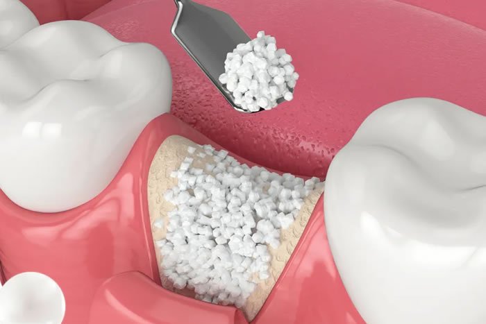
Bone powder (graft) selected according to the patient’s anatomical needs is placed in the cleaned defect area. To prevent the dispersed minerals from being washed away and to ensure that the gum tissue covering this area does not sag, a thin collagen-based membrane is placed over the powders. The membrane serves as a biological guide for cells to multiply in the proper layers. In the final stage, the reflected gum is returned to its original position and secured with fine sutures, allowing the area to heal.
Who Can Be Suitable for Bone Grafting?
Bone grafting is a procedure that stimulates the body’s self-healing mechanisms. Therefore, it is expected that the patient’s overall circulatory system and cell regeneration capacity are at healthy levels. In cases of children and early adolescents where jaw development is still ongoing, these types of volume increase surgeries are generally postponed to later ages when development has ceased, except for mandatory situations such as specific cyst surgeries.
In individuals with diabetes (sugar disease), osteoporosis, or disorders that affect the binding system, the patient’s medical doctor will confirm that clinical values are within normal ranges through consultation. Individuals whose medical values are kept stable can comfortably participate in these surgical procedures, provided they comply with oral hygiene rules.
How is a valuation made in Dental Bone Grafting?
The predictability of a surgical procedure is proportional to the accuracy of the obtained data. The physician determines the amount of graft material to be added to an edentulous area or around a tooth showing resorption not by estimation, but through detailed analysis of radiological sections. Whether the resorption in the jawbone is horizontal (width) or vertical (height) determines the structure of the membrane (barrier) to be applied and the type of bone powder to be used (synthetic, animal, etc.).
Additionally, the thickness and quality of the gum tissue over the surgery area are examined. When bone is added, it is essential to have healthy soft tissue present that is flexible enough to cover the new volume without creating tension. If the gum tissue is very thin, the planning of tissue grafting (soft tissue graft) may also be included in the process, strengthening the entire infrastructure in addition to bone surgery.
How is the Dental Graft Treatment Process Planned?Process planning; involves ensuring oral hygiene to eliminate inflammatory foci, selecting the biocompatible materials to be used, organizing the surgical appointment according to the hospital’s schedule, and designing the cellular healing (maturation) phase that will last for months.
The clinical routes in the İzmir Avrupadent branches operate through a phased discipline. If there is significant dental stone and gum infection in the area where grafting will be done or in neighboring teeth, this bacterial load can leak into the surgical site, leading to the rejection of the transferred minerals. To prevent this possibility, the oral flora is first sterilized through periodontal cleanings (scaling).
Once the preparations are complete, the physician creates an appropriate surgical schedule based on the nature of the procedure. If the procedure will be performed in a single localized area, a short appointment suffices, while extensive enhancements involving the entire jaw arch require a more detailed time frame. The patient is informed in advance about the dietary rules to follow after the surgery and the medical support aids they will use, clarifying every step of the process.
What Steps Are Followed in Dental Implant Application?
The ability of the tissue repair mechanism to function in the desired direction depends on the surgical steps being applied with biological respect. The clinical steps followed in a standard operation are as follows:
-
\t
- ▪ Anesthesia and Incision: After numbing the area with local agents, a thin incision is made in the gum to expose the underlying bone tissue.
\t- ▪ Cleaning the Site: All granulation (inflammatory) tissues on the bone are scraped away with special curettes to obtain a well-vascularized, healthy base.
\t- ▪ Material Application: The predetermined volume of sterile bone minerals is placed in cavities or fine sections in a concentrated manner.
- Barrier (Membrane) Coverage and Stitching: The area is closed with absorbable membranes to prevent the spread of added powders and the outer tissue should be stitched in a tension-free manner.
- ▪ Cleaning the Site: All granulation (inflammatory) tissues on the bone are scraped away with special curettes to obtain a well-vascularized, healthy base.
What Should Be Considered Before Dental Bone Graft?
Keeping the microbial population of the operating area at low levels in surgical interventions is essential to protect tissues from secondary infections. The patient should thoroughly brush their teeth to remove existing food residues before coming to the clinic. If there are prophylactic (preventive) antibiotics prescribed by the physician, they should not be neglected and used in accordance with the timings.
To maintain stability in heart rhythm and blood sugar levels due to the procedure being performed under local anesthesia, it would be beneficial to come to the appointment in a lightly nourished and stress-free state. If there is habitual use of tobacco that slows down cell renewal, limiting this intake at least a few weeks before the day of the operation is a fundamental supporter of clinical success.
What to Consider After Dental Bone Graft?
Since the procedure involves intervention under the periosteum, the formation of physiological swelling (edema) in the area of operation and the cheek is an expected reaction. Applying cold compresses during the first 24 to 48 hours after the procedure helps minimize this swelling. It is essential to use facial expressions with care to avoid tension in the stitched area and to prevent the graft pieces from shifting, thus avoiding actions like laughing or side stretching.
Sucking or drinking liquids through a straw can create a negative vacuum pressure inside the mouth, potentially dislodging the protective blood clot at the wound site and triggering bleeding. Nutrition should be maintained with easy-to-chew foods that do not exert pressure on the surgical area. Avoiding tobacco smoke until the primary healing phase of the tissues is complete is a critical factor in preserving treatment efficacy.
Which Dental Treatments Are Evaluated Before a Dental Bone Graft?
Volume bone augmentation is usually planned to prepare a foundation for another restorative or surgical procedure to be built on top. The fundamental treatment areas in dentistry where grafting is needed include:
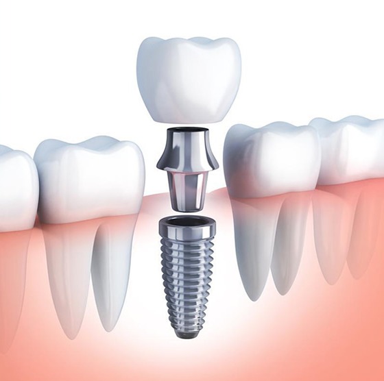
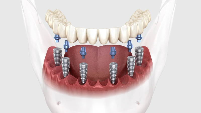
- Titanium Artificial Root (Implant) Applications: When there isn’t enough bone thickness surrounding the body to be placed in the jaw, to strengthen the area months before the implant or during the same session as the implant.
- Tooth Extraction Procedures (Socket Preservation): To prevent the bone in the empty socket left after tooth extraction from resorbing in the later stages, minerals are placed in that space simultaneously with the extraction.
- Aesthetic Bridge Restorations: To prevent the recession of the gum tissue beneath the porcelain bridges made in the frontal area and to mimic the natural tooth eruption profile, the cretin is filled.
What is the Relationship Between Bone Grafting and Implant Treatment?
When a titanium screw is placed in the jaw without at least one millimeter of healthy bone tissue surrounding it, it cannot withstand chewing forces in the long term and becomes exposed to infection by appearing visible on the outside. In previous years, due to the thinning of the bone structure over time in patients who lost their teeth, standard artificial roots could not be established in these areas.
Thanks to advancing bone powder (graft) technologies, thin or absorbed jawbones can be enlarged vertically (upwards) or horizontally (expansion). When these directed bone repair processes (YKR) are completed, the previously inadequately perceived jaw structure achieves an ideal and strong form that will securely envelop the implant.
How is Jaw Bone Deficiency Diagnosed?
When viewed from the outside, the jawbone only shows the mucosa covering it; it is quite difficult to accurately detect the amount of underlying resorption with the naked eye. However, initial data regarding how pointed or flat the bone is can be obtained from clinical pressure tests performed on edentulous areas. This manual examination is the initial step that needs to be confirmed with X-rays.
A definitive diagnosis is made using three-dimensional dental tomography. Based on this radiological data, the density of the bone (whether it is hard or spongy), the distance to the inferior alveolar nerve (mandibular canal), and how much the air spaces in the upper jaw (sinuses) have sagged are mapped out in detail. The obtained millimeter measurements scientifically indicate whether bone grafting is necessary.
What Diagnosis and Imaging Methods Are Used in Dental Implant Applications?
The predictability of surgical success is ensured by the clarity of the anatomical data obtained before the operation. The primary radiological devices that guide physicians in the clinic are:
- ▪ Dental Volumetric Tomography (CBCT): It reflects the volume, width, and neighboring anatomical spaces (sinuses, nerve pathways) of the bone in three-dimensional sections to determine the sizes of the pocket where the graft will be added.
- ▪ Panoramic Radiography: It provides a wide view of both the upper and lower jaws in a single image, allowing for the superficial detection of general resorption in tooth roots or cystic lesions.
- ▪ Periapical Films: With small film sizes focusing on a single tooth or extraction site, these films detail the bony trabeculae in that specific area.
What Materials Are Used in Dental Bone Grafting Applications?
The most suitable combination of materials is preferred by doctors according to the biological needs of the case. Bone (autograft) taken from another area of the patient’s jaw (for example, the back of a wisdom tooth) is the most valuable material for initiating cellular repair because it contains live cells. However, since taking the person’s own tissue will create an additional surgical site, it is often combined with other ready-made graft types.
| Type of Graft Material | Biological Origin and Properties |
|---|---|
| Xenograft (Animal) | Volume-maintaining powders obtained from sources like bovine (cattle), where proteins are eliminated and only skeletal minerals are left. |
| Allograft (Human-Derived) | Mineral matrices with a high potential for rapid regeneration to the body’s own bone, sourced from sterile tissue banks. |
| Alloplastic (Synthetic) | Inorganic filler materials chemically formulated in the laboratory, containing hydroxyapatite or tricalcium phosphate. |
| Autograft (From the Person Themselves) | Bone graft collected from the patient’s own body areas, such as the inside or outside of the mouth, igniting healing due to live cell integration. |
How Does the Healing Process Progress After a Tooth Bone Graft?
The closure of soft tissue incisions on the external surface and the removal of stitches is usually completed within the first 10-14 days, allowing the patient to return to normal life. However, the actual healing process is a cellular reconstruction that continues for months in the bone layer beneath the gum that cannot be seen. The graft added serves as a scaffold for the formation of new bone.
The body slowly sends its own osteoblast (bone-forming) cells into this scaffold (the powders). When sufficient vascularization is ensured, the added powders gradually dissolve, fully replaced by the patient’s own living bone. This biological transformation takes an average maturation time of between 4 and 8 months, depending on the region of the jaw and the volume of bone applied.
How Long Can the Dental Bone Graft Procedure Take?
The clinical stage of the procedure is a meticulous and concentration-intensive microsurgical step. The retraction of the gum tissue in the anesthetized area and the cleaning of the bed (bone) from infections takes a significant part of the duration. While bone powder is placed in the cleaned area, it may be necessary to tightly adapt the material to prevent spreading and to fix the membrane placed on top with tiny pins (nails).
Once all surgical procedures are completed, the stitches placed are done with great care (using very fine threads) to ensure that the tissue remains completely immobile. Since local anesthesia is in effect during the procedure, the patient does not feel clinical fatigue; the process is managed calmly in full compliance with the schedule planned by the dentist.
Why is Control Examination Important After Tooth Bone Graft?
The first follow-up after a surgical operation typically takes place in the 1st or 2nd week following the procedure. In this session, your doctor examines how well the gum tissue is healing. When sutures are removed, the wound area relaxes mechanically. This follow-up also plans when and with what firmness the patient will return to their home brushing practice.
The main significant control takes place approximately 4-6 months after surgery. If this procedure is designed as a basis for placing a titanium root (implant) in the future, the dental tomograms taken confirm whether the dust has been replaced by a homogeneous and dense layer of bone. Once the bone has reached sufficient density, the next surgical or prosthetic (crown) phase of the treatment plan can be confidently proceeded with.
Can Bone Grafting be Planned Together with Other Oral and Dental Treatments?
In clinical practice, procedures are often combined to save time and prevent tissue trauma. For example, if tooth extraction is necessary, the socket left behind is filled with bone powder immediately after the extraction to prevent the bone in that area from collapsing and narrowing in the coming months. Thus, when the implant is placed months later, there is no need for an additional bone procedure.
Similarly, when placing a titanium screw into the jawbone, if the bone wall on the lip side of the screw is very thin (not fully covering the screw), bone powder is packed into the remaining parts of the socket and covered with a membrane during the same procedure to ensure that the screw is completely embedded in the bone. This approach increases mechanical resistance and completely masks any gray metallic reflections that may occur in the gum.
How is Personalized Treatment Planning Done in Tooth Bone Graft Application?
Not every bone deficiency can be repaired with the same biological behavior. If the bone around the tooth stands as four walls and only the middle part has resorbed (if there is a box-shaped defect), the powders to be placed here easily adhere and quickly ossify. However, if the resorbed bone has disappeared like a flat wall (horizontally), it is difficult for the powders to adhere there. In such flat losses, the doctor constructs a tenting-like structure by securing the powders to the area with titanium pins and special plates.
Additionally, the material to be used is also personalized. While different sources of grafts are selected in areas where rapid resorption (absorption) is desired, powders with slow resorption that maintain their volume for a long time (as in sinus lifts) are prescribed to suit the biological characteristics required for the procedure.