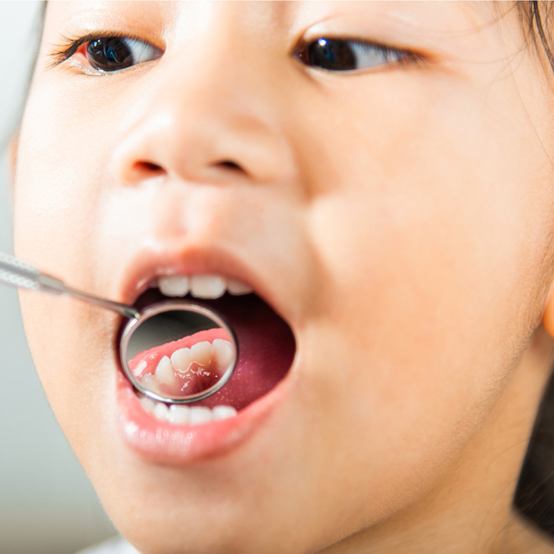
What Precautions Should Be Taken After Dental Filling in Children in Izmir?
The fundamental medical steps parents should pay attention to after a dental filling procedure in children in Izmir are as follows: if regional anesthesia was applied during the procedure, nothing solid should be given to the child for about two hours until the numbness completely wears off to prevent them from biting or injuring their lips, cheeks, or tongue. During this time, the child should be closely monitored, and to ensure the longevity of the filling, routines of brushing and dental flossing should be maintained with the same diligence without any excuses. Additionally, especially in the initial days, it’s crucial to avoid consuming extremely sticky (caramel, gummy bears, chewing gum) and extremely hard (ice, shelled nuts) foods that can shorten the lifespan of the filling. In cases of potential sensitivity, following the dentist’s recommendations is essential, and regular clinical check-ups should not be neglected, without falling into the misconception that the tooth will not decay again after treatment. The post-treatment period is at least as important as the medical intervention performed in the clinic, and the success of the filling is directly related to the conscious care and follow-up discipline the family provides at home.
Restorative (repair) dental treatments applied during childhood not only restore the anatomical form of the tooth; they also create a comprehensive medical shield that protects the child’s chewing function, speech ability, and overall body health from infections. However, the ability of the filling material that the physician meticulously places in a clinical setting to serve healthily throughout the child’s developmental stage depends on how accurately the post-procedure period is managed. Parents often think that once the dental filling is done, all problems are resolved and that the tooth is now completely safe. Health regulations and preventive dentistry principles emphasize that the sustainability of the treatment is possible through the family’s health literacy.
How Should Nutrition Be in the First Hours After the Filling Procedure?
Nutrition in the first hours after the filling procedure varies depending on the type of material used by the physician and whether anesthesia (numbing) was applied; in cases where anesthesia is used, in order to prevent the chewing reflex from harming the tissues, only warm liquids like water should be consumed until the numbness passes (approximately 2-3 hours), and solid food intake should be postponed absolutely.
In modern pediatric dentistry, composite or compomer fillings used achieve full hardness within seconds with special blue polymerization devices in the clinical chair. This means theoretically there is no need to wait for a time for the material to set or harden; the filling is completely ready for chewing when the child leaves the clinic. However, the main factor limiting nutrition is not the filling itself, but the local anesthesia applied for the procedure to proceed comfortably (regional numbness). This numbness prevents the child from fully feeling the muscles, lips, and tongue in that area. If the child attempts to eat during this numbness phase, they may not realize they are chewing their own lip or cheek, rather than the food. Therefore, in the first few hours, the child should only consume liquid foods that do not require chewing, such as water, buttermilk, or warm soup, and should avoid very hot or very cold drinks (as they could cause burns in the tissue).
Why Should Observation Continue Until the Effect of Local Anesthesia (Numbness) Wears Off?
Observing until the effect of local anesthesia wears off is a medical necessity to prevent children from exhibiting curiosity regarding the strange, swollen, and tingling feeling in their lips or cheeks, which leads them to lick, suck, or pull at that area and can result in serious injuries to the tissues from this physical trauma.
An adult can recognize the feeling of numbness logically and protect themselves, while young children (especially those in preschool age) perceive the numbness experienced after anesthesia as a very strange and playful situation. The child might think that their lower lip is drooping or thick; to test this feeling, they put their lip between their teeth and try to see how much they can bite without pain. Since they cannot adjust the biting force due to lack of pain, this situation can lead to what is frequently encountered in the clinical literature as “anesthetic trauma” if it goes unnoticed by the parent. Keeping the child under constant supervision of a parent or caregiver in the car, at home, or in nursery after they leave the clinic and encouraging them to “only let their lip sleep a little and not to touch it until they wake up” is the most crucial rule for safety after the procedure.
How to Understand Anesthetic Trauma (Lip and Cheek Biting) in Children and What Should Be Done?
Anesthetic trauma in children is understood by the presence of a significant swelling in the anesthetized lip or inner cheek area several hours or a day after the procedure, accompanied by a thick white-yellowish plaque (ulceration) and redness. While parents often panic by assuming this is an inflammation, this condition is merely a physical bite injury, and it should heal by keeping the area clean and using soothing gels as recommended by the physician.
Parents are most concerned about anesthetic trauma when looking for clinics. When the child wakes up on the night of the procedure or the next morning, one side of their lip may be severely swollen and the inner part might appear as a completely white ulcer. The parent may think that the instruments used by the doctor have caused an infection or that a new abscess has formed in the tooth. However, this white, ugly-looking wound is a cellular result of the child damaging their own tissue with their teeth (chewing) while under anesthesia. The tissue forms a white fibrin layer on top to heal itself. This wound is not an infection from the teeth and does not require antibiotic use. What needs to be done is to keep the wound clean, prevent the child from consuming acidic (lemon, tomato, etc.) foods that might irritate the wound, and wait for approximately 7 to 10 days for the tissue to regenerate and heal on its own.
| Condition / Symptom | Anesthetic Trauma (Lip/Cheek Bite) | Tooth-Related Abscess (Infection) |
|---|---|---|
| Onset Time | Within 1-2 days immediately after the procedure. | Usually appears gradually after weeks or months. |
| Appearance and Location | Wide, white-yellowish ulceration inside the lip or on the cheek. | Small, pimple-like (fistula) discharge at the exact root line of the gum. |
| Medical Approach | The area is kept clean, and spontaneous healing is expected. | Medical intervention requires treatment, canal therapy, or extraction. |
Is it normal to experience pain or sensitivity after a dental filling?
After a dental filling, mild sensitivity that lasts for a few days when pressure is applied to the tooth (while chewing) or when consuming cold foods is a reflection of slight tissue sensitivity that occurs when the cavity is cleaned (sensitivity of the pulp) and is considered medically normal; this sensation will completely disappear as the tissue heals.
Removing a tooth from a cavity is a surgical procedure at the microscopic level. The dentist removes the diseased tissue while getting quite close to the living center of the tooth (nerve chamber). This cleaning process and the chemical bonding reactions of the filling material placed in the tooth can create a temporary reaction in the nerve network inside the tooth (in the dentin canals). This is referred to in medical literature as “post-operative sensitivity”. If a child feels a slight sensitivity in the filled tooth while biting into a hard piece of food or drinking cold water in the initial days, it indicates that the vitality of the tooth is being maintained and that the tissue is in the healing phase. Generally, this condition diminishes over a few days and rarely continues for a few weeks. Dentists recommend avoiding extremely cold ice cream or very hot drinks for a few days to help ease this transition period.
What symptoms should prompt a follow-up visit to the dentist?
If the sensitivity worsens instead of diminishing day by day, if the child wakes up crying from sleep due to a throbbing pain starting, if a pimple-like swelling (abscess) appears on the gum, or if the child indicates that the filling touches the other teeth before they close their mouth (indicating height), they should promptly follow up with the dentist.
Although the success rate of clinical treatments is very high, in some cases, the dental pulp may not tolerate the bacterial infection caused by the decay. Even if the decay is cleaned and filled, the inflammatory response initiated by the bacteria in the nerve tissue may silently progress. If a filled tooth starts to experience severe pain on its own (without chewing anything) or if a fistula (a hole oozing pus) forms in the gum, it indicates that the dental nerve has died and that the filling procedure was insufficient, requiring a transition to root canal treatment (amputation/pulpotomy). Another significant clinical finding is the height of the filling. The child may not feel that they are completely closing their tooth due to numbness in the clinic. If they only mention that the tooth is making early contact (has become high) when they get home, the excessive mechanical chewing pressure exerted on that area may cause trauma to the tooth, so the dentist needs to reduce that height with a minor adjustment (grinding).
Is There a Risk of Newly Placed Baby Tooth Fillings Falling Out or Breaking?
There is always a possibility that newly placed baby tooth fillings may be pulled out of place by the child consuming overly sticky foods, such as caramel, gum, or jelly beans, or may physically break by chewing very hard materials like nut shells or ice, or due to the remaining solid walls of the tooth being very thin, leading to a risk of falling out or breaking during intense pressure.
Modern composite and glass ionomer restoration materials (fillings) used in dentistry chemically bond on a microscopic level to the tooth enamel and dentin tissue. Under healthy conditions, this bond is extremely strong. However, no matter how advanced the materials are, they cannot match the toughness and resilience of original and healthy tooth enamel. Especially after a large cavity has been cleaned out, if only very thin walls remain on the tooth, the filling tries to cling to these fragile walls. A gummy candy the child eats at school or home or a hard lollipop can create a suction effect that can pull the filling from the tooth. Alternatively, a strong impact (such as accidentally biting hard or hitting the tooth hard) can break the thin remaining wall of the tooth along with the filling. For this reason, parents should establish disciplined dietary rules regarding “sticky and extremely hard foods” for their children to extend the lifespan of filled teeth.
How Should Oral Hygiene Be Maintained at Home After Filling Treatment?
Post-filling treatment home oral hygiene should be meticulously maintained by brushing mechanically twice a day, in the morning and at night, with age-appropriate fluoride toothpaste under parental supervision, without falling into the medical misconception that the tooth is “now treated and will never decay again,” and by cleaning the interproximal areas (between two teeth) with floss to prevent plaque formation.
A dental filling procedure applied in the clinic is a successful repair that halts the spread of tooth decay; however, this repair is not a magical vaccine protecting the child from future cavities. The filling material itself does not decay and is not affected by acid. However, the microscopic fine line (margin) where the filling material ends and the child’s natural tooth enamel begins is where bacteria love to settle and adhere. If the child continues their old habits, frequently consumes carbohydrate-rich foods, and neglects to brush their teeth, bacteria will then start to erode the tooth right beside the filling, meaning they will begin to decay (burrow) the tooth at that margin. A filled mouth means a new hygiene period that requires more attention than before; it necessitates carefully brushing every evening to remove the soft plaque layer (biofilm) that accumulates on those margins.
What is Secondary (Subsequent) Cavity and Why Does it Occur in Filled Teeth?
A secondary cavity is the event of decay reoccurring just beneath or beside a filling as a result of bacteria leaking through the delicate margin lines where the filling material is bonded with previously filled teeth, due to insufficient brushing away from parental supervision and habitual excessive consumption of sweets (sugar intake).
The main scientific reason behind complaints such as “My tooth started hurting again a year after the filling was done” is often secondary decay. No matter how perfectly a restoration (filling) is made, the oral environment is a highly abrasive acid storm area, continually warm, moist, and subject to mechanical pressures. After consuming sugary food, acid production begins, and this acid targets the microscopic walls to which the filling is attached. Over time, bacteria enter through the millimetric leaks (micro-leaks) that form in that wall and create a new nest under the filling (in the invisible dark area). The tooth continues to decay from underneath. When the area beneath the filling is completely hollow, the filling can no longer find support and falls out. The only formula for preventing secondary decay is for the dentist to check these borders during routine examinations, and for the child to use impeccable brushing, ensuring that bacterial plaque does not settle in these areas at home.
| Characteristic | Primary (Initial) Decay | Secondary (Decayed) Decay |
|---|---|---|
| Formation Area | Natural tooth enamel or cavities that have never been treated. | Boundary lines where the filling has fused with the tooth enamel previously filled. |
| Visibility | Clearly visible as dull white spots or brown cavities from the outside. | Usually progresses secretly beneath the filling; more difficult to detect from the outside. |
| Primary Cause | Failure to brush the anatomical cavities and sugar consumption. | Microbial residues and weak interface hygiene on filling edges. |
What Role Does Dental Floss Play in the Care of Filled Teeth?
The use of dental floss plays an indispensable mechanical cleaning role in maintaining the borders of fillings and preventing the decay of neighboring teeth, as it cleans trapped food particles between fillings placed on the adjacent surfaces (interfaces) of the teeth, thus stopping bacterial accumulation and acid production in that enclosed area.
Parents’ brushing routines for their children generally cover the outer, inner, and top (chewing) surfaces of the teeth. However, the areas where childhood cavities and filling losses most frequently occur are the contact points where two teeth are closely pressed against each other. If a dentist has filled one tooth, there will be a very small space between that filling and the neighboring healthy tooth where it is physically impossible for toothbrush bristles to reach. When a child eats, food or carbohydrate particles get trapped in that gap. If these residues are not removed by pulling dental floss upwards, they will be turned into acid by bacteria thriving in an oxygen-free environment. The acid produced will erode the boundaries of the filling done by the dentist, creating secondary cavities and penetrating the neighboring healthy tooth. Therefore, in areas where fillings have been done (especially in the back molars), using suitable plant-based floss with parental assistance is a vital care step just as important as brushing.
How Do Post-Filling Follow-Up Procedures Work at Izmir Avrupadent Clinics?
In the clinics of İzmir Avrupadent, the follow-up processes after filling are managed with a system that considers the moment of treatment not as the end of the medical process but as its beginning; families are invited to the clinic with automatic appointment periods (recall system) varying between three to six months based on the child’s current caries risk profile, and the lifespan of restorations is visually/radiologically monitored.
A quality healthcare service requires standing behind the procedures performed and following them for many years. In our clinical organizations in İzmir, a child patient whose filling is completed and sent home is not left with the status of ‘healed and finished.’ The compliance of the filling with the child’s jaw growth, whether it has been exceeded or not, and whether there is any plaque (biofilm) accumulation on its edges is the responsibility of the physician. Our digital tracking system provides a transparent reminder to parents when it is time for the child’s check-up. During these brief check-up appointments, the edges of the fillings are examined with special instruments and lights, and necessary polishing operations are performed to remove discolorations on the surface if deemed necessary. Suspected minor leaks that could be overlooked are checked with low-dose mini x-rays, ensuring the child’s health is secured with digital data.
How Often Do Pediatric Dentistry Specialists Conduct Filling Checks?
Pediatric Dentistry specialists conduct filling checks every six months to monitor dental development in standard cases; in children who cannot establish a nutrition discipline, use systemic syrups, or are in a high caries risk group due to their dental structure, this period is reduced to three or four months for much closer medical supervision.
The frequency of medical follow-up (recall period) is based on a “personalized” protocol according to each child’s own biological and social map. If a child coming to the clinic brushes their teeth perfectly twice a day with parental assistance, does not have a nighttime feeding, and does not consume sugary snacks, the physician knows that this child’s filling is not at risk and proceeds with the standard 6-month check-up schedule. However, if there have been numerous cavities detected in the past, if they breathe through their mouth due to lip structure or adenoids, or if a child cannot tolerate brushing due to special needs (autism, cerebral palsy, etc.), there is a very high likelihood of cavity leakage around the filling. The specialist doctor calls these children every 3 months for short periods, applies professional fluoride varnishes around the fillings, and performs a chemical renewal of the barrier that prevents bacterial attachment.
How Do Filled Primary Teeth Fall Out When Permanent Teeth Erupt?
When filled primary teeth complete their growth in the mandible of the permanent teeth beneath them and erupt correctly upward (when they fall out), due to the natural biological pressure exerted by the erupting permanent tooth, they gradually dissolve their roots at the cellular level. A primary tooth that has no remaining root will shake out as a whole along with the filling material on it.
One of the concerns frequently expressed by parents is the fear that a filling placed in a baby tooth will get stuck in the child’s mouth and block the incoming permanent (adult) tooth. Biological processes show that this fear is entirely unfounded. The biocompatible filling material placed by the dentist only occupies the “crown” area (the visible upper part) of the tooth; it does not extend into the root structure of the tooth or into the bone. As the child’s growth timeline progresses (for example, around ages 10-11), the permanent tooth waiting in the alveolus moves upwards. This movement activates cells called “osteoclasts” that dissolve the roots of the baby tooth. When the root is completely dissolved, only the upper crown part (and the filling inside it) remains of the tooth. The tooth normally becomes loose and naturally falls out while the child is eating, smoothly making way for the permanent tooth.
How Should the Child’s Psychological Adaptation After Filling Be Supported?
The psychological adaptation of the child after the filling should be supported by presenting the experience in a clinical setting as a success story, ensuring that the dentist is never used as a “punitive figure” or “intimidation tool”, positively reinforcing the courage shown by the child with encouraging words from the parents, and fostering a sense of pride in the new filling.
Having a child sit in the dental treatment chair and patiently wait there is a significant psychological event for their small world. The attitude of the doctor and parents after the procedure shapes the child’s perception of future healthcare institutions (dental phobia). Often, parents opt for a highly misleading and damaging way of communicating, saying things like, “Look, if you don’t brush your teeth, I’ll take you back to that doctor, they will use needles again.” This sentence codes the doctor as a punisher and the clinic as a torture center. Instead, saying, “Today, the doctor put magical star dust on your tooth, how brave you were, I’m proud of you. Now let’s protect this beautiful tooth by brushing it” is the correct pedagogical approach that encourages the child to take ownership of that tooth and willingly go to the next check-up.
What Changes in Diet Support the Lifespan of Fillings?
In the dietary regimen; completely cutting out ready-made fruit juices and carbonated drinks that can dissolve the microscopic boundaries where the filling adheres to the tooth, avoiding overly sticky candies like caramel and jelly beans that may pull the filling from its place (creating a vacuum effect), and increasing the intake of tooth-friendly foods that quickly neutralize acid (buffering) like cheese, yogurt, and water significantly supports the lifespan of the filling.
The oral environment is essentially a chemical and physical testing chamber for filling materials. A child’s diet is the primary factor that determines how long the material used by the dentist in the clinic will last. Consumed acidic beverages (cola, packaged fruit juice) can not only damage the dental enamel but also affect the surface structure of the filling material at a microscopic level (can lead to wear). More dangerously, these acids can erode the bonding interface that joins the filling to the tooth, creating a pathway for bacterial infiltration. Additionally, children often consume candies by directly applying them over fillings on their back molars. A highly adhesive candy or caramel can create a strong vacuum effect when combined with chewing force, physically detaching the filling from the tooth. Therefore, after filling treatment, families are strongly advised to avoid the culture of “frequent snacks” and to create a main meal-focused, sugar-free nutrition program.
Does Nighttime Teeth Grinding (Bruxism) Damage Fillings?
Nighttime teeth grinding (bruxism) occurs with uncontrolled and extremely intense muscle contractions of the jaw, exerting immense mechanical pressure on the tooth surfaces and fillings, which can lead to cracks in the filling material, edge chipping, or total wear down alongside the tooth’s natural tissue, causing significant damage to the fillings. This situation is examined during Pediatric Dentistry follow-ups.