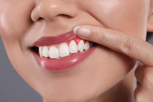
What is Periodontology?Periodontology; is a branch of basic dentistry that involves the examination of the health of the soft tissues surrounding the teeth, the special fibers (periodontal ligament) that connect the tooth root to the jawbone, and the jawbone supporting the tooth, as well as the diagnosis and clinical treatment of inflammatory diseases that occur in these structures.Our teeth are not directly found inside the jawbone; there are fine fibers that function like a cushion between the bone and the tooth root. All this supporting structure is covered by a pink and firm layer of gum tissue, protecting against external factors. The science of periodontology deals not only with the visible part of the tooth but also with the health of these invisible foundational structures that hold the tooth in the mouth. Weakness in the foundation can lead to tooth loss, regardless of how healthy and intact the visible part of the tooth is.
In the Avrupadent clinics located in Izmir, periodontology approaches are based on the principle of protecting oral health as a whole. Gum diseases often progress silently, with early stages being difficult to notice without discomfort. The treatment procedures applied in this field aim to stop the destruction in the supporting tissues of the tooth and to reconstruct the necessary biological infrastructure so that the patient can maintain chewing function with their own teeth throughout their lifetime.


What Diseases Does Periodontology Concern?
The diseases periodontology deals with; superficial inflammations affecting only the gum (gingivitis), deep tissue destructions where the inflammation has reached the jawbone (periodontitis), gum recessions, gum overgrowths, and asymmetries of the soft tissues inside the mouth.Gum diseases usually begin with a simple plaque buildup and progressively affect deeper tissues over time. The main disease states diagnosed in clinical examinations that require periodontal treatment are as follows:
Why Do Gum Diseases Occur?
The main reason for gum diseases is the accumulation of microbial dental plaque (bacterial plaque) at the junction of the teeth and gums, which is due to insufficient oral hygiene, and this plaque hardens over time into tartar.The oral cavity naturally hosts millions of bacteria. Food residues, proteins, and bacteria combine to form a colorless, sticky film known as bacterial plaque on the teeth. If this plaque is not removed through daily brushing and flossing, it hardens into tartar by settling with the minerals in saliva. The porous surface of tartar provides a foundation for more bacteria to adhere, and the toxins (acids) released by these bacteria irritate the gum tissue, triggering infection.
Although the primary causative bacteria is present, there are secondary risk factors that accelerate the progression of the disease. The use of tobacco products is the biggest contributor to gum diseases as it suppresses the immune system. Systemic disorders such as diabetes, hormonal fluctuations during pregnancy, genetic predisposition, stress, and poor dietary habits are also among the factors negatively affecting gum health.
What Are the Symptoms of Gum Diseases?
The symptoms of gum diseases; bleeding while brushing or spontaneous bleeding, redness and swelling in the gums, a persistent bad smell in the mouth, gums pulling away from the teeth, and in advanced stages, gaps or looseness in the teeth.Healthy gums have an orange peel appearance, are dull, tight, and light pink in color; they definitely do not bleed during brushing. Diseases usually start with very mild symptoms, which can be neglected by patients for a long time. According to clinical observations in Europe, the most common warning signs that lead individuals to consult a dentist are as follows:
- Bleeding noticed during brushing, when biting into a hard food, or in the morning upon waking on the pillow.
- The natural pink color of the gum tissue losing its tone, turning dark red or purplish.
- The gum tissue feels soft and swollen when touched, accompanied by a pain sensation.
- Black triangular spaces forming between the teeth, resembling the elongation of dental sizes (roots emerging out).
- The presence of inflammation (pus discharge) from the spaces known as gum pockets and persistent bad breath that doesn’t go away with brushing.
How are Gum Diseases Diagnosed?
Diagnosis process; the specialist doctor (periodontist) clinically evaluates the color, shape, and bleeding status, measures the depth of gum pockets with millimeter instruments called periodontal probes, and determines the amount of bone loss using panoramic/periapical X-rays.The extent of gum diseases cannot be understood just by visual inspection. The most reliable way to detect the damage in the bone surrounding the tooth is through a periodontal probing procedure. The dentist gently places a measuring instrument with a blunt end and millimeter markings between the tooth and the gum. In a healthy mouth, this depth is between 1-3 millimeters. If this value exceeds 4 millimeters, it indicates that bacteria have begun to cause bone loss and that a pathological “gum pocket” has formed.
During the probing, the tissue’s bleeding response is also recorded. The obtained numerical data is supported by digital X-ray images. The radiological images clearly show how much of the jawbone supporting the tooth roots has deteriorated. All these objective findings are combined to determine the stage of the disease.
How is Periodontal Treatment Planned?
Treatment planning; is done in stages according to the stage of the disease. Initially, non-surgical approaches (cleaning of tooth stones, smoothing of root surfaces) are employed to control inflammation, and if necessary, advanced surgical procedures are performed, and ultimately the process is monitored with maintenance (protective) treatment.The periodontal approaches in the İzmir Avrupadent clinic require a step-by-step progression of discipline. In the first session, the source of the patient’s disease is explained, and personalized brushing and cleaning training is provided. If the patient does not apply proper oral care, it will not be possible to maintain the long-term effects of any treatment performed in the clinic. The hygiene habits maintained at home are the first step of the treatment.
At the clinical stage, the gums above the tooth boundaries and the tartar just below are cleaned using ultrasonic devices (scaling). If the disease has affected the bone level (periodontitis), under local anesthesia, the gum pockets are accessed, and the microbial deposits and inflamed tissues on the tooth roots are scraped out and flattened with special instruments (curettage). A few weeks after the initial treatment phase is completed, the healing status of the tissues is re-evaluated; if deep pockets have closed, the maintenance phase is planned, and if not, the surgical phase is entered.
In What Cases Are Gum Treatments Applied?
Situations where treatments are applied; range from simple gum bleeding to advanced bone loss that leads to loose teeth, and from retraction of the gum resulting in sensitivity or irregular gum boundaries that disrupt the aesthetics of the smile (gummy smile).Dentistry is not just about repairing a broken tooth; if the supporting bone around the tooth is not healthy, no filling or porcelain can serve its purpose. Therefore, periodontal treatments are applied with both health and aesthetic expectations in mind. The main conditions that require periodontal intervention in clinical practice are as follows:
What Methods Are Used in Periodontal Treatments?
Clinical methods used; include non-surgical scaling (tooth tartar cleaning) and root planing procedures, periodontal surgeries (flap operations) for deep tissue injuries, bone/tissue graft (powder) applications, and surgical methods such as aesthetic gingivectomy.The stage of the disease determines the technique to be used. In early stage gingivitis cases, ultrasonic devices are used in dentistry to remove tartar from the tooth surface by vibration and polish the surface with polishing procedures. If inflammation has progressed to the depths of the gums, affecting the root surface, root planing is performed with hand instruments (curettes) under local anesthesia. In this procedure, microbial deposits on the root surface are scraped away to create a clean area for the tissue to reattach firmly to the tooth.
In cases where basic treatments are insufficient due to the presence of deep bone pockets, the “Flap Surgery” comes into play. In this surgical method, the gum is slightly separated from the bone with a small incision. The doctor cleans the inflamed tissue and the deformity in the bone by visual inspection. If there is bone resorption, bone graft (powder) and membrane are placed in those areas to stimulate the regeneration of the lost bone, and the tissue is stitched back together.
Why is Early Evaluation Important in Gum Diseases?
The importance of early evaluation; complete clinical recovery can be achieved with simple cleaning and hygiene methods before bone resorption begins or in the initial stages, while if it is delayed, tooth losses or advanced surgical interventions become unavoidable.Gingivitis (the initial stage) is a condition that is completely reversible. With just a few sessions of cleaning and the correct brushing technique, the gum can fully regain its healthy form. However, if intervention is not made at this stage, the disease progresses to periodontitis. Bone resorption that starts in the jawbone is irreversible; that is, new bone will not spontaneously grow in place of the resorbed bone. At this stage, what the doctor can do is stop the bone resorption and halt the progression of the disease.
In cases where the supporting bone is lost, teeth gradually begin to loosen, cannot withstand chewing forces, and despite having no fractures, they have to be extracted. Even placing an implant in place of these extracted teeth becomes difficult, as the bone tissue that would support the implant has deteriorated due to gum disease. The only way to prevent this chain reaction of destruction is early intervention.
How is a Periodontal Examination Performed?
Periodontal examination; involves the observation of the color, shape, and consistency of the gum tissue, measuring the gap (pocket) between the tooth and gum with special probing tools in millimeters, determining the bleeding index, and checking the amount of tooth mobility through finger examination.Unlike a routine dental examination, a periodontal examination focuses on understanding the hidden structures beneath the tooth. Your dentist will visit each of your teeth individually and take periodontal probing measurements from 6 different points of each tooth. These measurements indicate how deep the gum pocket is. The obtained values are recorded on the patient’s clinical form (periodontal mapping) to ensure that tissue healing is monitored with measurable data in the subsequent stages of treatment.
During the examination, the amounts of recession in the gum tissue, the exposure levels of the tooth roots, and any bone loss at the fusion points of the roots (furcation) are identified. At the same time, the patient’s oral hygiene habits, the amount of plaque accumulation are evaluated to create a personalized oral care program.
How is an Evaluation Done Before Gum Treatment?
Pre-Procedure Evaluation; this is not limited to a simple oral examination but includes a detailed medical analysis of the individual’s systemic diseases (diabetes, cardiovascular conditions, hypertension), regular medications used (such as blood thinners), smoking habits, and genetic predispositions.Oral health cannot be considered independent of general health. Especially in the presence of uncontrolled diabetes (sugar disease), even a minor inflammation in the mouth can lead to rapid bone loss and delayed wound healing. Therefore, in the Izmir Avrupadent protocols, the patient’s blood sugar levels are closely monitored to ensure stability for the success of the treatment process.
Another critical point is the medications used. Some hypertension medications, epilepsy medications, or immunosuppressants used after organ transplants can cause severe gum growth (hyperplasia). In such cases, merely cleaning the dental calculus does not solve the issue; your doctor will evaluate the process thoroughly by consulting with your healthcare provider (and making changes to medication if necessary).
How Does the Gum Treatment Process Progress?
The treatment process; starts with mouth hygiene education, ultrasonic dental calculus cleaning, and, if necessary, root planing procedures in deeper areas. Healing of the tissues is awaited; surgical intervention is performed for non-healing pockets, and the process is monitored through periodic maintenance checks.Periodontal treatment is a marathon that measures the biological responses of the tissues, rather than a one-time procedure. The initial step, root planing procedures, is performed in accordance with the density of calculus present in the patient’s mouth, requiring between 2 and 4 sessions, with half of the jaw (quadrant) being anesthetized. At this stage, the infected layer on the tooth roots is scraped away to expose the healthy root surface.
After basic procedures, a healing period of approximately 3-4 weeks is expected. Following this period, the patient is re-evaluated. If the gums have tightened, bleeding has stopped, and pocket depths have returned to normal limits (1-3 mm), the phase of “maintenance” (protection) is entered without the need for surgery. If infected deep pockets (5 mm and above) still exist, the doctor will plan a local flap operation (gum surgery) to clean the inflammation directly under the area.
What to Pay Attention to After Periodontal Treatment?
Things to be careful about after treatment; strictly adhere to the specific brushing techniques indicated by the physician, consistently use interdental brushes and dental floss, avoid products that delay wound healing, and use the prescribed antiseptic mouthwashes for the specified duration.The gums may be slightly sensitive and mildly swollen for the first few days following root planing or surgical procedures. Additionally, since calculus on the root surfaces has been removed, the teeth may exhibit temporary sensitivity to hot and cold. During this period, using sensitivity-relieving toothpastes recommended by the doctor will provide relief.
After the treatment, the tissues may heal and the gaps between the teeth might become more discernible. This indicates that the gum’s attachment has receded (it has returned to a healthier condition); however, these newly opened gaps are conducive areas for bacterial plaque accumulation. For this reason, in addition to standard brushing, the use of appropriately sized inter-dental brushes is crucial for maintaining clinical improvements.
What Should Be Considered to Protect Gum Health?
The golden rules for protecting your health; brush at least twice a day using a soft-bristled toothbrush with fluoride toothpaste (with a brushing motion from gum to tooth), do mechanical cleaning daily with dental floss, and visit your dentist regularly every six months.A toothbrush cannot reach the interdental areas where the teeth contact each other anatomically. Gum diseases and cavities mostly start in the surfaces that this toothbrush cannot reach. Therefore, using dental floss or a water flosser is not merely a detail dependent on preference, but an essential part of oral hygiene.
Using a hard toothbrush does not clean teeth better; instead, it can lead to gum recession (mechanical trauma) and enamel wear (abrasion). Brushing with soft-bristled brushes and using circular massage motions without applying excessive pressure supports tissue health. However, without cleaning done with professional clinical devices, plaque and microscopic calculus may form that cannot be removed by just brushing; therefore, routine polishing procedures performed by a dentist prevent the accumulation.
How Can Gum Diseases Affect Tooth Structure?
Inflammatory processes do not directly damage the tooth’s own tissue (enamel or dentin); however, since they destroy the supporting structures of the tooth, they indirectly disrupt the position and integrity of the tooth. The impacts of the disease on the teeth are as follows:
\t- Displacement and Spacing: As the jawbone erodes, the teeth experience loss of support and begin to open up like a fan, moving forward and backward due to the effects of chewing pressure.
\t
- Root Exposures: When the gum recedes, the weak root surface not protected by the enamel is exposed; these areas are much more sensitive to acid attacks and can quickly develop root caries.
\t
- Increased Sensitivity: The dentin channels in the exposed root surface react sharply to cold, hot, or sweet foods.
- Mobility (Loosening) and Loss: Just like trees being uprooted in soil erosion, teeth that lose their bone support gradually loosen over time, leading to pain during chewing and eventually being lost.
What are the Diagnosis and Imaging Methods Used in Periodontal Treatment?
Diagnosis methods; include clinical periodontal probing (depth measurements) as well as panoramic X-rays that show the general structure of the jaws, periapical films that provide a clearer depiction of the bone levels of specific teeth, and 3D dental tomograms (CBCT) for advanced surgical planning (such as bone grafting, etc.).The visible area is only a small part of the iceberg. The real damage occurs beneath the gum tissue, in the bone structure. Thanks to periapical (small) X-rays, the dentist can assess how far the bone boundary surrounding the tooth has receded and determine whether the bone loss is horizontal or vertical in nature.
Particularly in cases of bone loss involving roots that penetrate between the jaws (furcation defects) or in cases planned for regenerative surgery (bone formation), two-dimensional X-rays may be insufficient. In such cases, dental volumetric tomography allows for a cross-sectional examination of tissues millimeter by millimeter, with the bone structure being three-dimensionally mapped to determine the surgical route.
Why is Control Examination Important in Gum Diseases?
The clinical importance of control examinations; the natural tendency of periodontal diseases is to reemerge (recurrence). No matter how well the patient brushes, calculus formation can continue; without regular maintenance (care) phases, the disease begins to silently progress.The patient’s file is not closed after periodontal treatment; it is included in a lifelong program called “Maintenance Treatment” (Supportive Periodontal Treatment). Depending on the individual’s risk profile (smoking status, systemic health, calculus formation rate), the physician determines control appointments at intervals of 3, 4, or 6 months.
In these checks, the gum pockets are measured again with a probe. If there is an area showing signs of infection (bleeding), a small ultrasonic cleaning or laser application is performed only in that area to stop the disease before it reaches the bone. For individuals who regularly attend check-ups, the time that natural teeth remain in the mouth dramatically increases and the risk of tooth loss is minimized.
Can Periodontal Treatments Be Planned Alongside Other Dental Treatments?
Clinical principles necessitate that procedures be performed together; in fact, prior to almost any procedures (implants, porcelain crowns, orthodontics) that will be performed in dentistry, it is essential to establish the health of the gum tissue, which is the foundation for the application, through periodontology.A comprehensive (multidisciplinary) approach is essential in oral structure. For example, if a zirconium crown or laminate will be made within the scope of smile design for a patient, the gum must not be bleeding or inflamed before the measurement is taken. If measurements are taken while the gums are inflamed and the porcelain is attached, once the gums heal and retract later, the edges of the porcelain may become exposed, leading to an aesthetic disaster. Therefore, periodontal health is maintained before aesthetics.
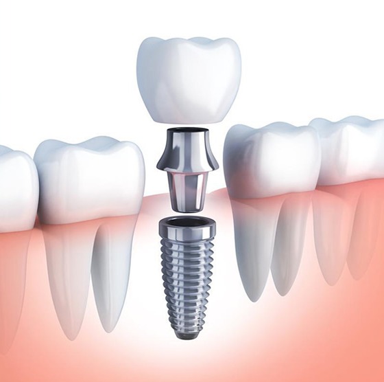
Similarly, in a patient undergoing wire treatment (orthodontics), the bone support must be complete. Trying to move a tooth in an inflamed bone can lead to the rapid loss of that tooth. When planning implant surgery, the mouth must be completely cleaned to prevent the bacteria flora in the mouth from contaminating the area around the implant (peri-implantitis) and eliminate the risk of infection.
How is Periodontal Treatment Planned Specifically for the Individual?
In personalized planning; medical protocols are modified according to the individual’s age, genetic structure, gingival biotype (thickness), systemic disorders (such as diabetes), and disease stage, creating a completely unique map for that patient that includes everything from antibiotic use to the type of surgical graft.Everyone’s oral anatomy and healing potential are different. In individuals with thick gingival biotypes, gum recession is rare while pocket deepening is common; in individuals with thin gingival biotypes, rapid gum recessions (recession) occur. Experts at Izmir Avrupadent base their treatment on these structural differences, designing procedures such as laser pocket cleaning for thick gums and taking a piece from the palate to reinforce (connective tissue graft) for thin gums.
The same disease table requires different treatment protocols for a smoking individual and a non-smoking individual. Because smoking disrupts the bloodstream, it slows down the expectation of clinical improvement; in such patients, the process is optimized by avoiding surgical operations or ensuring that the patient completely quits smoking before and after the operation.
Our teeth are not directly found inside the jawbone; there are fine fibers that function like a cushion between the bone and the tooth root. All this supporting structure is covered by a pink and firm layer of gum tissue, protecting against external factors. The science of periodontology deals not only with the visible part of the tooth but also with the health of these invisible foundational structures that hold the tooth in the mouth. Weakness in the foundation can lead to tooth loss, regardless of how healthy and intact the visible part of the tooth is.
In the Avrupadent clinics located in Izmir, periodontology approaches are based on the principle of protecting oral health as a whole. Gum diseases often progress silently, with early stages being difficult to notice without discomfort. The treatment procedures applied in this field aim to stop the destruction in the supporting tissues of the tooth and to reconstruct the necessary biological infrastructure so that the patient can maintain chewing function with their own teeth throughout their lifetime.
What Diseases Does Periodontology Concern?
Gum diseases usually begin with a simple plaque buildup and progressively affect deeper tissues over time. The main disease states diagnosed in clinical examinations that require periodontal treatment are as follows:
Why Do Gum Diseases Occur?
The oral cavity naturally hosts millions of bacteria. Food residues, proteins, and bacteria combine to form a colorless, sticky film known as bacterial plaque on the teeth. If this plaque is not removed through daily brushing and flossing, it hardens into tartar by settling with the minerals in saliva. The porous surface of tartar provides a foundation for more bacteria to adhere, and the toxins (acids) released by these bacteria irritate the gum tissue, triggering infection.
Although the primary causative bacteria is present, there are secondary risk factors that accelerate the progression of the disease. The use of tobacco products is the biggest contributor to gum diseases as it suppresses the immune system. Systemic disorders such as diabetes, hormonal fluctuations during pregnancy, genetic predisposition, stress, and poor dietary habits are also among the factors negatively affecting gum health.
What Are the Symptoms of Gum Diseases?
Healthy gums have an orange peel appearance, are dull, tight, and light pink in color; they definitely do not bleed during brushing. Diseases usually start with very mild symptoms, which can be neglected by patients for a long time. According to clinical observations in Europe, the most common warning signs that lead individuals to consult a dentist are as follows:
- Bleeding noticed during brushing, when biting into a hard food, or in the morning upon waking on the pillow.
- The natural pink color of the gum tissue losing its tone, turning dark red or purplish.
- The gum tissue feels soft and swollen when touched, accompanied by a pain sensation.
- Black triangular spaces forming between the teeth, resembling the elongation of dental sizes (roots emerging out).
- The presence of inflammation (pus discharge) from the spaces known as gum pockets and persistent bad breath that doesn’t go away with brushing.
How are Gum Diseases Diagnosed?
The extent of gum diseases cannot be understood just by visual inspection. The most reliable way to detect the damage in the bone surrounding the tooth is through a periodontal probing procedure. The dentist gently places a measuring instrument with a blunt end and millimeter markings between the tooth and the gum. In a healthy mouth, this depth is between 1-3 millimeters. If this value exceeds 4 millimeters, it indicates that bacteria have begun to cause bone loss and that a pathological “gum pocket” has formed.
During the probing, the tissue’s bleeding response is also recorded. The obtained numerical data is supported by digital X-ray images. The radiological images clearly show how much of the jawbone supporting the tooth roots has deteriorated. All these objective findings are combined to determine the stage of the disease.
How is Periodontal Treatment Planned?
The periodontal approaches in the İzmir Avrupadent clinic require a step-by-step progression of discipline. In the first session, the source of the patient’s disease is explained, and personalized brushing and cleaning training is provided. If the patient does not apply proper oral care, it will not be possible to maintain the long-term effects of any treatment performed in the clinic. The hygiene habits maintained at home are the first step of the treatment.
At the clinical stage, the gums above the tooth boundaries and the tartar just below are cleaned using ultrasonic devices (scaling). If the disease has affected the bone level (periodontitis), under local anesthesia, the gum pockets are accessed, and the microbial deposits and inflamed tissues on the tooth roots are scraped out and flattened with special instruments (curettage). A few weeks after the initial treatment phase is completed, the healing status of the tissues is re-evaluated; if deep pockets have closed, the maintenance phase is planned, and if not, the surgical phase is entered.
In What Cases Are Gum Treatments Applied?
Dentistry is not just about repairing a broken tooth; if the supporting bone around the tooth is not healthy, no filling or porcelain can serve its purpose. Therefore, periodontal treatments are applied with both health and aesthetic expectations in mind. The main conditions that require periodontal intervention in clinical practice are as follows:
What Methods Are Used in Periodontal Treatments?
The stage of the disease determines the technique to be used. In early stage gingivitis cases, ultrasonic devices are used in dentistry to remove tartar from the tooth surface by vibration and polish the surface with polishing procedures. If inflammation has progressed to the depths of the gums, affecting the root surface, root planing is performed with hand instruments (curettes) under local anesthesia. In this procedure, microbial deposits on the root surface are scraped away to create a clean area for the tissue to reattach firmly to the tooth.
In cases where basic treatments are insufficient due to the presence of deep bone pockets, the “Flap Surgery” comes into play. In this surgical method, the gum is slightly separated from the bone with a small incision. The doctor cleans the inflamed tissue and the deformity in the bone by visual inspection. If there is bone resorption, bone graft (powder) and membrane are placed in those areas to stimulate the regeneration of the lost bone, and the tissue is stitched back together.
Why is Early Evaluation Important in Gum Diseases?
Gingivitis (the initial stage) is a condition that is completely reversible. With just a few sessions of cleaning and the correct brushing technique, the gum can fully regain its healthy form. However, if intervention is not made at this stage, the disease progresses to periodontitis. Bone resorption that starts in the jawbone is irreversible; that is, new bone will not spontaneously grow in place of the resorbed bone. At this stage, what the doctor can do is stop the bone resorption and halt the progression of the disease.
In cases where the supporting bone is lost, teeth gradually begin to loosen, cannot withstand chewing forces, and despite having no fractures, they have to be extracted. Even placing an implant in place of these extracted teeth becomes difficult, as the bone tissue that would support the implant has deteriorated due to gum disease. The only way to prevent this chain reaction of destruction is early intervention.
How is a Periodontal Examination Performed?
Unlike a routine dental examination, a periodontal examination focuses on understanding the hidden structures beneath the tooth. Your dentist will visit each of your teeth individually and take periodontal probing measurements from 6 different points of each tooth. These measurements indicate how deep the gum pocket is. The obtained values are recorded on the patient’s clinical form (periodontal mapping) to ensure that tissue healing is monitored with measurable data in the subsequent stages of treatment.
During the examination, the amounts of recession in the gum tissue, the exposure levels of the tooth roots, and any bone loss at the fusion points of the roots (furcation) are identified. At the same time, the patient’s oral hygiene habits, the amount of plaque accumulation are evaluated to create a personalized oral care program.
How is an Evaluation Done Before Gum Treatment?
Oral health cannot be considered independent of general health. Especially in the presence of uncontrolled diabetes (sugar disease), even a minor inflammation in the mouth can lead to rapid bone loss and delayed wound healing. Therefore, in the Izmir Avrupadent protocols, the patient’s blood sugar levels are closely monitored to ensure stability for the success of the treatment process.
Another critical point is the medications used. Some hypertension medications, epilepsy medications, or immunosuppressants used after organ transplants can cause severe gum growth (hyperplasia). In such cases, merely cleaning the dental calculus does not solve the issue; your doctor will evaluate the process thoroughly by consulting with your healthcare provider (and making changes to medication if necessary).
How Does the Gum Treatment Process Progress?
Periodontal treatment is a marathon that measures the biological responses of the tissues, rather than a one-time procedure. The initial step, root planing procedures, is performed in accordance with the density of calculus present in the patient’s mouth, requiring between 2 and 4 sessions, with half of the jaw (quadrant) being anesthetized. At this stage, the infected layer on the tooth roots is scraped away to expose the healthy root surface.
After basic procedures, a healing period of approximately 3-4 weeks is expected. Following this period, the patient is re-evaluated. If the gums have tightened, bleeding has stopped, and pocket depths have returned to normal limits (1-3 mm), the phase of “maintenance” (protection) is entered without the need for surgery. If infected deep pockets (5 mm and above) still exist, the doctor will plan a local flap operation (gum surgery) to clean the inflammation directly under the area.
What to Pay Attention to After Periodontal Treatment?
The gums may be slightly sensitive and mildly swollen for the first few days following root planing or surgical procedures. Additionally, since calculus on the root surfaces has been removed, the teeth may exhibit temporary sensitivity to hot and cold. During this period, using sensitivity-relieving toothpastes recommended by the doctor will provide relief.
After the treatment, the tissues may heal and the gaps between the teeth might become more discernible. This indicates that the gum’s attachment has receded (it has returned to a healthier condition); however, these newly opened gaps are conducive areas for bacterial plaque accumulation. For this reason, in addition to standard brushing, the use of appropriately sized inter-dental brushes is crucial for maintaining clinical improvements.
What Should Be Considered to Protect Gum Health?
A toothbrush cannot reach the interdental areas where the teeth contact each other anatomically. Gum diseases and cavities mostly start in the surfaces that this toothbrush cannot reach. Therefore, using dental floss or a water flosser is not merely a detail dependent on preference, but an essential part of oral hygiene.
Using a hard toothbrush does not clean teeth better; instead, it can lead to gum recession (mechanical trauma) and enamel wear (abrasion). Brushing with soft-bristled brushes and using circular massage motions without applying excessive pressure supports tissue health. However, without cleaning done with professional clinical devices, plaque and microscopic calculus may form that cannot be removed by just brushing; therefore, routine polishing procedures performed by a dentist prevent the accumulation.
How Can Gum Diseases Affect Tooth Structure?
Inflammatory processes do not directly damage the tooth’s own tissue (enamel or dentin); however, since they destroy the supporting structures of the tooth, they indirectly disrupt the position and integrity of the tooth. The impacts of the disease on the teeth are as follows:
-
\t
- Displacement and Spacing: As the jawbone erodes, the teeth experience loss of support and begin to open up like a fan, moving forward and backward due to the effects of chewing pressure.
- Root Exposures: When the gum recedes, the weak root surface not protected by the enamel is exposed; these areas are much more sensitive to acid attacks and can quickly develop root caries.
- Increased Sensitivity: The dentin channels in the exposed root surface react sharply to cold, hot, or sweet foods.
- Mobility (Loosening) and Loss: Just like trees being uprooted in soil erosion, teeth that lose their bone support gradually loosen over time, leading to pain during chewing and eventually being lost.
\t
\t
What are the Diagnosis and Imaging Methods Used in Periodontal Treatment?
The visible area is only a small part of the iceberg. The real damage occurs beneath the gum tissue, in the bone structure. Thanks to periapical (small) X-rays, the dentist can assess how far the bone boundary surrounding the tooth has receded and determine whether the bone loss is horizontal or vertical in nature.
Particularly in cases of bone loss involving roots that penetrate between the jaws (furcation defects) or in cases planned for regenerative surgery (bone formation), two-dimensional X-rays may be insufficient. In such cases, dental volumetric tomography allows for a cross-sectional examination of tissues millimeter by millimeter, with the bone structure being three-dimensionally mapped to determine the surgical route.
Why is Control Examination Important in Gum Diseases?
The patient’s file is not closed after periodontal treatment; it is included in a lifelong program called “Maintenance Treatment” (Supportive Periodontal Treatment). Depending on the individual’s risk profile (smoking status, systemic health, calculus formation rate), the physician determines control appointments at intervals of 3, 4, or 6 months.
In these checks, the gum pockets are measured again with a probe. If there is an area showing signs of infection (bleeding), a small ultrasonic cleaning or laser application is performed only in that area to stop the disease before it reaches the bone. For individuals who regularly attend check-ups, the time that natural teeth remain in the mouth dramatically increases and the risk of tooth loss is minimized.
Can Periodontal Treatments Be Planned Alongside Other Dental Treatments?
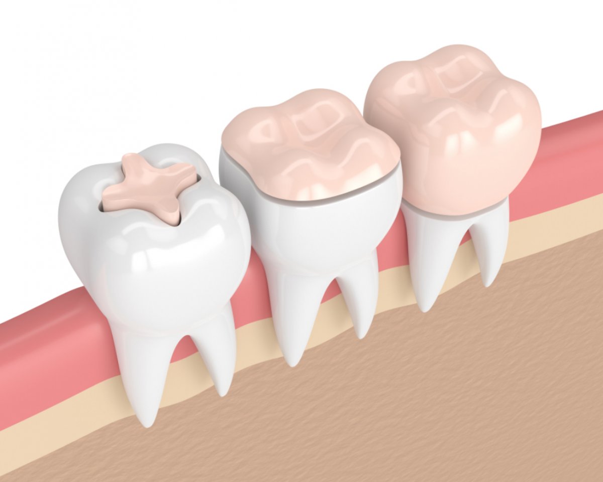
A comprehensive (multidisciplinary) approach is essential in oral structure. For example, if a zirconium crown or laminate will be made within the scope of smile design for a patient, the gum must not be bleeding or inflamed before the measurement is taken. If measurements are taken while the gums are inflamed and the porcelain is attached, once the gums heal and retract later, the edges of the porcelain may become exposed, leading to an aesthetic disaster. Therefore, periodontal health is maintained before aesthetics.
Similarly, in a patient undergoing wire treatment (orthodontics), the bone support must be complete. Trying to move a tooth in an inflamed bone can lead to the rapid loss of that tooth. When planning implant surgery, the mouth must be completely cleaned to prevent the bacteria flora in the mouth from contaminating the area around the implant (peri-implantitis) and eliminate the risk of infection.
How is Periodontal Treatment Planned Specifically for the Individual?
Everyone’s oral anatomy and healing potential are different. In individuals with thick gingival biotypes, gum recession is rare while pocket deepening is common; in individuals with thin gingival biotypes, rapid gum recessions (recession) occur. Experts at Izmir Avrupadent base their treatment on these structural differences, designing procedures such as laser pocket cleaning for thick gums and taking a piece from the palate to reinforce (connective tissue graft) for thin gums.
The same disease table requires different treatment protocols for a smoking individual and a non-smoking individual. Because smoking disrupts the bloodstream, it slows down the expectation of clinical improvement; in such patients, the process is optimized by avoiding surgical operations or ensuring that the patient completely quits smoking before and after the operation.