Why is Digital Planning Important in Aesthetic Dentistry?
The restorative processes that reconfigure the asymmetries, form irregularities, and color differences in the structures of the mouth, teeth, and jaw in accordance with biological principles have evolved into a new dimension with technological revolutions in the medical field. Aesthetic Dentistry, based on the detailed planning capabilities of contemporary clinical disciplines, has transitioned from solely relying on the physician’s manual skills and the technician’s visual perception to becoming a predictable science guided by mathematical data and computer-assisted algorithms. The digitization in dental practice minimizes human error margins at every stage, from measurement acquisition to model production and from smile design to the milling of porcelain blocks, allowing for the creation of a virtual copy of the outcome even in the initial stages of treatment.
The facial anatomy of individuals, lip movements, tooth alignment, and periodontal (gum) structure shows significant differences. In traditional methods, the harmony of these variables typically becomes clear as treatment progresses or when the prosthetic is fitted; however, with digital planning, all these biometric parameters are projected on the screen during the very first examination session. By superimposing the patient’s facial photographs with three-dimensional scans of the mouth in a computer environment, it allows for the prior determination of how well the ceramic restorations to be produced will conform to the patient’s expressions and bony contours. The guide we have prepared examines, from an objective perspective, which devices are used in digital workflows, the medical advantages of optical scanners that replace traditional measuring materials, and how this data is processed for the benefit of the patient in accordance with European clinic standards.
How Does the Digital Planning Process Begin with Intraoral Scanners?
The first and most critical step in restorative processes is the accurate replication of the patient’s existing oral tissues in the laboratory. In traditional methods, silicone-based impression pastes are filled into metal or plastic trays and placed in the patient’s mouth, waiting to set. This can trigger a gag reflex for patients and can be a discomforting action. In the first step of digital workflow, these trays and pastes are eliminated. Instead, a pen-sized camera (intraoral scanner) is slowly moved over the teeth to capture high-resolution images of all tissues.
The scanner calculates the smallest indent on the tooth enamel, the junctions of the gum tissue, and the distances to adjacent teeth with micron-level precision. The thousands of visual data obtained are combined in the scanner’s software within seconds to create a three-dimensional (3D) colored digital model. The dentist examines the material loss in the teeth, the occlusal relationship, and the levels of the gum tissue by rotating this model 360 degrees on the screen along with the patient. The digitization of data prevents the risk of deformation that could occur during the physical transfer of measurements.
What Are the Differences Between Traditional Measurement Methods and Digital Measurement?
In classical dentistry, physical measurements taken are exposed to various temperature and humidity changes after being removed from the mouth. Depending on the structure of alginate or silicone materials, microscopic contractions or expansions may occur during the hardening reaction of the material poured into the mold after the measurement is taken. These dimensional changes are a factor affecting the fit of the porcelain to be produced (its edge sealing capability). On the other hand, digital measurements obtained with optical scanners do not undergo volumetric changes as they do not contain physical substances; the measurement in the mouth maintains the same accuracy as the measurement displayed on the screen, down to one thousandth of a millimeter.
However, communication speed is also one of the major differences between the two methods. The shipping of a physical impression to the laboratory may take days for the technician to prepare the model, while a digital impression (files in STL or PLY format) can be instantly sent to a laboratory anywhere in the world via email or cloud systems with a single click. The table below includes an evaluation of both impression concepts in terms of medical and patient comfort:
| Evaluation Parameter | Traditional (Silicone/Alginate) Impression | Digital (Optical Scanner) Impression |
|---|---|---|
| Patient Comfort | Pastes that take up space in the mouth can trigger the gag reflex. | It is quite comfortable since only a small camera is used. |
| Dimensional Stability | Microscopic distortions can occur depending on heat and time. | Since the data is digital, it does not undergo volumetric or physical changes. |
| Transmission Time to the Laboratory | It requires time for physical transportation by courier or cargo. | It is sent in seconds over the internet connection. |
| Archiving and Storage | Physical storage requires storage space for the received models. | It is preserved as digital data for a lifetime on cloud servers. |
How are CAD/CAM Systems Integrated into Restoration Production?
Computer Aided Design (CAD) and Computer Aided Manufacturing (CAM) concepts are advanced technology products adapted from industrial production to medical procedures. In aesthetic dentistry planning, when the doctor takes a digital measurement, the CAD phase begins as it is displayed on the technician’s screen in the laboratory. The technician uses the mouse pointer on virtual models to draw the outer contours of the new tooth, the peaks, and the contact surfaces with adjacent teeth. During this design process, the software provides warnings regarding the breakage risk of porcelain by showing colorful maps of where the occlusion forces will contact.
Once the design is approved, the data is directed to CAM devices. CAM devices are adapted versions of industrial CNC machines. Blocks made of pure zirconium or glass ceramics (lithium disilicate) that are fabricated with a homogeneous structure, containing no air voids or structural weaknesses, are placed inside the device. Diamond mills on robotic arms mill out the virtual design from these blocks with millimeter precision. This process, freed from human hand vibrations or cast errors, helps restorations fit into the tooth like a puzzle piece.
How Does Digital Photography and Facial Analysis Affect Planning?
Having a flawless shape of a single tooth inside the mouth does not mean it will create an aesthetic entirety when placed on a person’s face. The width, length, and positioning of the teeth should establish an organic connection with the individual’s skeletal facial structure. To establish this connection, various angles of high-resolution photographs should be taken in a clinical studio environment with proper light sources and DSLR (digital single-lens reflex) cameras while the patient is at rest, talking, and in full smile positions.
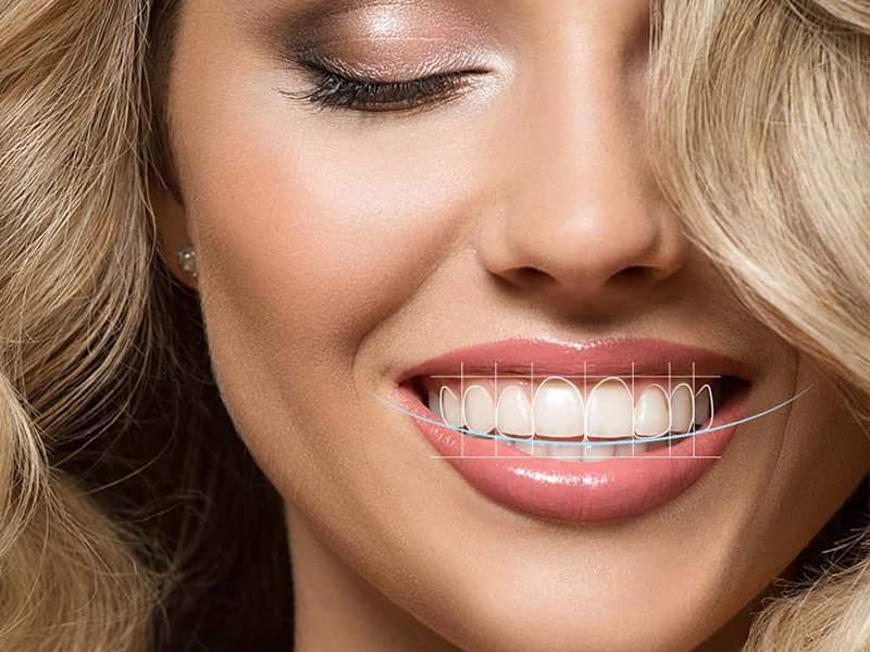
These two-dimensional photographs are superimposed onto three-dimensional dental models obtained from intraoral scanners in specialized smile design software. A horizontal line (pupillary line) passing through the patient’s pupils and a vertical line (midline) dividing the face in half are determined on the software. The incisal edges of the new teeth to be designed are positioned parallel to this pupillary line, and the junction point of the two front teeth will be aligned with the vertical midline. Additionally, the curvature of the lower lip during smiling guides the shape of the upper teeth. The integration of digital photography ensures that the design addresses not only the inside of the mouth but the entire facial geography clinically.
What Benefits Does Viewing the Treatment Result in Virtual Environment Provide for the Patient?
The main concern of patients in medical and aesthetic procedures is the possibility of not liking the outcome after an irreversible procedure has been performed. In traditional methods, the patient could only have a general idea when the porcelain teeth arrived from the laboratory and were tried in their mouth. The application of digital planning to the medical literature, referred to as “Mock-up” (the physical trial of a virtual design), is a communication tool that completely eliminates this uncertainty.
The tooth forms approved together with the patient on the computer screen are transferred to a temporary model mold via 3D printers. During the clinical appointment, a tooth-colored acrylic-based temporary material is filled into this mold and placed on the patient’s teeth (without cutting or grinding the teeth). With the solidified material in seconds, the patient physically tests the clarity of the targeted outcome in front of a mirror, its harmony with the lips, and its effect on speech. Any points that are not approved or wish to be modified (such as “make the teeth a bit shorter” or “make the edges more rounded”) are noted, and the tooth cutting procedure only begins after this agreement is reached.
What is the Role of 3D Printers in Digital Workflow?
3D printing technologies (Additive Manufacturing), which have rapidly expanded their application areas in dental clinics and laboratories, complement CNC milling machines. While milling machines produce by carving a ceramic block (subtractive method), 3D printers manufacture by hardening special liquid resins layer by layer using ultraviolet light or lasers (additive method). When there is a need for a physical jaw model to work on the digitally obtained measurements in the laboratory, these models are printed with 3D printers instead of being cast in a mold.
Additionally, temporary crowns that are placed to prevent aesthetic loss during aesthetic procedures or “surgical guide plates” that assist the physician during gingival surgeries are produced with these printers with micron-level precision. With advancing technology, medical R&D efforts are ongoing towards printing even long-term permanent restorations with 3D printers, and these devices have secured their place among indispensable equipment in digital workflows.
How is the Harmony of Pink and White Aesthetics Achieved with Digital Software?
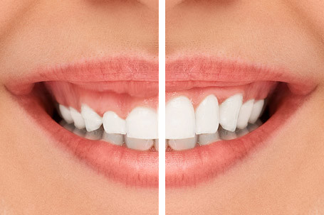
The visual harmony within the mouth depends not only on the shape of the white teeth but also on the symmetry of the pink gum tissue that surrounds those teeth like a frame. Asymmetrical appearance of the gums while smiling or the perception of some teeth being very short due to excess tissue is detected through the analysis of the digital software’s ‘zenith point’ (the apex of the gum). In the planning of Aesthetic Dentistry, the tips of adjacent teeth are connected by a virtual line on the screen of the software.
If the gum of one tooth is lower than that of another, the physician determines how much that tooth needs to be extended on the software. This digital determination guides how much the gum (gingivectomy) will be shaped using diode laser or cautery during the surgical phase. Digitally matching the boundaries of the pink background without interfering with the proportions of the white teeth ensures that the produced ceramic restorations remain within a biological balance.
How is Systemic Alignment and Closure (Occlusion) Analysis Examined in a Digital Environment?
For an aesthetic restoration to retain its shape in the mouth for many years, it depends not only on its beautiful appearance but also on its ability to withstand chewing forces. The human jaw is a dynamic and mobile system; when speaking or swallowing, the teeth continually come into contact with each other. In traditional methods, closure control was attempted to be understood by placing carbon papers in the mouth. However, this method could not precisely measure the timing and intensity of the chewing force.
During the digital planning phase, three-dimensional scans are taken not only of the upper and lower teeth but also of the patient’s jaw closure (bite registration) using intraoral scanners. The software marks points that contact early in red or orange when the jaws close on top of each other. If the designed porcelain tooth meets the other teeth earlier and with greater force, the design is adjusted millimetrically in the computer environment for balance. This level of precise calculation of the closing mechanics (occlusion) supports the mechanical longevity of the porcelain even in patients with bruxism.
What Standards Protect Digital Data in Avrupadent Clinics?
Data management in medical processes is not just an infrastructure that supports the patient’s current treatment but also future potential medical needs. Traditional impression models are vulnerable to damage, distortion, and storage issues; however, digital models obtained from optical scanners (STL or PLY files) can be preserved in databases without ever undergoing any volumetric alteration. According to European standards, facial analyses, color selections, and CAD designs of the patient are stored in special archive systems while adhering to personal data security protocols.
The greatest medical advantage offered by this digital archiving emerges in the event of a mechanical accident years later. For instance, if a porcelain crown or laminate is broken due to trauma, the patient may not need to return to the clinic for a new impression. The physician can send the file related to that patient in the archive to the laboratory, requesting the exact replica of the broken tooth (same form, same size, and contact points) to be milled in CAM devices. Digital archiving is a clinical safety network that elevates the sustainability of treatment.
What Are Frequently Asked Questions?1. Will there be a feeling of nausea when taking digital impressions?
Since no pastes or large spoons are placed inside the mouth, and only a pen-sized thin camera is used to scan the teeth, the nausea reflex is not triggered, making it a very comfortable process.
2. Does digital planning involve radiation?
Intraoral optical scanners do not use x-rays (radiation); they capture thousands of frames per second with LED or laser-based light sources, thus there is no risk in applying them even to pregnant individuals.
3. Will the design on the computer match the final result?
CAD/CAM systems use three-dimensional data from computers to produce by carving with robotic milling tools, with the design on the screen being a micron-level copy of the shape of the physical porcelain that is fitted into the mouth.
4. Do intraoral scanners harm the gums?
Since the tip of the scanner makes optical readings just above the dental surface without making direct hard contact with the tissues, it does not cause any physical damage to the gums or enamel.
5. Can digital dentistry be applied to all age groups?
It can be used in all age groups, from pediatric (children) patients who can remain still during the procedure to older individuals, without considering medical limitations on obtaining measurements.
6. Are the porcelains produced with CAD/CAM systems durable?
The factory-made ceramics or zirconium blocks used in these systems have a homogeneous structure, as they do not contain air bubbles or weak points like those in casting methods, and they possess an extraordinary high resistance to wear.
7. How long does the digital measurement process take?
Scanning the entire lower jaw, upper jaw, and the bite relationship typically takes about 3 to 5 minutes, depending on the dentist’s experience and the patient’s mouth opening.
8. Can changes be made to the teeth during the virtual design phase?
Yes, changes can be made to the length, shape, and edge angles of the teeth within seconds according to the patient’s request, as the design is entirely processed on the computer, allowing for alternatives to be produced.
9. Can digital scanning be done on pregnant individuals?
Since it does not contain any radiation, harmful chemical agents, or risk of ingestion, the screening process is medically safe unless there are other surgical barriers restricted by the obstetrician during pregnancy.
10. Is measuring paste used in digital workflows?
Digitalized clinical procedures do not require traditional measuring pastes like silicone, alginate, or polyether; all data is obtained optically.
11. Are permanent teeth produced with 3D printers?
Nowadays, 3D printers are mostly used for printing working models, surgical templates, and temporary prostheses; permanent zirconium or porcelain crowns are primarily manufactured in CAM devices (milling machines).
12. How long does it take to send the design to the laboratory?
Once the scan is completed and the data package (STL/PLY file) is created, it allows the technician to start working instantly by dropping onto the digital screen in seconds through cloud systems.
13. How does the error margin of digital measurements compare to traditional measurements?
Traditional measurement errors due to the expansion of material related to blood or dentin mixture are compensated for by software algorithms in digital measurements, so the precision of fit (marginal seal) is much higher.
14. Is digital planning also used in procedures like clear aligners?
Not only in porcelain veneers and aesthetic procedures, but also for the virtual planning of tooth movements in clear aligner treatments correcting tooth misalignments, this scanning data forms the essential medical infrastructure.
15. Can digital data be stored for future reuse?
All measurements, design, and closure data related to patients can be stored undamaged in digital archives and can contribute to the reproduction of the same tooth with zero error in the event of a potential porcelain breakage years later.