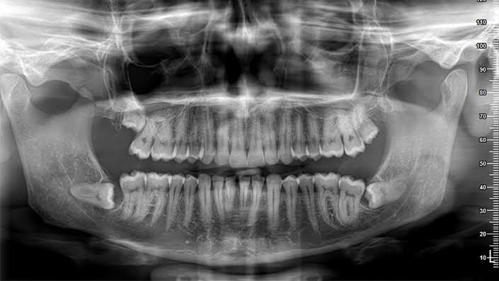
3D Jaw and Dental Tomography (technically known as Cone Beam Computed Tomography or CBCT) is an advanced imaging method used to obtain three-dimensional and detailed images of the jawbone and teeth. This method allows for a clear examination of many details from the bone structure in the jaw to the tooth roots, joint areas, and sinuses. Thus, it is a reliable technology that significantly facilitates diagnosis and treatment planning in the field of dentistry, reducing the risk of complications.
What is 3D Jaw and Dental Tomography?
3D Jaw and Dental Tomography is a special imaging method that allows for the three-dimensional evaluation of dental and jaw structures, obtained with a specific X-ray device. By eliminating the two-dimensional limitations of traditional X-rays, it provides much more comprehensive information for dentists and patients.
The foundation of this technology lies in the rotation of a conical beam of X-rays around the patient and the reconstruction of numerous 2D projections in the computer. As a result of the procedure, the closest cross-sectional and three-dimensional visuals of the tooth, bone, and related structures to reality are obtained. It gives precise and clear information especially in cases such as dental implant applications, jaw bone evaluations, and impacted tooth analyses. Additionally, it increases patient comfort due to the very short scanning times.
Unlike the massive medical tomographies in general health, this method focuses solely on the oral and jaw area. Thus, a three-dimensional data set is created with lower radiation, allowing for detailed examination. In short, 3D Jaw and Dental Tomography is a technology specially developed for dentistry, providing valuable information in diagnosis and planning processes.
In Which Cases is 3D Jaw and Dental Tomography Preferred?
3D Cone Beam Computed Tomography is a preferred imaging method when more complex or critical details regarding the jaw and teeth are required. Generally, it is primarily used in the following types of cases. For example, evaluating the volume and density of bone for implant applications, determining the location of nerves in jaw surgery planning, identifying the exposure of impacted teeth in orthodontic treatments, diagnosing joint disorders, and analyzing advanced endodontic problems are all highly beneficial.
While 2D panoramic X-rays are often sufficient in dental practices, there are cases where this two-dimensional data is inadequate. For instance, extra root canals or narrow canals can be overlooked during root canal treatment. In such cases, 3D data significantly increases accuracy. Additionally, when there is a need to examine the bone structure in the temporomandibular joint (TMJ), 3D Cone Beam Computed Tomography offers the best solution.
What is the Difference Between 3D Cone Beam Computed Tomography and 2D X-ray?
The biggest difference between 3D Cone Beam Computed Tomography and 2D X-rays is the level of detail added by the third dimension. Two-dimensional panoramic or periapical X-rays provide an overlapping view of bone and tooth structures. Particularly, adjacent anatomical structures can sometimes obscure each other. However, in 3D tomography, hidden lesions, tooth roots, or bone loss can be much more easily detected due to cross-sectional images.
Moreover, significant scaling or exposure errors can occur in 2D X-rays. In 3D Cone Beam Computed Tomography, however, measurements can be made very close to reality (1:1 ratio). This provides a significant advantage in procedures where millimetric accuracy is important, such as implant placement and orthodontic movement planning.
On the other hand, the amount of radiation is higher compared to 2D X-rays. However, thanks to technological advancements, many devices today have options such as “low dose” or “pediatric mode” to significantly reduce unnecessary radiation risks. Nevertheless, unnecessary use should be avoided, and 3D tomography should only be preferred when there is a real need for diagnosis and treatment planning.
How is 3D Jaw and Tooth Tomography Applied?
3D Jaw and Tooth Tomography is performed using a conical beam X-ray source and detector that rotates around the patient’s head. During the procedure, the patient stands or sits in a fixed position. The device captures hundreds of two-dimensional images with a single rotation; these images are combined with special software to create a single three-dimensional data set. This data set allows your doctor to examine any slice he or she desires and to make accurate measurements.
Although the scan duration can vary depending on the device and settings, it generally lasts between 5 and 40 seconds. A short scanning time increases patient comfort and minimizes blur caused by movement. The digital data obtained after scanning can be processed and analyzed instantly. This allows for quick treatment planning.
Today, many devices have different “field of view (FOV)” options available. For example, if there is an issue concerning only a few teeth, a small area can be selected for a low dose. However, when it is necessary to examine the entire jaw or both jaws together, a broader area is scanned. This flexibility is crucial in reducing unnecessary radiation risks with a personalized approach for each patient.
What Advantages Does 3D Jaw and Tooth Tomography Offer?
3D Cone Beam Computed Tomography offers numerous advantages in diagnosis and treatment planning through three-dimensional imaging. Primarily, it allows for the examination of anatomical structures without overlapping. It facilitates the identification of small details such as fractures in the root canals or the presence of additional canals. Additionally, accurately determining the volume of bone and the position of nerves in implant surgery significantly reduces surgical risks.
Another advantage is that measurements can be made with much greater accuracy. In implant planning, the height and thickness of the bone are measured with millimetric precision, thus reducing the likelihood of encountering unwanted surprises. In orthodontics, the position of impacted teeth or the relationship between the jaws is assessed very closely to reality.
Moreover, 3D data can be integrated with other elements in digital dentistry (for example, intraoral scan data, facial scans). This allows for the creation of a complete digital replica of the patient’s mouth and planning in virtual environments. The ability to examine the relationships between teeth and jaws from every angle helps facilitate a faster and more predictable treatment process.
Is 3D Cone Beam Computed Tomography Safe?
3D Cone Beam Computed Tomography is generally a safe method when applied with appropriate indications and dose settings. The radiation level is much lower compared to medical CT scans. Additionally, thanks to “low dose protocols” in many devices today, the amount of radiation can be further reduced. However, it is still a higher dose compared to 2D panoramic X-rays or periapical films. Therefore, it should always be carefully assessed whether the scan is truly necessary.
Particularly in children and adolescents, due to higher radiation sensitivity, extra caution is taken. Whenever possible, it is ensured to avoid using 3D tomography in these age groups or to choose a smaller field of view to target the lowest dose. Additionally, patients are advised to remove any metal accessories (ear piercings, glasses) before the procedure, and to wear protective thyroid collars if possible.
Within the framework of the ALARA (As Low As Reasonably Achievable) principle, the special needs of the patient should be taken into account, and 3D tomography should be used when necessary. This approach ensures both the safety of the patient and the success of the treatment.
What Are the Limitations of 3D Jaw and Tooth Tomography?
3D Jaw and Tooth Tomography is an excellent resource for detailed examination of the teeth and jaw bones, but it has some limitations. One of the main disadvantages is its inability to clearly show soft tissues. In cases where soft structures such as muscle tissue, joint discs, or gums need to be evaluated, different imaging methods such as MRI are required. For example, problems with the disk structure of the jaw joint cannot be detected through CBCT, but it shows bony disorders very well.
Another limitation is the artifacts caused by beam hardening due to metal restorations. If there are many metal fillings, crowns, or implants in the mouth, unwanted lines or dark areas may appear in the images. Although manufacturers have developed software algorithms to reduce these types of artifacts, completely eliminating them is not easy.
Moreover, in detecting small fractures, 3D tomography may not always be superior to 2D X-rays. In some cases, due to factors such as resolution and artifacts, tiny interproximal fractures may be missed or appear misleading. Therefore, the routine use of 3D tomography for “fracture scanning” is not recommended.
Finally, looking at the radiation dose, it is incorrect to use 3D tomography unnecessarily in simple cases where it is not really needed. This increases both the patient’s costs and the radiation burden. Therefore, it is most appropriate to use it in cases where it is genuinely required as assessed by the specialist.
How Does 3D Jaw and Tooth Tomography Integrate into the Digital Treatment Process?
3D Jaw and Tooth Tomography seamlessly integrates with today’s digital dentistry applications, advancing treatment planning to a significant level. This integration is particularly evident in implant surgery. Tomographic data is combined with digital intraoral scanning results to study the gum tissue and bone structure in virtual environments. Then, the diameter, length, and angle of the desired implant are determined; the distance to nerve pathways or adjacent tooth roots is adjusted in the most accurate manner. After this plan is digitally approved, surgical guides are produced with 3D printers to ensure millimeter accuracy in surgery.
A similar approach is also applicable in the field of orthodontics. Factors such as the position of impacted teeth, jaw constriction, and jaw joint position are analyzed in a digital environment. Again, the 3D Jaw and Tooth Tomography images are superimposed with the patient’s dental models, allowing detailed evaluation of tooth roots, bone boundaries, and areas where teeth can move. Thus, wire treatment or clear aligner planning can be done much more precisely.
In endodontics, when complex canal structures or root fractures are detected, it becomes easier to locate canal entrances with guides prepared using 3D data. These innovations that increase the success of treatment can also shorten the duration of treatment.
Additionally, digital planning and design tools (CAD/CAM systems) can achieve ideal fit in advanced prosthetic applications such as upper jaw, lower jaw, or facial prostheses by utilizing 3D tomography data. This allows the patient’s dental, jaw, and facial morphology to be gathered on a single digital platform, aiming for near-perfect aesthetics and function.
Thanks to all these integrations, 3D Jaw and Dental Tomography has opened the way for faster, precise, and personalized treatments in today’s dentistry. The unnecessary trial and error during treatment has diminished, significantly increasing patient satisfaction.
What Does 3D Jaw and Dental Tomography Promise for the Future?
3D Jaw and Dental Tomography promises much more comprehensive and patient-friendly features in the future, with the ongoing advancements in technology.
One of the most striking developments is AI (artificial intelligence) supported imaging analysis applications. These applications automatically scan the taken tomographies to quickly detect potential lesions or abnormalities. For example, it may become possible to automatically highlight cystic formations in the jawbone, canal blockages, or apical lesions with the help of algorithms. This saves time for clinicians while reducing the risk of missing potential issues.
Another significant innovation is 4D imaging methods that include the temporal dimension. Some manufacturers are working on systems that can monitor jaw movements in real time. This allows for the analysis of the bone dynamics during chewing or joint movements. Especially for temporomandibular joint (TMJ) disorders or chewing difficulties, a functional perspective is expected.
There is also a continuous progress in reducing radiation dose. More precise detector technologies and advanced software algorithms aim to generate similar quality visuals using much lower X-rays. This situation will particularly make 3D Jaw and Dental Tomography applications safer for sensitive groups such as children and young adults.
In the future, it is possible to reduce the size of devices and increase the prevalence of office-type portable systems. Thus, even small clinics having access to 3D imaging will facilitate the widespread adoption of this technology and make it a standard practice.