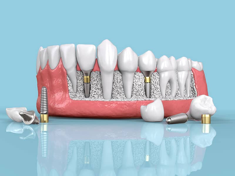
What Examinations Are Conducted Before Implant Treatment?
Prosthetic and surgical processes planned to compensate for tooth deficiencies are not just a series of procedures encompassing the moment of operation; the most critical phase of this process consists of comprehensive examinations conducted before treatment. The successful outcome of surgical procedures characterized as implantology is dependent upon the detailed analysis of the patient’s oral physiology and systemic health status with precise data. The pre-operative assessment phase requires a multidisciplinary approach that combines both the clinical experience of the dentist and modern imaging technologies.
Before the decision to place a titanium screw into the jawbone, doctors perform a wide-ranging screening from bone density in the area to the thickness of the gum, including the patient’s chronic diseases and the medications they use. The collection of this data allows for the preemptive identification of anatomical variations, assessment of potential medical risks, and tailoring surgical steps based on these parameters. In this content we have prepared, we will detail the clinical evaluations conducted by doctors before the surgical operation, the radiological tests performed, and the history-taking phases (patient medical history).
What Does a Dentist Check During the First Clinical Examination?
During the first clinical examination, the dentist checks the condition of the existing teeth in the oral cavity, the cavities, the color and consistency of the gums, the width of the spaces created by missing teeth, and the overall health of the soft tissues. At this stage, the dentist manually assesses whether the oral flora is prepared for surgical intervention using special mirrors and probes (measuring instruments).
Clinical evaluation begins the moment the patient sits in the dentist’s chair. The doctor primarily observes extraoral (outside the mouth) structures such as asymmetry, facial contours, and jaw joint movements. Then, by entering the mouth, the thickness of the gum tissue in the area where the planned procedure will take place and whether the neighboring teeth have tipped into this space is examined. If there are tipped teeth towards the gap, the distance for the prosthesis to be inserted might be reduced; this situation may lead to additional orthodontic or prosthetic interventions in treatment planning.
Additionally, old fillings, worn bridges, or minor fractures in the mouth are recorded individually. Since the mouth is evaluated as a whole, the focus is not only on the tooth-less area; sources of infection in other teeth are also examined to ensure the overall chewing balance is maintained. As a medical rule, surgical procedures are deemed inappropriate by medical guidelines if there is an active focus of bacteria (abscess, deep fracture, etc.) in the mouth.
Why Is the Hygiene Status of the Mouth Before Implantation Important?
The hygiene status of the mouth before implantation is examined to prevent the accumulation of dental calculus and bacterial plaques from creating an infection risk in the surgical area. In mouths with active gum inflammation (gingivitis or periodontitis), the process of titanium root anchoring to the jawbone may be disrupted due to bacterial activity, making the hygiene index extremely important.
Periodontal health (gum and surrounding tissue health) is a fundamental requirement for any dental surgery. During the examination, the dentist measures the depth of the pockets between the tooth and the gum using a device called a periodontal probe, which has millimeter markings. In a healthy gum, this depth ranges from 1-3 mm, whereas in cases of inadequate hygiene, the depth increases and dental calculus progresses beneath the gum into the bone. A surgical intervention performed in this bacterial environment prepares the ground for a medical condition known as “peri-implantitis,” which leads to the erosion of the bone surrounding the titanium root.
Patients with inadequate oral hygiene are not given immediate appointments. In the initial stage, a detailed dental calculus cleaning (scaling) and, if necessary, root surface smoothing (root planing) are planned. The patient is informed about the proper brushing techniques and the use of interdental brushes. After an average period of a few weeks of this hygiene motivation and tissue healing, when the gums turn pink, firm, and free of bleeding, the appropriate groundwork for the surgical phase is prepared.
What Imaging Techniques Are Preferred in Radiological Assessments?
In radiological assessments, panoramic radiographs, which generally show the overall condition of the jaws, and three-dimensional Dental Volumetric Tomography (CBCT), which provides detailed millimetric information about the bone structure, are generally preferred. While panoramic radiographs provide a broad overview, tomography allows for a cross-sectional analysis of the thickness of the bone and the adjacent anatomical spaces.
Panoramic radiography, a two-dimensional imaging technique, is a standard component of the initial examination. This X-ray allows for the assessment of the overall resorption level in the jawbone, the root lengths of existing teeth, and the positions of cysts or impacted teeth (e.g., wisdom teeth) detected on a single film. However, since it is two-dimensional, it provides data on the height of the bone but does not give clear information about its “width” (thickness).
Since titanium materials are cylindrical, a certain amount of healthy bone tissue on the cheek and tongue side is needed when placing them in the middle of the jawbone. Advanced imaging techniques are used to measure the thickness of the bone in this direction. The table below compares the medical evaluation metrics of two main radiological methods used in examinations:
| Imaging Method | Medical Data Provided | Purpose of Use in Examination |
|---|---|---|
| Panoramic X-ray (2D) | Overall appearance of the jaws, condition of tooth roots, vertical bone level, large cysts. | Initial diagnosis, assessment of overall oral health, and preliminary planning. |
| Dental Tomography (CBCT – 3D) | The millimetric thickness of the bone (width), three-dimensional location of nerve canals, sinus depth. | Surgical mapping, open calculation, and clear determination of the need for bone graft (powder). |
| Periapical X-ray | Very detailed and close-up images covering only 2-3 teeth. | Close analysis of regional root infections or a specific single area. |
What Data Does Three-Dimensional Jaw Tomography (CBCT) Provide?
Three-dimensional jaw tomography (CBCT) provides the vertical height and horizontal thickness of the jawbone, the millimetric location of the inferior alveolar nerve (nervus mandibularis), and the rate of sagging of the maxillary sinus cavities in the upper jaw. This data is a critical roadmap to avoid damaging anatomical structures during surgical procedures.
In regions with bone deficiency, the bone tissue shows a trend of thinning from the outside in over the years. Even if bone height appears adequate in panoramic X-rays, it can be noticeable in tomographic sections that the bone is as thin as a knife’s edge. The CBCT device separates the jaw into hundreds of microscopic sections and projects them onto a computer screen. The physician examines the bone from every angle by rotating these sections through the relevant software.
The most important anatomical reference in surgeries of the posterior part of the lower jaw is the mandibular nerve, which provides sensation to the lip and jaw. Since placing the screw very close to this nerve can lead to loss of sensation, the nerve canal is digitally marked on the tomography, and a safe medical distance is maintained. Similarly, in standard planning procedures like those of Avrupadent clinical protocols, the distance of the sinus air cavities in the upper jaw is also measured with these scans; if the distance is insufficient, a sinus lifting operation is added to the treatment schedule.
How Does Bone Density Measurement Shape the Treatment Plan?
The bone density measurement determines the primary stability (initial retention strength) of the titanium screw to be placed within the bone and the biological healing period, thereby shaping the treatment plan. Classifying the quality of the bone directly affects the speed of the drilling instruments that the physician will use during the operation and the waiting time for osseointegration.
Radiological scans show not only the shape of the bone but also the density of the trabecular (spongy) tissue within its internal structure. In medical literature, the density of the jawbone is classified from D1 to D4. D1 type bone is very hard and dense like hardwood; it is usually found in the anterior parts of the lower jaw. D4 type bone, on the other hand, is soft and porous like Styrofoam; it is commonly encountered in the posterior regions of the upper jaw.
When creating a cavity in a hard bone like D1, medical drills are used at very low speeds and with plenty of sterile serum to prevent the bone from overheating and its cells from being damaged (necrosis). In a type D4 soft bone, the cavity is made slightly narrower than required for the screw to hold tightly to the bone, allowing the screw to embed itself by compressing the bone with its threads. The density also affects the patient’s waiting time after treatment; since the cells in the soft bone wrap around the titanium for a longer duration, the waiting period is medically extended.
Why Is the Patient’s General Health History (Anamnesis) Investigated?
The patient’s general health history (anamnesis) is questioned to identify systemic disorders, chronic diseases, and allergies that directly affect the safety of the surgical intervention and the healing process. This phase ensures the prior management of factors that could complicate dental operations, such as coagulation issues or systemic disease.
The health information forms filled out by the patient during the examination process are not merely procedural details but medical requirements. Surgeries previously undergone, conditions requiring hospital admission, or history of radiation therapy or chemotherapy are recorded through these forms. For example, in a patient who has received radiation therapy (treatment of cancer) to the head and neck area, the blood supply to the jawbone is reduced, which decreases the potential for bone cells to renew themselves. This may affect wound healing during the procedure, thus requiring a different medical assessment.
Similarly, in individuals with suspected pregnancy, radiological screenings are avoided initially, and surgical procedures may be postponed based on the gynecologist’s direction. In the anamnesis phase, the doctor’s direct communication with the patient reveals discomfort that the patient may forget or consider unrelated to dental practice (for example, thyroid problems), thereby strengthening the medical safety net.
Which Systemic Diseases Require a Consultation with a Medical Doctor?
For systemic diseases such as cardiovascular diseases, uncontrolled diabetes (sugar disease), osteoporosis (bone loss), autoimmune disorders, and kidney or liver failure, a consultation (written opinion) from a relevant specialist is requested. This allows for the effects of the operation on the patient’s overall health to be managed collaboratively with the medical doctors.
In individuals who have undergone heart valve surgery or have arterial blockages, the use of antibiotics is required beforehand to prevent bacteria that may enter the bloodstream during oral surgical procedures from reaching the heart, a process known as “infective endocarditis prophylaxis.” The physician will consult with the hospital’s cardiologist to specifically plan this antibiotic regimen for the patient. Similarly, the amount of adrenaline in the anesthetic solutions used for patients with a bypass history is adjusted according to the cardiologist’s guidance.
In diabetic patients, if insulin use or diet keeps blood sugar regulated, the process operates normally, similar to that of an individual without diabetes. However, in patients whose blood sugar fluctuates constantly (unregulated), the susceptibility to infections increases, and the cellular healing mechanism slows down. In these cases, surgical procedures cannot be initiated without approval from an endocrinologist or internist. Medical consultations are among the fundamental medical safety procedures that protect the overall integrity of the patient and ensure the process is guided by scientific data.
Is It Necessary to Perform a Blood Test Before Implantation?
Blood tests are not routinely required from everyone before implantation; however, if the patient’s medical history indicates diabetes, bleeding disorders, liver issues, or vascular system diseases, it is a medical necessity to have a blood test to measure relevant parameters (HbA1c, INR, hemogram, etc.). These tests are indicative for surgical decision-making.
Individuals with diabetes often undergo an HbA1c (Hemoglobin A1c) test requested by the physician after examination. This test reflects the patient’s average blood sugar level over the last three months. A result that significantly exceeds reference values indicates a risk of poor wound healing and inflammation in the surgical area, so it is expected that the diabetes is stabilized by the healthcare providers beforehand. Similarly, for individuals experiencing clotting issues, PT (Prothrombin Time) or INR tests may be requested to analyze whether any bleeding that may occur during surgery can be controlled.
In cases diagnosed with osteoporosis and using intensive medication, specific blood values or calcium levels indicating bone metabolism may be examined. For individuals with liver dysfunction, knowing the medical values in advance is important because there may be issues in metabolizing antibiotics and pain relief medications prescribed after surgery, allowing for proper dosage adjustments by the physician.
How Do Medications Affect Pre-Treatment Evaluation?
Medications used, especially anticoagulants and osteoporosis medications (bisphosphonates), directly impact pre-treatment evaluation as they alter the bleeding profile and bone healing. During the examination phase, the daily doses of these medications are recorded, and a consultation is obtained from the prescribing physician for dosage adjustments before the operation.
Due to the risk of heart attack, arrhythmia, or a history of blood clots, many patients are using blood thinners. The use of these medications, when a surgical incision will be made, may complicate the control of bleeding that occurs during the operation, such as seepage. The dentist will medically plan whether the medication should be stopped a few days before the procedure or whether to switch to a lower-risk alternative drug by communicating with the specialist who prescribed the medication. Since stopping the medications on the patient’s own initiative carries serious life risks, all arrangements will be made under the doctor’s supervision.
Bisphosphonate medications taken intravenously or orally to increase bone density may suppress the cellular turnover in the jawbone. In individuals using these medications heavily, when intervention is made to the bone, the healing mechanism may not work, and a medical condition known as “jaw bone necrosis” (loss of bone vitality) may occur. The doctor will evaluate how long the patient has been using this medication and in what dosage during the examination phase to decide whether the procedure can be performed or whether a break is necessary.
How is Digital Aesthetic Design and Analysis Conducted?
Digital aesthetic analysis is conducted during the examination phase by transferring the patient’s dental, lip, and facial proportions into a computer environment using intraoral optical scanners and high-resolution photographs. This way, the position of the titanium root to be placed is planned in advance to meet the aesthetic expectations of the prosthesis that will be produced at the end of the treatment.
Especially in anterior tooth deficiencies, the most important part of the examination is analyzing the aesthetic expectations. The dentist replicates the current three-dimensional shape of the teeth using intraoral scanners. Later, clinical photographs are taken that show the position of the lips when the patient is speaking and smiling. These data are combined in software to virtually design the final state of the missing tooth.
The prepared virtual prosthesis is based on the “Top-Down” planning concept. That is, the dentist first draws the final form of the tooth to be made, then integrates the jawbone tomography into this design and places the screw virtually in a manner that it will support exactly from the center of that tooth. This digital analysis method, which is frequently used in modern clinical examinations like those in Avrupadent processes, prevents errors in opening and aesthetic asymmetries when transitioning to the prosthetic phase.
What Issues Does Jaw Closure and Joint Examination Prevent?
Jaw closure (occlusion) and joint examination analyze the way the lower and upper teeth contact each other, preventing excessive chewing loads beyond the capacity of the new prosthetic tooth. At the same time, it ensures the early detection of potential shifts in the jaw joint or forces that could cause damage like nocturnal teeth grinding (bruxism).
Although the titanium material placed in the toothless area is tightly attached to the jawbone, it does not have a “periodontal ligament” (the cushioning ligament) that provides flexibility like natural teeth do. Therefore, the forces generated during chewing are transmitted directly to the bone. During the initial examination, the way the patient closes their jaw and how the teeth contact each other during lateral chewing movements are carefully monitored. If there is only a closure anomaly that places excessive load on that area, this can lead to the porcelain breaking over time or damage to the bone surrounding the screw.
Additionally, if there is a habit of teeth grinding or gnashing (bruxism) during sleep, this condition can be detected during joint examination through hypertrophy (enlargement) of the jaw muscles and wear on the teeth. When this type of medical diagnosis is made, treatment planning involves using rigid acrylic night plates during sleep or medical relaxing applications aimed at the chewing muscles (such as chewing muscle injections, etc.) to protect the underlying structures.
Why is Gum Thickness (Biotype) Examination Important?
The examination of gum thickness (biotype) is vital for assessing the quality of the soft tissue in the surgical area, thus determining the adequacy of the biological barrier that will protect the neck part of the titanium root from bacteria. The thin structure of the gum should be identified during the examination phase, as it may lead to aesthetic gray reflections and tissue retractions over time.
The gum tissue inside the mouth does not have the same characteristics in everyone. In medical terminology, thick and fibrotic tissue is referred to as “thick biotype,” while transparent and delicate tissue is called “thin biotype.” During the initial examination, the dentist tests the structure of the gum with a periodontal probe. Tissues with a thick biotype are much more resistant to surgery, heal quickly, and tightly wrap around the neck part of the implant, isolating it from external factors.
However, if a thin biotype is in question, it can reflect a noticeable metallic gray color from the outside, and these tissues show a tendency for retraction over the years due to micro-traumas. If this clinical condition is detected during examination, an additional surgical step is included in the treatment schedule, which involves the transfer of a small piece of soft tissue taken from the hospital’s palate to the relevant area, referred to as a “connective tissue graft.” Thus, the gum is thickened, ensuring both aesthetic harmony and long-term tissue defense.
Frequently Asked Questions (FAQ)
1. How long does the initial examination take?
The first appointment, which includes a general oral examination, a review of the medical history, and radiographic assessments, typically lasts between 30 to 45 minutes. If additional tests such as tomography or digital scanning are necessary, this duration may extend somewhat.
2. Is a tomography scan mandatory?
While not mandatory for every case, performing a three-dimensional analysis becomes a medical necessity, especially in areas of the lower jaw where anatomical structures (such as sinus cavities and nerve canals) pose risks, and in cases with limited bone volume. The physician determines the need for tomography based on the data from the panoramic radiograph.
3. Is there a risk of radiation in tomography?
Dental tomography (CBCT) devices focus on a much narrower area (only the jaw region) compared to medical tomographs used in hospitals, which results in a significantly lower radiation dose. The detailed surgical guidance provided outweighs the medical benefits of this minimum dose taken.
4. I am using a blood thinner; should I mention this during the examination?
You should inform your physician about all medications you are taking, including prescribed or over-the-counter drugs and herbal supplements. Since blood thinners can alter the coagulation process during surgery, it may be necessary to adjust the dosage prior to the operation in consultation with your physician.
5. Should I come fasting before the examination?
There is no need to come on an empty stomach for the first appointment, where only examination and radiological imaging will be done. In fact, coming with your routine diet for clinical examinations prevents possible drops in blood sugar. However, if a surgical appointment is scheduled in the following days, the doctor will instruct you on your eating status according to the type of anesthesia.
6. If my jawbone is resorbed, will it be evident during the examination?
Yes, the panoramic X-rays or three-dimensional tomographies taken show the losses (resorption) in the vertical height and horizontal width of the jawbone in millimetric values. The doctor diagnoses whether there is a need for additional bone powder (graft) application by examining these scans.
7. Is a panoramic X-ray sufficient on its own?
In standard cases where the bone structure is regular and the distances are adequate, a panoramic X-ray can be used as a guide. However, a panoramic X-ray is two-dimensional; it does not show the thickness (width) of the bone. In complex surgical areas, tomography support is medically required to determine the thickness.
8. If there is suspicion of pregnancy, can an X-ray be taken?
During pregnancy or in cases where there is suspicion of pregnancy, if there is no medical emergency (severe abscess, cyst, etc.), X-ray examinations are generally postponed until after childbirth. In urgent cases, with the consent of the obstetrician and the gynecologist, low-dose protective X-rays can be taken with a lead apron.
9. I have diabetes, will extra tests be requested during the examination?
Diabetes is a systemic disease that directly affects wound healing. During your examination, your doctor may want to evaluate your HbA1c test result, which shows your average blood sugar levels over the past three months, or your recent glucose measurements, and may request approval (consultation) from an endocrinology specialist if necessary.
10. How is a digital measurement taken?
The device known as an intraoral scanner, which is pen-sized and equipped with an optical camera, collects data by moving over the surfaces of the teeth. These devices capture thousands of photographs of the structures inside the mouth within seconds and create a three-dimensional, color virtual model that accurately represents reality in the computer environment.
11. Can it be determined from an X-ray that my bone density is low?
Tools such as dental tomography (CBCT) software are used to analyze the density of the spongy tissue in the bone. The doctor can determine the degree of hardness of the jawbone (D1-D4 scale) by evaluating digital sections, which can define the technical approach during surgery.
12. Are gum diseases visible in X-rays?
The color of the gum and soft tissue inflammation are not directly visible in X-rays; these are detected during the doctor’s visual examination of the mouth. However, if gum disease progresses and starts to erode the underlying jawbone, this damage (periodontal bone loss) can be clearly observed in radiological scans.
13. Does the dentist perform blood tests?
In dental clinics, blood sampling is usually not performed. The dentist informs the patient of the necessary medical tests (such as Hemogram, INR, HbA1c, etc.) based on the examination results. The patient must submit the results of these tests performed at a medical center or hospital to the dentist for inclusion in their clinical file.
14. I have jaw joint pain, should I mention it during the examination?
Yes, temporomandibular joint (jaw joint) discomfort affects the treatment planning for teeth grinding during sleep. Mentioning this condition ensures that the dentist includes night splints or joint treatments in the plan to balance the chewing forces to protect the titanium implant post-surgery.
15. Is the procedure (surgery) performed on the day of the examination?
If the patient’s systemic health is stable, bone measurements are sufficient, and there is enough time for digital planning in the clinic, the procedure can be performed on the same day with the dentist’s approval. However, if there is an infection or if medical consultations or blood tests have been requested, the surgical appointment will be rescheduled for a later date.