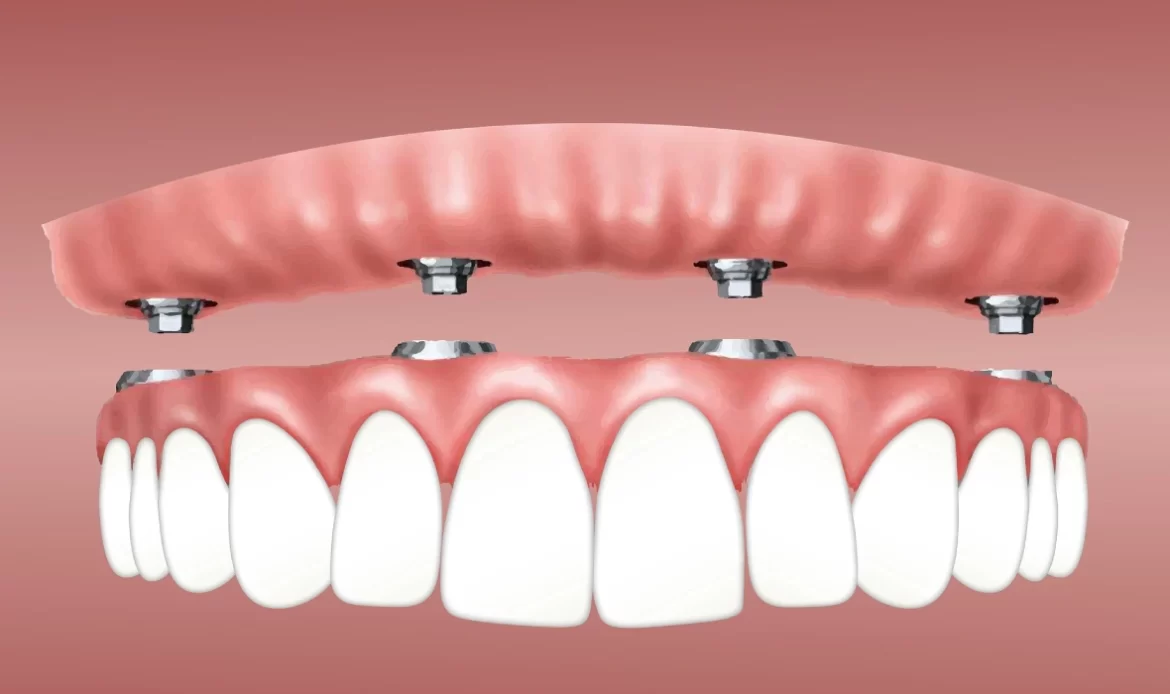
How is implant treatment applied in cases of multiple tooth loss?
The loss of multiple teeth in the mouth due to trauma, severe fractures, or periodontal damage leads to a significant reduction in chewing function and disrupts the physiological balance in the jawbone. The procedures applied in implantology, which are intended to support such multiple deficiencies medically, aim to reorganize the lost tissues according to their anatomical structure. While traditional methods often plan removable ( detachable) prosthetics or long bridges that require extensive cutting of healthy teeth for replacing multiple teeth, current medical approaches involve creating fixed units supported by the jawbone using titanium materials.
In cases where multiple teeth are missing, the treatment plan is specifically tailored according to the location of the gaps within the mouth, the existing vertical and horizontal volume of the jawbone, and the condition of the adjacent healthy teeth. In side-by-side deficiencies, the biomechanical integration of bridge systems with titanium screws is ensured, while each area in the spaced gaps distributed in different regions of the jaw is evaluated according to its anatomical characteristics. In this comprehensive content, the clinical steps followed in compensating for multiple tooth losses, radiological calculations, stages of bone tissue integration, and the details of prosthetic phases will be examined from a medical perspective.
Is it necessary to place a screw in every gap in cases of adjacent multiple tooth loss?
In medical tables where multiple teeth have been lost in a side-by-side arrangement, placing a separate titanium implant for each missing tooth is not a medical necessity. In dental practice, in an area where three adjacent teeth are missing, it is generally preferred to position a titanium root implant at the starting and ending points of the gap. A body (pontic) representing the missing third tooth is then added between these two support points to form a bridge prosthesis. This method prevents the need for excessive medical procedures on the patient’s jawbone and preserves healing tissue. Additionally, optimizing the number of parts to be embedded within the bone helps maintain the normal blood supply level in the jaw anatomy. This system, determined by calculating anatomical factors and chewing forces, provides long-term biomechanical balance from a medical perspective, meeting the functional needs of the patient.
The body tooth placed between the two implants does not directly apply mechanical pressure on the lower jawbone; it transfers the load to the titanium carriers on its right and left. In specific cases where bone volume is insufficient or in long gaps involving four to five adjacent teeth, the number of support points may be increased based on the radiological assessment by the dentist. This medical strategy reduces the size of the surgical area while simultaneously allowing for the creation of physiological gaps (embrasures) that facilitate easier cleaning of the prosthetic superstructure inside the mouth.
How are Bridge Prostheses with Titanium Roots Combined in Multiple Tooth Deficiencies?
Bridges applied in complex tooth deficiencies are specially designed in the laboratory to be supported only by titanium roots and are integrated with the jaw structure. In traditional bridges supported by natural teeth, the healthy teeth on the edges of the gap are reduced (enamel is prepared) to be used as legs for the bridge, whereas in this method, the supporting legs of the bridge consist of artificial materials placed in the jawbone. After the biological osseointegration (bone retention) process is completed, medical intermediaries called “abutments” are screwed onto the titanium roots. The zirconium or porcelain bridge body prepared by technicians is precisely fitted onto these intermediaries and bonded with medical agents (cements) or directly secured with screw systems. This way, the mechanical stress generated during chewing is transmitted directly to the bone via the titanium legs without reflecting on the natural teeth, preserving the integrity of the oral tissues.
The underside of the body of these bridges, prepared in the laboratory stage, is designed to maintain slight contact with the gum (ovate pontic). This design not only provides the appearance as if the tooth is growing out of the gum naturally but also creates a physiological form that minimizes the accumulation of food debris beneath the bridge. The metal or zirconium infrastructure of the bridge system is produced with rigidity (hardness) that does not allow flexing between the two or three screws it is supported by, laying the groundwork for achieving chewing efficiency standards.
What Methods Are Followed When Three or More Teeth Are Lost in the Back Region?
When three or more molars (large back teeth) are lost in the very back areas of the jaw, a clinical condition called “free-end edentulism” arises, as there are no remaining natural teeth to support a bridge prosthesis in that area. To medically compensate for this condition, titanium supports are planned to be placed in the back area, following steps to restore function to the region. In traditional methods, only removable hook-like (partial) prostheses can be made when there are no supporting teeth in the back; however, when using implantology procedures, it becomes possible to create a fixed bridge restoration thanks to the two artificial roots placed in this area. Considering the density of the jawbone in the back area and the occlusal forces exerted, the diameters of the medical components to be applied here are generally selected in wider forms and a pressure-resistant infrastructure is organized.
One of the medical conditions frequently encountered when this method is applied in the upper jaw’s back regions is the sagging of the maxillary sinuses (air cavities). Over time, as tooth losses occur, the sinus floor displaces downwards, reducing the height of the jawbone. In radiological examinations where this anatomical change is detected, a “sinus lifting” (sinus floor elevation) operation is performed before placing material in the area, and bone powder (graft) is added to the newly created space. After the relevant graft integrates with the patient’s own bone structure, titanium supports are placed in the back area for planning a fixed bridge.
How is a Treatment Map Drawn When There Are Intervals in the Gaps of the Jaw?
If there are multiple independent (spaced) edentulous areas on the right, left, or frontal region of a jawbone, the treatment plan will be drawn by assessing each gap according to its own anatomical conditions. The dentist analyzes the bone height, the distance to adjacent teeth, and the gingival biotype of each missing area through three-dimensional dental tomography (CBCT) images. In cases of multiple and scattered deficiencies, mechanically connecting distant gaps with a single long bridge is not an appropriate approach. Instead, each edentulous gap is planned individually or restored with short bridges if they are next to each other. The dentist conducts multidirectional medical planning to balance the occlusal plane of the entire dentition based on the positions of the existing healthy teeth in the mouth. The surgical intervention for each region can be performed in the same clinical session, but taking into account the patient’s overall health condition and the duration of the procedure, it can also be divided into different sessions.
In the rehabilitation of spaced gaps, the periodontal (gum) health of the existing natural teeth is also meticulously examined. If there is bone loss or infection in the teeth surrounding the gaps, priority is given to treating these teeth to stabilize the oral flora. In standardized process management, such as European dental clinic protocols, digital design software is utilized in the laboratory phase to ensure the color and shape uniformity of the crowns to be placed in different regions, planning for even distant prosthetics to provide a homogeneous aesthetic appearance in the mouth.
How is the Load on the Jawbone Calculated in Multiple Implantology Applications?
In multiple applications, calculating the vertical and horizontal mechanical loads on the jawbone is a clinical necessity for ensuring long-term biological stability. During the chewing cycle, when the lower and upper jaws come into contact, the force produced by the facial muscles (masseter and temporal muscles) is transmitted directly through the prosthetic structures to the titanium roots and subsequently to the jawbone. This force calculation takes into account the patient’s facial anatomy, the angle of the jaw, the presence of involuntary parafunctional habits such as teeth grinding (bruxism) during sleep, and whether there are natural teeth or prosthetics in the opposing jaw. If the load capacity is not correctly calculated and the number of supports is kept insufficient, micro-movements may start within the system over time, loosenings may be observed in the connecting screws, and stress-related resorption (marginal bone loss) may occur in the surrounding jawbone.
The physician performing these calculations through medical software optimizes the length and diameter of the materials to be placed in the edentulous area in such a way as to distribute the incoming force over a broad surface. Bridge systems applied in multiple deficiencies work on the principle of “splinting” (connecting to one another). The mechanical resistance exhibited against chewing forces by the connected structures is significantly higher compared to individual structures. Thus, a sudden and sharp pressure reaching a certain point is transmitted through the body to other support points, minimizing the regional stress on the bone.
Can Multiple Tooth Extractions and Titanium Placement Be Done in the Same Session?
In clinical cases where multiple teeth are irretrievable within the jaw, the extraction of teeth and the placement of titanium screws into the jawbone can be performed in the same clinical session. This medical procedure, known as “immediate placement”, depends on the condition that there is no acute abscess, cyst, or active infection in the root area of the extracted teeth. Additionally, the bone walls surrounding the socket (the area where the tooth was extracted) must have sufficient thickness, and the screw to be placed should be able to achieve primary stability (initial retention force) that can hold tightly to these bone walls. When radiological data indicates that these conditions are favorable, the physician extracts the problematic teeth using atraumatic techniques (without damaging surrounding tissues) and, after cleaning the resulting sockets with sterile solutions, positions the artificial roots in the same session. This approach reduces the number of times the patient needs to sit in the surgical chair, shortens the treatment timeline, and helps to maintain anatomical form by preventing the onset of bone resorption after extraction.
During this multiple procedure performed in the same session, if there is a gap between the socket of the extracted tooth and the titanium component placed, these anatomical gaps are filled with bone graft (powder) to support the healing process. The surgical area is closed with soft tissue sutures. Throughout the cellular regeneration process in the region, the patient adheres to the nutritional and hygiene rules specified by the physician, going through the medical healing period.
How Do Users of Partial Dentures Transition to a Fixed System?
In order for individuals using partial removable dentures (commonly known as hook or clasp dentures) to transition to fixed systems supported by the jawbone, the current state of the jaw anatomy is examined in detail through radiological scans. When removable dentures are used for many years, due to the pressure exerted by the denture on the tissue and the burden placed on the natural teeth where the clasps grip, resorption in the jaw crests and mobility (rocking) in the teeth can occur. During the transition process, the physician measures the bone thickness in edentulous areas to plan how many medical supports will be placed in these areas. If the jawbone has sufficient volume, titanium screws are placed in the edentulous areas, and after the biological integration process is completed, fixed zirconium or porcelain bridges prepared in the laboratory are attached to these screws. This allows the patient to escape from the removable hook dentures that need to be taken out of the mouth every day for cleaning while eating or speaking, transitioning to a fixed chewing system in the form of their own natural teeth.
If the jawbone has significantly resorbed due to prolonged use of the removable denture, direct placement is not performed. Initially, bone powder (graft) and membrane applications are performed to medically increase the height and width of the jaw crest (ridge). During the several months required for this grafting procedure to integrate with the bone, the patient can continue using their existing removable denture by supporting it with soft liners. Once the bone matures, the surgical phase begins, laying the foundations for the fixed restoration.
Can Natural Teeth and Implants Be Connected in Cases of Multiple Tooth Loss?
In bridge prosthetics applied for multiple tooth deficiencies, the connection of one foot to a natural tooth while the other foot is attached to a titanium screw placed in the jawbone is a biomechanical approach that is rarely preferred and generally avoided in current dental protocols. The root of the natural tooth is connected to the jawbone by microscopic elastic fibers known as the “periodontal ligament” and has a small allowance for elastic (micro movement) within the bone during chewing. However, titanium materials fuse with bone at the cellular level (osseointegration) and remain completely rigid (stable) in the jaw because they do not contain any elastic fibers around them. When a single, solid bridge is attached to these two structures, which have completely different elastic capacities, the natural tooth tries to flex when chewing pressure is applied, while the titanium structure remains stable. This situation can lead to the disintegration (desemantation) of the structure on the natural tooth side of the bridge over time, buildup of stress in the bone surrounding the titanium screw, and even fractures in the prosthetic body.
Due to this biomechanical incompatibility, it is aimed that the feet of the planned bridges in the rehabilitation of multiple deficiencies have the same physical properties. That is, a bridge is designed to be isolated in such a way that it is supported either solely by natural teeth or solely by artificial titanium roots. The restoration of the edentulous area runs independently over its own supporting units without establishing any connection to the adjacent healthy tooth.
How Do Digital Measurement Processes Work for Multiple Gaps in Clinics?
The use of digital technologies in the restoration of multiple gaps in clinics enhances the precision of measurement and planning phases. After the osseointegration process is completed, intraoral optical scanners come into play instead of traditional silicone impression materials when moving on to the prosthetic manufacturing phase. The dentist scans the patient’s oral cavity with special cameras that capture thousands of frames per second, transferring a three-dimensional colored model of the upper and lower jaw to the computer screen. Special medical pieces known as ‘scan bodies’ are attached to the titanium roots placed in the multiple gaps, allowing the screws’ openings and depths within the bone to be copied millimetrically to the digital model. This type of digital workflow adopted by institutions like Avrupadent eliminates potential material-related flexibility or distortion errors that might occur during the impression phase, enabling the prosthesis to arrive from the laboratory with much higher compatibility.
Scanning data is transmitted instantly to the dental laboratory over the internet. Dental technicians use CAD (Computer-Aided Design) software to virtually illustrate the anatomy of multiple missing teeth, contact points with adjacent teeth, and the occlusion relation with the opposing jaw. Once the design is approved, CAM (Computer-Aided Manufacturing) devices carve zirconium or metal blocks to produce the porcelain framework in three dimensions. The digital process reduces the time spent in the patient’s chair while supporting the anatomical accuracy of the treatment outcomes.
In Multiple Restorations, Is Zirconium or Metal-Backed Porcelain Preferred?
The selection of the upper structure material to be used in multiple restorations (long bridge systems); it is shaped according to the position of the edentulous area in the mouth, the length of the bridge, and the functional requirements of the patient. In cases of multiple tooth loss that includes anterior regions (incisors and canines), aesthetic expectations and translucency take precedence, so ceramics with a zirconium framework are generally planned. Zirconium does not create a metallic gray reflection at the gum line due to its white color and provides an aesthetic profile close to natural enamel. Moreover, it is structurally beneficial for supporting the health of the surrounding gum due to its very high biological compatibility.
However, in the back areas (regions of few teeth) and in multiple deficiencies that cover long spans such as three or four units, the mechanical resistance and fracture strength that must be shown against chewing forces become the main criteria. In these cases, porcelain bridges with a metal framework made from hard alloys like chrome-cobalt or titanium are frequently preferred due to their resistances. Nowadays, aesthetic and resistance balance can also be achieved in back areas with the development of monolithic (single-piece) zirconium blocks. The physician decides on the most suitable material for the case from a medical point of view by evaluating the patient’s jaw closure relationship (occlusion) and the spacing of the applied support points.
How is Gum Shaping Achieved in Adjacent Implementations?
In areas where adjacent titanium screws are located, shaping of the gum tissue (especially the formation of the pink triangular papillae between the teeth) is one of the most critical points in surgical and prosthetic planning to ensure aesthetic and biological integrity. When teeth are extracted, the triangular gingival structure between the teeth is flattened along with the surrounding bone. When two titanium roots are placed side by side, the space between them must be left according to medical standards (generally a minimum of 3 millimeters); otherwise, bone cannot be nourished in this narrow area, and the gingiva above cannot take shape. After the surgical healing is completed, the doctor applies temporary acrylic bridges to the area before proceeding to the porcelain stage. By providing special bumps on the lower parts of these temporary teeth, mechanical pressure is applied to the flattened gingival tissue, and over time, this tissue takes shape according to the pressure and extends into a correct triangular form between the teeth.
This dynamic shaping process is referred to as “soft tissue manipulation.” When the gum tissue reaches the desired consistency and natural wavy form (scallop), the final measurements are taken, and permanent ceramic teeth are produced according to this new form. If the patient’s gingival biotype (thickness) is very thin, a thin connective tissue graft taken from the palate during the surgical phase can be transferred to this area to increase the volume of the gum tissue. Thus, dark-colored gaps (black triangles) between adjacent porcelain teeth are prevented, ensuring a completion that meets medical and aesthetic standards.
What Are Frequently Asked Questions?
1. I have a missing third tooth, will three screws be placed?No, generally in cases where three adjacent teeth are missing, only two medical screws are placed in the jawbone and a body that represents the missing third tooth is added, designing a three-unit bridge prosthesis. 2. Does the surgical operation take a long time in multiple procedures?The duration of the operation varies depending on the number of materials to be used and whether bone powder is required. Under standard conditions, placing two or three units planned side by side takes approximately 45 minutes to 1 hour of clinical time. 3. Can multiple implants be placed immediately after extraction?If there is no acute inflammation or cyst in the root area of the extracted teeth and if bone thickness is adequate, both extraction and placement of titanium roots can be performed in the same clinical session. 4. How many supports are placed in a jaw with all teeth missing?In cases of complete toothlessness, depending on the anatomical structure of the jaw and the concept the doctor will apply (All-on-4, All-on-6, etc.), generally, 4 to 6 supports can be planned for one jaw, and if bone volume permits, up to 8 supports can be placed. 5. Will the patient be awake during the procedure?Yes, the procedures are carried out under local anesthesia of the area, similar to standard dental procedures. Since nerve transmission in the relevant area is blocked, the patient remains awake throughout the process; however, managing the procedure is comfortable. 6. Can I switch from a removable denture to a fixed tooth?If the vertical and horizontal volume of your jawbone is suitable, or can be made suitable with the addition of bone powder, it is possible to transition from hook-type prosthetics to fixed porcelain or zirconium bridges by placing titanium supports. 7. Can a bridge be made by connecting natural teeth to artificial roots?Since the response of these two structures to the forces of flexion and torsion within the bone differs, in current dental approaches, connecting titanium roots to natural teeth is biomechanically not preferred. 8. Can patients with heart or diabetes undergo multiple treatments?Individuals with controlled (regulated) diabetes, hypertension, or heart conditions can safely participate in this medical process with the approval of relevant medical doctors through examination and blood tests (consultation). 9. Will I be toothless during the waiting period?During the months of waiting for bone healing, temporary movable prosthetics or temporary fixed plastic crowns that do not put pressure on the surgery area are planned to support the patient’s chewing and speaking functions, filling the aesthetic gap. 10. Are zirconium bridges applied in cases of multiple deficiencies?Zirconium’s tissue compatibility and light permeability are especially advantageous in areas with deficiencies that encompass the smile line. However, in the back regions covering very long deficiencies, the dentist may suggest different metal alloys considering the fracture resistance. 11. How is multiple treatment planned if there is jawbone resorption?When bone height or thickness is reduced, direct placement is not performed. The volume of the area can be increased with additional surgical procedures such as bone graft (powder) and sinus lifting, after which the tissue’s maturation is awaited before proceeding to the main process. 12. How should daily care be for multiple applications?In addition to the standard tooth brushing routine, the medical recommendation is to use specially designed interface brushes, bridge sub-threads, or water flosser to prevent food accumulation in the lower part of bridge bodies. 13. How long does bone integration (osseointegration) take?The establishment of the cellular network around the used parts varies according to bone structure but typically takes an average of 2-3 months for the lower jaw and 3-4 months for the upper jaw. If there are additional graft procedures, this period may be extended. 14. Does tobacco use affect the multi-healing process?Tobacco products narrow the capillaries in the mouth, reducing blood flow to the tissues. This condition is likely to slow down the rate of cell renewal, so doctors recommend limiting tobacco use during the operation period. 15. Will there be an aesthetic change in jaw structure after treatment?In cases of multiple tooth loss, over time, the lip and cheek contour can become fuller and more naturally shaped, completing the aesthetic formation, thanks to the mechanical support provided by the new ceramic teeth placed in the jawbone. |