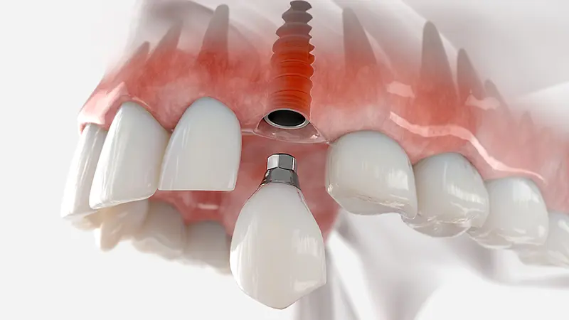
What are Implant-Supported Solutions for Total Tooth Loss?
Total edentulism, the clinical condition where all natural teeth in the mouth are lost, is a medical condition that fundamentally alters the chewing function, which is the mechanical starting point of the digestive system. In completely toothless jaws, serious physiological changes in bone anatomy, facial muscles, and aesthetic proportions begin with the loss of structures that transfer chewing pressure to the tissues. The discipline of Implantology, developed for the rehabilitation of this medical condition, offers the patient a functional and structural support unit by placing titanium structures in place of lost tooth roots.
In traditional dentistry, cases of total edentulism were generally rehabilitated with tissue-supported complete dentures (plates). However, these appliances, which only receive support from the gums and palate, can show mobility due to the tongue movements in the lower jaw and the muscle dynamics in the upper jaw. In current medical protocols, the goal is to secure these dentures using a specific number of titanium screws positioned within the jawbone or to create fixed bridge systems that cannot be entirely removed by the patient. In this context, supported solutions planned according to jaw anatomy in cases of total edentulism, surgical planning principles, prosthetic materials, and the management of treatment processes will be examined with a scientific and objective approach.
What Anatomical Changes are Observed in the Jaw Bone in Cases of Total Edentulism?
In cases of complete edentulism, vertical and horizontal volumetric reductions (resorption) are observed in the alveolar bone due to the loss of all mechanical stimuli. With the increased activity of osteoclastic (bone-resorbing) cells, the bone crests become blurred, flattening and leading to structural collapses in the lower third of the jaw.
Tooth roots stimulate the cells within the jawbone microscopically during chewing. This stimulation mechanism ensures that the bone remains vital and maintains its volume. When all teeth in a denture are lost, the body’s need to send calcium and minerals to this area decreases. The body’s physiology metabolizes this inactive tissue by reabsorbing (resorbing) it. Particularly in the first months following tooth extractions, the rate of cellular loss is high, leading to a progressive atrophy (shrinkage) pattern over the years.
The reflections of anatomical changes are not limited to the bone but also directly affect the extraoral (facial) profile. The lip muscles, losing their bony support, begin to collapse inward, reducing the distance (vertical dimension) between the tip of the nose and the tip of the chin. This situation creates folds resembling early signs of aging in the profile appearance. Additionally, in severe cases where the lower jaw bone has significantly eroded, the nerve passing through the bone (mandibular nerve) may come very close to the oral mucosa, causing nerve pressure and seepage during chewing for patients using traditional denture prosthetics.
What Functional Issues Does Implantology Address in Complete Tooth Loss?
In cases of complete tooth loss, implantology applications respond to the reduced chewing capacity due to movement of traditional dentures, speech disorders related to loss of retention, and psychological hesitations. By establishing bone-supported fixed systems, it aims to realize the chewing cycle within physiological limits.
Classic total dentures (palates) primarily rely on the passive balance of the cheek and tongue muscles along with the retention of the lower jaw. The pressure applied to chew food can cause the prosthesis to slide over the tissues, reducing chewing efficiency. Systems supported by titanium roots restrict the movement of the prosthesis. This allows patients to consume hard foods rich in vegetables and proteins (such as meat, apples, nuts), alleviating the digestive burden in the gastrointestinal (stomach-intestine) system.
When examined from a phonetic perspective, classic palate dentures covering the entire hard palate diminish the sense of taste and alter the pronunciation of certain consonants. In fixed or titanium-supported movable systems, the upper palate area can be largely or completely left open. This structural design allows the patient’s tongue to freely contact the palate vault, preventing the obstruction of taste receptors and helping to shape phonetic sounds appropriately.
How Many Artificial Roots Should Be Planned for Fixed Complete Jaw Prosthetics?
The number of artificial roots to be placed for fixed complete jaw prosthetics is generally planned to be between 4 and 8 for a single jaw, depending on the density of the jawbone, the width of the jaw arch, and the condition of the opposing teeth. This number, determined by anatomical measurements, is based on the principle of equal distribution of mechanical load.
In medical planning, the biomechanical requirements vary for each case. Anatomically, the upper jawbone has a more spongy (cancellous) structure compared to the lower jaw. Therefore, to support a fixed bridge system in the upper jaw, it is recommended that the force distributed to the bone surface is allocated to more support points. Placing 6 or 8 units in a wide and densely bone-structured average upper jaw is considered a standard approach.
The lower jaw structure consists of a cortical (hard and dense) layer that is thick. This hardness ensures that the medical parts placed adhere much more firmly to the bone. Therefore, 4 or 6 support points are usually sufficient to carry a fixed prosthetic system in the lower jaw. The table below summarizes the planned number of medical supports and biomechanical necessities according to the jaws:
| Application Area | Planned Unit Count | Anatomical and Biomechanical Necessities |
|---|---|---|
| Lower Jaw (Mandible) | 4 to 6 Units | The bone density is high, primary stability (initial adhesion) is easily achieved. Four or six support points show sufficient mechanical resistance for load distribution. |
| Upper Jaw (Maxilla) | 6 to 8 Units | The bone structure is softer, and sinuses are located in the posterior areas. The number of supports is increased for a more balanced transmission to the soft bone where the load is placed. |
| All-on-4 Concept (Lower/Upper) | Only 4 Units | It is a special concept where a biomechanical bridge is established with two flat, two opened units when anatomical barriers like nerves or sinuses are present in the posterior areas. |
How Does the All-on-4 Concept Provide a Biomechanical Process in Completely Edentulous Jaws?
The All-on-4 concept works by placing two titanium units at a vertical angle in the anterior part of the jaw, and two units at an angle of 30-45 degrees in the posterior parts to avoid anatomical voids. This angled placement provides extensive biomechanical support in a polygonal form to the fixed prosthesis while eliminating bone deficiencies in the posterior areas.
Particularly in patients who have remained edentulous for many years, severe bone resorption is observed in the posterior regions of the jawbone. The downward sagging of the maxillary sinuses in the upper jaw and the proximity of the mandibular nerve to the surface in the lower jaw medically restrict the placement of standard vertical implants in these areas. Traditional planning required additional surgical procedures that could take months, such as bone graft (powder) and sinus lifting, to overcome these obstacles. The All-on-4 (Four-System) concept reduces the need for these additional surgeries by targeting healthy bone immediately in front of anatomical barriers.
Positioning the medical components placed at the back in a backward-opening manner extends the length of the implant within the bone, while shifting the point of emergence in the chewing plane (on top of the prosthesis) further back. Thus, a single (one-piece) fixed bridge that is screwed between the two anterior and two posterior support points, creates a trapezoid (trapezium) model that spreads the load over a wide area. In this biomechanically balanced system, in appropriate cases, it is possible to screw in temporary fixed prostheses on the day of the operation (early loading).
In Which Bone Structures is the All-on-6 Method Preferred?
The All-on-6 method is a preferred approach in wide jaw arches where the vertical and horizontal volumes of the jawbone are suitable for supporting six artificial roots, especially where chewing forces are calculated to be high. The six support points help distribute stress placed on the skeletal structure of the prosthesis over a larger area, thereby increasing the resistance of the system.
In scenarios where the jaw arch is very wide or the opposing arch has the patient’s own natural teeth, the occlusal (chewing) forces on the prosthesis are quite high. Meeting the rigid and uncontrolled forces applied by the opposing natural teeth with only four units can create long-term mechanical stress. When the physician determines that the bone volume in the posterior regions is sufficient through three-dimensional radiological analyses, they include an additional two support points in the plan to optimize the force distribution.
One of the medical advantages of the All-on-6 system is that the bridge body can be extended further towards the back sections (the cantilever distance decreases). The two additional supports function as a carrier column near the position where the larger molars are located at the back. Thus, the risk of bending or breaking at the ends of the prosthesis is minimized. This method, which supports chewing stability, can also be made applicable to marginal bone structures by being supported by bone grafting procedures.
How are Removable Dentures Made Implant-Supported?
Removable dentures are made implant-supported by installing generally 2 to 4 titanium units in the anterior regions of the jawbone and connecting them with “retainer attachment pieces.” The sockets placed in the inner part of the prosthesis pass over these attachments (click system) to stabilize the prosthesis to the mucosa.
This medical approach is defined in the literature as “implant-supported overdenture” (superior prosthesis). It is particularly preferred by physicians in medical cases where the patient is elderly or where the bone tissue is insufficient for a fixed bridge prosthesis. The anterior part of the jaw (between the canine teeth) is the least affected by bone loss and the anatomical area with the highest bone density. The small number of units to be placed in this area provides primary retention without the need for complex bone graft surgeries.
The attached prosthetic systems hold the prosthesis in place but some of the chewing force is still transmitted to the gum tissue at the back (tissue and implant-supported system). The prosthesis can be removed from the mouth by the patient for daily cleaning purposes; however, it does not move during speech or stretching while in the mouth. Compared to fixed systems, it constitutes a functional medical option due to requiring less surgical site, the practicality in treatment planning, and ease of cleaning.
What Are the Structural Differences Between Bar Retained Systems and Locator Systems?
Bar retained systems connect titanium roots with a thick metal bar (bar) to distribute the load, whereas locator systems carry independent retainers on top of each root. While bar systems distribute forces evenly across the entire body, locator systems function with point connections.
The choice of connection piece in the design of overdenture prostheses (removable) is determined according to the number of units placed and the angle of the jawbone. The table below presents the clinical features and structural differences of both systems side by side:
| Feature | Bar Retained Systems | Locator (Locator/Ball) Systems |
|---|---|---|
| Biomechanical Connection | All support points are rigidly soldered/connected by a metal bar inside the mouth. | Each support unit carries its own independent click-retainer head. |
| Force Distribution | The chewing load is transmitted uniformly to all titanium roots through the bar system. | The chewing force is locally reflected onto the tissues where the prosthesis sits and onto each unit. |
| Open Tolerance | The bar system compensates for disparities when the screws cannot be placed perfectly parallel to each other. | In non-parallel placements (open situations), the retaining elastics can be quickly dislodged when attaching and removing the prosthesis. |
| Internal Oral Volume | Due to the metal bar occupying a certain space, it may require the prosthesis to be produced slightly thicker inside. | Since they are smaller and independent pieces, they allow for prosthesis design in narrow areas. |
Why is the Use of Surgical Guides (Template Plates) Recommended in Complete Edentulous Treatments?
In complete edentulous treatments, the use of a surgical guide (template plate) is recommended to achieve a flawless procedure with incisions (flaps) not made extensively in the gingiva, planned in a computer environment with three-dimensional tomography data. This method minimizes post-operative edema and tissue trauma by increasing anatomical precision.
Since there is no natural tooth to reference in a completely edentulous arch, great care is required regarding the distance and angle of the medical components to be placed. In classical methods, the clinician exposes the gingiva completely, observes the bone with the naked eye, and plans according to the condition of the surgical field at that moment. In digital dentistry applications, however, the patient’s intraoral scan is combined with bone tomography in specialized software days before the procedure.
In the virtual environment, all titanium implants are virtually placed in the areas where the bone is most dense. Subsequently, based on this design, a transparent surgical plate is printed using three-dimensional (3D) printers. On the plate, there are metal sockets that guide the surgeon’s drills. On the day of the operation, this plate is fitted into the patient’s edentulous jaw, and the surgeon completes the procedure by accessing through these small holes. This closed method (flapless surgery), which requires little or no stitches, is an advanced level medical protocol that supports cellular healing speed in completely edentulous cases.
How Are Completely Edentulous Cases with Very Insufficient Bone Volume Managed?
Completely edentulous cases with very insufficient bone volume are managed using the transfer of bone blocks taken from the jaw exterior, wide-diameter synthetic bone grafting (powder implantation), or special long implant systems such as zygomatic that extend to the cheekbones. This way, severely atrophied jaws are medically supported.
In patients with advanced resorption, who have used removable dentures for years, surgeries involving ‘cortical augmentation’ (thickening of the jaw ridge) are planned. In the upper jaw, volume is created by extensive lifting of the sinus cavities. If the bone framework in the upper jaw has thinned and grafting is not expected to succeed, much longer titanium pieces called ‘Zygomatic’ (for example, 40-50 mm) are directly fixed into the patient’s cheekbones (zygoma) through the mouth. Since the cheekbone exhibits no resorption and has a very hard structure, this area serves as a perfect mechanical anchor point.
In cases of advanced resorption, the bone layer under which the inferior alveolar nerve passes may be significantly thinned. In such clinical scenarios, specially designed ‘short’ or ‘extra short’ implants are positioned in the short sections above the nerve line of the jawbone, minimizing the risk of damaging the nerve, or in specific situations deemed suitable by the physician, the nerve’s position may be slightly shifted surgically.
How are Temporary Implants Integrated into the Post-Surgical Process?
Temporary implants are integrated on the same day with screws if the primary retention values of the titanium parts placed are clinically found to be sufficient (immediate loading). If the retention value is borderline, conventional removable temporary implants that do not apply pressure are used until the bone matures.
In cases of complete edentulism, a toothless waiting period is not medically preferred to support patients’ social and psychological lives. Once the surgical procedure is completed, the physician measures the stabilization of the parts within the bone (ISQ or torque values). If implants are firmly anchored in the bone with high values, acrylic-based fixed temporary teeth that are pre-prepared in the laboratory or produced on the same day are screwed onto the supports through intermediary parts. The patient can enter the clinic toothless and leave in the evening with fixed temporary teeth.
However, if the bone structure is very soft (for example, D4 type bone) or if dense bone powder (graft) has been used, immediately applying mechanical load on the screws can hinder the osseointegration process. In this case, the titanium roots are completely covered with gum tissue, stitches are placed, and the healing of the tissue is awaited. After an average of one week, the removable prosthesis used by the patient before the operation is lined with soft tissue material (soft liner) to ensure adaptation to the jaw. This removable temporary prosthesis supports aesthetics and light chewing function during the waiting period that lasts for months.
What Prosthetic Materials Can Be Planned for Complete Jaw Restorations?
In complete jaw restorations, titanium base acrylic teeth, monolithic zirconia bridges, or ceramic (porcelain) arrangements with metal substructures can be planned as prosthetic materials. The choice of material is made based on whether the teeth in the opposing jaw are natural or prosthetic, differences in bone interior distances, and chewing pressure.
Acrylic hybrid prostheses with titanium substructures are preferred, especially in cases of significant bone loss when the pink gingiva part needs to be kept longer. The acrylic (plastic-based) material absorbs chewing forces, transmitting a softer force to the underlying screws, and repairs can easily be made in a laboratory setting at the moment of breakage. Additionally, the titanium bar framework adds high structural resistance to the system.
Monolithic zirconia blocks are among the commonly planned complete jaw restoration materials in contemporary digital dentistry. Due to being milled from a single zirconia block with CAD/CAM systems, their fracture resistance is very high. Surface defects that can occur in porcelain are not seen in monolithic zirconia. The non-porous surface significantly contributes to the oral health by reducing bacterial plaque retention.
What Are the Surgical Planning Differences Between the Upper Jaw and the Lower Jaw?
The surgical planning differences between the upper jaw and lower jaw stem from the trabecular (honeycomb-like) density of the bone tissue and the position of adjacent anatomical areas. While more units and a longer cellular waiting period are planned in the upper jaw, the location of the nerve in the lower jaw determines the surgical boundaries.
The maxilla (upper jaw), fixed to the skull, is an anatomical structure that contains air cavities such as sinuses and nasal passages. The existence of these cavities and the structural softness of the bone (D3-D4 type bone) can complicate the primary retention of titanium screws. For this reason, the surface characteristics of the parts used in maxillary surgery can be selected in special forms (such as hydrophilic) that will increase cell attachment, and a maturation period of generally 6 months is given.
The mandible (lower jaw) is movable and is subjected to intense muscle activity during chewing. Its bone structure is very hard (D1-D2 type bone). This hardness provides a more favorable biological ground for early loading (immediate denture placement) protocols by increasing initial retention strength. However, the most critical distinction in planning the lower jaw is the mandibular nerve canal that runs through the posterior sections. In tomographic data, the distance of the nerve to the jaw ridge (crest peak) is calculated in millimeters; materials of 10 mm, 8 mm, or shorter lengths are added to the surgical map based on this distance.
What Should the Maintenance Routine Be for Implant-Supported Prostheses in Complete Tooth Absence?
The maintenance routine for supported prostheses applied in complete tooth absence should include washing the undersides of bridges with an oral irrigator (water flosser), the use of special thick dental floss (superfloss), and daily brushing steps. The absence of natural teeth does not eliminate the need for cleaning; bacterial plaque control is a critical necessity for tissue health.
A fixed bridge prosthesis that covers the entire jaw rests on titanium support points placed in the jawbone. Food remnants that accumulate beneath the contact points (pontic undersides) of this bridge, when combined with saliva, form dental plaque. If these plaques are not cleaned, they can lead to a condition called “peri-implantitis,” which causes inflammation of the gum and bone around the titanium screw. If the inflammation progresses, the bone surrounding the artificial root may erode, jeopardizing the stability of the entire system.
Cleaning only the front and back surfaces of the teeth is not sufficient. Devices that spray water under pressure support mechanical cleaning by providing pressurized water under the bridges. Individuals using movable (removable or bar) prostheses should remove their prostheses from the mouth every night or at the end of the day and wash them externally with liquid soap and special brushes; parts of the retainer metal that remain inside the mouth should also be brushed with a soft toothbrush. Regular clinical examinations every six months, professional cleaning of plaque accumulation, and radiological checks should not be neglected.
What Clinical Protocols Are Followed for Complete Edentulism Cases Within Avrupadent?
Complete edentulism cases within Avrupadent are monitored through multidisciplinary medical consultations, three-dimensional digital tomographic (CBCT) analyses, standard sterilization cycles, and patient-specific prosthetic design protocols. Physicians manage the process based on general scientific principles focusing on the patient’s jaw anatomy and medical history.
The treatment period begins with a detailed examination of the oral cavity and an analysis of the patient’s overall systemic condition (medications used, diabetes, cardiovascular history). In Avrupadent procedures, virtual surgical planning is conducted not only with two-dimensional X-rays but also with three-dimensional dental tomography, which reveals the millimetric volume of the bone. To avoid damaging the anatomical structure, the distances to sinuses and nerves are mapped through these programs.
In surgical procedures, a medical environment is prepared under strict sterilization rules to ensure infection control. During the cellular healing period after the operation, radiological and clinical follow-ups are conducted through an appointment system. In the prosthetic fabrication phase, digital measurements are taken with intraoral scanners to create aesthetic laboratory designs that are compatible with opposing jaw closure and facial proportions. This protocol chain is monitored to maintain medical standards and clinical predictability.
Frequently Asked Questions (FAQ)
1. How long does implant treatment take for complete tooth loss? The surgical operation for a single jaw takes approximately 1-2 hours. Biological healing in the lower jaw takes about 2-3 months, while in the upper jaw, it is expected to take 3-4 months. The prosthetic laboratory procedures are completed within a few weeks. 2. If all my teeth are extracted, can fixed teeth be placed on the same day? If there are no acute infections in the areas where teeth have been extracted, and the bone density is at suitable levels for primary stability, fixed temporary teeth can be placed on the day of the operation using All-on-4 or All-on-6 concepts. 3. I cannot use my removable denture, can I switch to an implant-supported denture? Yes, if the vertical and horizontal thickness of the jawbone is found to be suitable through clinical analyses (or supported with graft), your prosthesis can be converted to a stable non-moving form with 2-4 titanium units placed in the edentulous area. 4. Why are posterior areas placed with open placements in the All-on-4 system? In the case of lower jaw implants, the back parts (30-45 degrees) are directed towards the healthy bone at the front to avoid damaging the hanging maxillary sinus cavities in the upper jaw and to overcome these anatomical obstacles. 5. What is a zygomatic implant and when is it used? They are special titanium systems that extend beyond the jawbone in very specific medical situations where the upper jawbone is severely resorbed and cannot be supported by bone powder (graft), anchoring to the higher hard zygomatic bone. 6. How many implants are sufficient for completely edentulous jaws? To stabilize removable dentures, 2-4 implants are sufficient, while for establishing a fixed bridge system, typically 4-6 units in the lower jaw and 6-8 units in the upper jaw are adequate to meet the bone mechanics. Planning is done specifically for each patient. 7. Can complete tooth loss treatment be applied to elderly patients? Chronological age does not set an upper limit for this procedure. If the patient’s overall systemic health condition can tolerate surgical intervention, medical treatments can also be planned for individuals of advanced age. 8. Should implant-supported removable dentures be removed at night? Yes, it is clinically recommended to remove and clean removable dentures with clasps or bars before sleep to allow the underlying mucosa (gum tissue) to breathe, rest, and reduce the risk of intraoral infections. 9. How is the underside of fixed complete jaw dentures cleaned? Since the protein’s outer layer can potentially cause food residue, mechanical cleaning should be performed regularly every day using special interdental brushes (superfloss), and pressurized water jet devices. 10. Can a treatment plan be made for cases of complete toothlessness with bone loss? In cases where the bone volume has weakened, direct procedures cannot be performed; however, the anatomical volume of the region can be increased with synthetic or organic bone powder grafting. After tissue maturation (4-6 months), transition to the main medical procedure can be ensured. 11. Is there a difference in chewing between fixed prosthetics and removable prosthetics? Fixed systems provide very close, high bite force to natural teeth since they transfer chewing force directly to bone through artificial roots. Removable systems transmit some of the force over the mucosa but are much more stable compared to old classic dentures. 12. Why can more implants be planned in the upper jaw than in the lower jaw in complete toothlessness? The upper jaw (maxilla) has a spongy and soft anatomical structure (trabecular). In order to enhance stability by distributing the chewing force that comes to a soft surface over a larger area, the number of support points in the upper area is increased. 13. Can the color and size of the teeth be adjusted during the trials? During the trial sessions in the prosthetic laboratory phase, the arrangement, color, size of the teeth, lip support, and phonetic (sound production) status are examined together with the patient. Desired aesthetic revisions can be applied before the final medical fixture/bolting process. 14. Do materials placed in the jawbone cause problems in tomography or MRI? Titanium metals are non-ferromagnetic (non-magnetic) in structure, so they do not create any physiological problems for the patient in Magnetic Resonance (MR) or hospital tomography, nor do they generate signals in security detectors. How is the process managed for completely edentulous individuals with diabetes? In order to support cellular wound healing, the patient’s average blood sugar level over the last three months (HbA1c test) is examined. When values are drawn to reference ranges in consultation with a physician, the operation can be performed in accordance with standard sterilization rules. |