Why is the jawbone important in implant treatment?
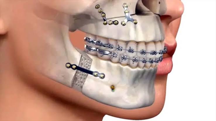
The fundamental basis of implantology procedures used to compensate for dental deficiencies through medical means is the jawbone. As a biological structure (alveolar ridge), the jawbone not only encases the roots of teeth or artificial titanium materials, but also serves as a distribution mechanism for the massive mechanical pressures that occur during the chewing process. In this context, the clinical planning of the medical procedures to be applied depends directly on the volume, mineral density, and cellular regeneration capacity of the bone. Unlike other parts of the skeletal system, the jawbone is a reactive anatomical form that maintains its volume while teeth are present and rapidly changes shape when teeth are lost.
Placing titanium screws into the jaw is not a pure and simple mechanical screwing process. On the contrary, it is a biological integration process expected to occur at a microscopic level between a metal surface and living bone cells. When bone tissue is insufficient, adequate biomechanical resistance to carry the upcoming porcelain teeth cannot be achieved. In this guide, the anatomical structure of the jawbone, which is within the scope of medical interventions, the cellular changes experienced after tooth loss, variations in density, and clinical evaluation processes will be detailed in accordance with scientific parameters.
What Anatomical Changes Occur in the Jawbone After Tooth Loss?
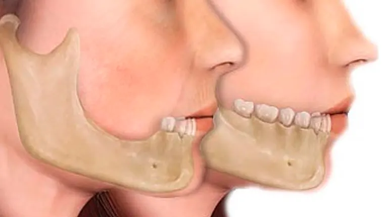
When a tooth is lost, mechanical signaling to the jawbone ceases. Bone cells that do not receive this signal begin to lose volume as a physiological response, leading to resorption. This situation causes the jaw ridge to shrink both horizontally and vertically, resulting in structural changes in facial anatomy.
A natural tooth is held in the jawbone socket (alveolus) by the fine periodontal fibers surrounding its root. Each chewing motion exerts microscopic pressure on the tooth bone. This pressure keeps the bone-forming cells (osteoblasts) active, ensuring the continuity of the calcium cycle in the area. With the extraction of the tooth, this vital stimulus is completely cut off. The body starts to resorb the bone tissue in this area at the cellular level, perceiving it as no longer functional. Bone-resorbing cells (osteoclasts) become more active in the region.
According to medical data in the literature, during the first six months following tooth extraction, a significant narrowing of horizontal bone thickness is observed. In subsequent years, the bone continues to resorb vertically (in height) as well. This anatomical resorption progresses differently in the lower jaw compared to the upper jaw. In cases of tooth loss in the upper jaw, the bone does not merely resorb upwards from within the mouth, but also, the sinus air spaces in the upper area sag downwards, reducing the bone volume from two directions. In the lower jaw, as the bone erodes, the mandibular nerve canal that passes inside the bone and provides sensation to the lip moves closer to the surface. These cellular changes in jaw anatomy are medical criteria that directly determine the limits of surgical planning.
How Does the Titanium Root Retain in the Jawbone (Osseointegration)?
Osseointegration is the biological bonding process at the microscopic level between the surface of the implanted titanium material and the jawbone cells. It works by the bone-forming cells migrating to the porous areas on the titanium, creating new bone islands. This biological retention provides the stability necessary for the prosthesis to withstand chewing forces.
Titanium is a medical metal that is compatible with human physiology, corrosion-resistant, and not rejected by the tissue as a foreign material. When the screw is placed inside the jawbone during the procedure, the mechanical compression force applied by the surgeon (primary stability) ensures that the screw remains in place initially. However, the actual long-term clinical success is dependent on secondary stability (osseointegration), which is linked to biological healing. Immediately after the procedure, a blood clot forms in the area, and the inflammatory healing phase begins.
The growth factors and bone-forming cells that arrive at the area through the bloodstream specifically establish themselves on the sandblasted and roughened titanium surface. These cells, which settle into the microscopic pits, mature over time and mineralize, becoming hard bone tissue. Once this cellular network is established, the titanium screw behaves almost like an organic part of the bone itself. Doctors recommend not placing occlusal (chewing) loads on the area for several months, depending on the jaw structure, to achieve the desired density of this cellular union.
How Does Jawbone Density (Bone Quality) Impact Implantology Planning?
Jawbone density directly affects the initial retention resistance (primary stability) of the artificial root and the cellular healing process. Bone quality is classified in medical literature ranging from D1 to D4. While retention is high in dense and hard bones, a longer healing period is a medical necessity in spongy and soft bones.
Just as everyone’s skeletal structure is different, the bone quality in different areas of the jaw is also not homogeneous. The hard layer that surrounds the outer surface of the bone is called cortical bone, while the inner structure rich in pores and blood vessels is referred to as trabecular (spongy) bone. The density of the jawbone is clinically classified into four main groups according to the ratios of these two layers in the region. The level of density determines many details, from the rotation speed of the drilling (milling) tools to be used by the dentist to the spaces between the titanium structures to be placed.
| Bone Type | Structural Properties | Impact on Surgical Planning |
|---|---|---|
| D1 Type Bone | It is very hard, similar to hardwood. It is almost entirely composed of thick cortical bone (Generally the anterior region of the mandible). | The initial retention strength is very high, but since its vascularization is low, the milling process is performed slowly and with plenty of cooling fluid to prevent the cells from overheating. |
| D2 Type Bone | It consists of a thick outer cortical layer and a dense spongy inner layer. | From a clinical perspective, it is the most favorable bone type. Its vascularization and hardness ratio is balanced, and the healing process progresses ideally. |
| D3 Type Bone | It has a thin outer cortical layer and a wide porous spongy inner structure (Typically the anterior region of the maxilla and the posterior area of the mandible). | Due to its soft structure, instead of making wide holes with drills, instruments that compress the bone are used to medically increase the density. |
| D4 Type Bone | It is very soft like Styrofoam. The cortical layer is almost nonexistent, and the internal structure is very porous (Typically the posterior region of the maxilla). | It is difficult to ensure initial retention. Specially designed threaded materials are selected, and the longest waiting period is planned for osseointegration. |
What Are the Fundamental Differences Between the Upper Jaw and Lower Jaw Bone Structures?
The upper jaw (maxilla) has a more porous, soft, and spongy bone structure, while the lower jaw (mandible) is dense, thick, with cortical layers and a hard anatomy. These structural differences alter the milling techniques used during surgical procedures, waiting periods, and the encountered anatomical spaces (sinus or nerve canal).
The maxilla is a complex bone fixed to the skull skeleton, containing the nasal cavity and air chambers known as the maxillary sinuses. To reduce weight in this area of the body, its bone structure carries a trabecular (cancellous) characteristic. While implantology procedures are applied to the upper jaw with soft bone tissue, the maturation of osseointegration around the placed piece requires more time compared to the lower jaw.
The mandible is a structural unit that has a movable joint (temporomandibular joint) and is exposed to the intense chewing forces of facial muscles. To adapt to this high mechanical stress, it has thick and hard tissue with a significant calcium deposit. The largest anatomical structure in the lower jaw is not the density of the bone but the canal through which the “inferior alveolar nerve” (lower jaw nerve) passes. When procedures are performed on the jawbone, the millimetric distance to this nerve is detected radiologically, and the length of the material to be selected is planned not to exceed this distance.
What Medical Procedures Are Applied in Case of Inadequate Jaw Bone Volume?
In clinical scenarios where the jawbone’s volume is insufficient, additional surgical procedures such as bone grafting, the use of membranes, and sinus floor elevation (sinus lifting) are employed. Through these operations, the physiological foundation where the titanium implant will be placed is medically supported and augmented vertically and horizontally.
As a result of long-term tooth loss or periodontal (gum) diseases, the width or length of the bone may become thinner than the diameter of the titanium screw to be placed. The bone tissue that remains outside or covers the area with a thin layer cannot provide long-term biomechanical support during function. At this point, bone augmentation (volume enhancement) comes into play. During the procedure, allografts (bovine) and synthetic or autogenous bone grafts (powders) taken from another area of the patient are placed in the thinned area. To prevent these added particles from dispersing and soft tissue cells from leaking into the area, the outer portions are covered with special barrier membranes.
The grafted area is used as a mold by the body. Over the months, blood vessels penetrate into this graft, and the patient’s own bone cells absorb these powder particles, leaving their place for new, living jawbone. In cases where the sinuses have sagged in the upper jaw, the mucosa that is pushing down the sinus cavity is gently lifted, and the cavity below is filled with graft (sinus lifting). These additional procedures expand the treatment spectrum by aligning the anatomical limits of the jawbone with medical requirements.
What Are the Effects of Systemic Diseases on Jawbone Metabolism?
Systemic diseases such as osteoporosis, diabetes, and thyroid disorders affect the renewal rate and mineral density of the jawbone. Imbalances in blood sugar or the use of medications that alter bone metabolism are parameters that should be closely monitored through medical consultations, as they can change the capacity of cells to adhere to the titanium surface.
Diabetes (sugar disease) is a systemic disease that disrupts microcirculation (capillary blood supply). The jawbone requires rich blood support, and thus oxygen and defense cells, to heal after an intervention. When diabetes is uncontrolled, the intra-bone blood flow decreases, the activity of osteoblast cells is suppressed, and the risk of infection increases. This situation prolongs the bone healing period. Therefore, before the procedure, the patient’s HbA1c (three-month blood sugar) level should be drawn to reference ranges to ensure medical safety.
Osteoporosis (bone loss) is a decrease in bone mineral density throughout the body. The jawbone can also be affected by this condition. However, the more critical factor is the bisphosphonate-type medications used for osteoporosis treatment. These medications slow down bone destruction while also locking the bone cycle (renewal). When a patient using bisphosphonates undergoes intervention on the jawbone, the bone may fail to self-repair, leading to necrosis (tissue death). Therefore, a detailed anamnesis of the medications used during the examination phase should be obtained, and consultation (opinion) from orthopedic/endocrinology physicians should be sought regarding the feasibility of the procedure.
What Radiological Imaging Methods Are Used in the Examination of the Jawbone?
In the examination of the jawbone, two-dimensional panoramic radiographs and three-dimensional dental volumetric tomography (CBCT) devices are used. Tomography is a fundamental tool that allows for the digital planning of surgical procedures in a virtual environment by showing the millimetric thickness, height, and anatomical nerve pathways of the bone in cross-section.
Panoramic X-rays, which are standardly taken in dental clinics, display all teeth and the jaw structure in two dimensions (height and width) on a single film. They provide sufficient data to establish a general diagnosis and to see horizontal bone levels. However, to place cylindrical medical screws in the center of the jawbone, knowing the “thickness” (the third dimension) of the bone is also essential.
At this point, Dental Volumetric Tomography (CBCT) comes into play. These devices operate with a significantly lower dose of radiation compared to medical tomographs taken in hospitals and reflect the jawbone in microscopic sections on a computer screen. The physician measures the thickness of the bone in the region without teeth in millimeters in the cheek and tongue directions. Based on this data, the dimensions of the piece to be used during surgery are clarified. The table below shows the comparison of these two methods used in medical assessments:
| Assessment Criteria | Panoramic X-ray (2D) | Dental Tomography (3D – CBCT) |
|---|---|---|
| Bone Thickness Measurement | Does not show. Only presents the vertical dimension. | Provides the buccolingual (cheek-tongue) thickness of the bone at a millimeter level. |
| Identification of Anatomical Structures | Presents the nerve canal and sinuses as a general outline in two dimensions. | Shows where the nerve canal passes through the bone in three dimensions (with depths). |
| Surgical Guide (Planning) | Does not allow for virtual surgery and the production of surgical templates (plates). | Supports virtual planning to be done digitally by transferring to special software. |
What is the Role of Jawbone in the Formation of an Aesthetic Gum Profile?
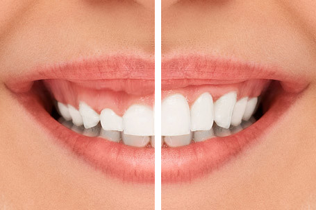
The gum tissue shapes itself following the form of the underlying jawbone. If the bone level is at a natural height between the teeth, the gum papillae (triangular parts) aesthetically maintain the form. When the bone erodes, the gum tissue also retracts significantly, and the foundation of pink aesthetics relies on the support of the underlying bone.
During the smile, it is not only the appearance of the white porcelain teeth that determines the aesthetic perception, but also the symmetry and health of the pink gum tissue surrounding these teeth (pink aesthetics). There is a rule in the medical literature called “biological width”; the gum typically tends to position itself at a specific distance (approximately 2-3 mm) from the underlying jawbone. If the jawbone has eroded vertically due to tooth loss, the gum tissue will also recede downwards in correspondence with this erosion.
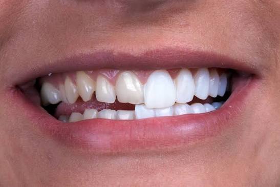
Especially in the anterior region with tooth losses, the primary cause of the black spaces (dark triangles) formed around the inserted porcelain tooth is the resorption of the nearby bone tissue (interdental septum) in that area. When the volume and supportive structure of the jawbone are preserved or surgically augmented (grafted), the overlaying soft tissue gains a fuller form, wrapping the prosthesis like an aesthetic frame. Therefore, aesthetic outcomes cannot be viewed independently of the jawbone formation.
How Does Chewing Pressure Support the Physiology of the Jawbone?
The mechanical pressure created during the act of chewing is transmitted to the jawbone through the tooth roots or artificial titanium roots. This mechanical transmission stimulates the cells within the bone, actively maintaining the local mineralization cycle and medically supporting that the bone volume in the area remains within physiological limits.
Wolff’s Law, which is a fundamental principle in orthopedics and skeletal physiology, states that bones reshape themselves (remodeling) according to the mechanical loads they are subjected to. Just like the muscles of individuals who exercise, bones also strengthen by increasing their calcium deposits when under load. When the load is removed, the body thins the bone to conserve energy. The physiology of the jawbone is fully subject to this law.
In individuals using movable denture prostheses that rest only on the gum tissue, chewing pressure reflects not directly on the jawbone but rather superficially on the gum surface. This causes bone loss to persist for years because it does not stimulate the internal structure of the bone. However, artificial roots placed in the jawbone through implantology practices directly transmit masticatory (chewing) forces to the bone body. This mechanical transmission sends a signal to the osteoblast cells within the bone, indicating ‘there is a need for a function’ and medically supports the slowing of bone atrophy (resorption) and the preservation of form in that area.
How is Bone Assessment Conducted in Avrupadent Clinical Processes?
Bone assessment in Avrupadent clinical processes is carried out with a multidisciplinary approach, involving the collection of the patient’s overall health history and three-dimensional tomography analyses. The density and volume of the bone are measured millimetrically through digital software, resulting in a medical map tailored to each individual’s anatomy.
During the initial clinical examination, the patient’s systemic discomforts, and any medications affecting bone metabolism, are recorded to create a detailed health history. Periodontal (gum) examinations are conducted, and sources of infection in the mouth, if any, are identified. A key principle for a healthy bone assessment is to maintain a non-infected flora.
In the process of taking measurements, a digital model of the jawbone is created using three-dimensional Dental Tomography (CBCT) data. During the planning phase, the locations of nerves, sinuses, and adjacent tooth roots within the bone are marked, and the directions and dimensions of the medical materials to be used are virtually placed based on this data. In necessary cases, surgical guide plates are produced over these virtual designs, aiming to conduct the operations within the predetermined medical limits.
Frequently Asked Questions (FAQ)
1. How is the progression of jawbone resorption monitored? The positioning of titanium supports that will apply mechanical pressure to the area of missing teeth sends physiological stimuli to the bone cells, slowing down the rate of resorption in that area and supporting volume preservation. 2. How long does the bone powder (graft) procedure take? The surgical placement of the graft is performed within the same session. However, to ensure that these particles biologically integrate with your body and transform into a solid jawbone, a maturation period of 4 to 6 months is expected, depending on the cellular structure. 3. Does having low bone density prevent treatment from being performed? Low density does not necessitate the cancellation of the procedure; however, the surface characteristics of the selected medical materials will differ, and the ossification period (waiting time) is medically planned to be longer for non-standard cases. 4. How does smoking affect the jawbone? Tobacco products narrow the capillaries that nourish the jawbone, reducing the amount of oxygen and immune cells that reach the area. This situation medically increases the risks of cellular integration as it slows down wound healing. 5. Why is a longer wait expected for the upper jawbone? The upper jaw (maxilla) has a spongy and porous structure, requiring more biological time for bone cells to envelop the titanium surface and achieve clinical stability compared to the denser structure of the lower jaw. 6. Does a panoramic X-ray show bone thickness? No, panoramic X-rays provide a two-dimensional (width-height) view of the bone’s height and the status of existing teeth. Three-dimensional tomography is needed to measure the bone thickness in the cheek-tongue direction. 7. Does titanium damage the jawbone? Pure titanium and its medical alloys are among the materials with the highest biological compatibility with human cells. They do not possess toxic or reactive properties, so they do not harm bone tissue; rather, they are embraced by bone cells. 8. Can procedures be performed on patients with osteoporosis? Yes, it can be done, but the dosage and duration of bisphosphonate medications used for osteoporosis are crucial. Since these medications slow bone regeneration, a medical consultation with the relevant specialist is necessary before the procedure. 9. Is bone powder (graft) animal-derived? Medical grafts can generally be sourced from either bovine, synthetic, or products created in a laboratory. All of these grafts are medical products that have been sterilized at very high temperatures, completely free from proteins, leaving only the mineral scaffolding. 10. When does jawbone development complete? In human anatomy, the skeletal system and jawbone development typically stabilize around the age of 18. Whether growth continues is confirmed radiologically by doctors using hand-wrist X-rays or special graphs. 11. When does bone loss begin after tooth extraction? Bone resorption begins at the cellular level in the weeks following a tooth extraction. Especially in the first 6-month period, the narrowing of the bone’s horizontal thickness (volume loss) reaches the highest physiological rates. 12. How do sinus cavities alter jawbone planning? When teeth are extracted from the upper jaw at the back, the air cavities (sinuses) above sag downward, constricting the height of the bone. To counter this, surgical procedures are planned in which the sinus membrane is pushed up and filled with graft (sinus lifting). 13. How is the diameter of the piece to be used determined according to bone structure? The diameter of the titanium piece to be selected is calculated by the doctor based on radiological tomography measurements, ensuring that at least a 1-1.5 mm thick healthy bone wall is left on the cheek and tongue sides of the existing bone. 14. Do pieces placed in the bone cause issues during MRI scans? Titanium metals are biological materials that do not have magnetic properties (non-ferromagnetic). Therefore, they do not create a physiological problem for patients during Magnetic Resonance (MR) or hospital tomography, and they do not emit magnetic signals in security devices. 15. Which foods support jawbone metabolism? A balanced intake of foods rich in vitamin D, vitamin K, calcium, and phosphorus (dairy products, green leafy vegetables, fish) also supports the repair and mineralization processes of jawbone cells, just as it does throughout the body. |