What is evaluated during the first examination in aesthetic dentistry?
The restorative disciplines that reconstruct the deformities, asymmetries, and tissue losses in the oral and facial region within a medical framework require an extremely detailed preparatory phase. Although the first appointment, where individuals present aesthetic demands to the clinics, is often perceived as merely a stage of conversation where complaints are listened to; for medical professionals, this session is the center of the data collection and analysis process that determines the biological infrastructure for all the procedures to be performed. No prosthetic or restorative application built on any foundation other than a healthy one can be sustained in the long term. Therefore, the initial contact point where visual expectations are compared with the limits of medical feasibility serves as the compass for the entire treatment roadmap.
Aesthetic Dentistry principles guide this initial assessment phase, encompassing a wide range from micro-level (the enamel structure of a single tooth) to macro-level (the overall skeletal balance of the face). The physician analyzes the patient’s lip elasticity during smiling, the activity of chewing muscles, and the thickness (biotype) level of the gums, basing every detail from the chemical properties of the materials to the tooth’s millimetric form on these analyses. In this guide, we detail which medical procedures the patient can expect during the initial examination, the importance of radiological and photographic records, the impact of jaw closure on aesthetics, and the medical planning protocols in Avrupadent processes in an objective manner.
What is the Main Purpose of the First Examination in Aesthetic Dentistry?
The primary goal in planning a medical procedure is to address aesthetic concerns while preserving the existing physiological health of the tissues and treating any existing diseases if present. When a patient presents with complaints about the misalignment or discoloration of front teeth, the first aspect the physician focuses on is not just the color of that tooth, but the overall condition of the supporting jawbone and gums. The first appointment is more about exploring the question, “Can the existing anatomical foundation support this aesthetic burden?” rather than simply asking, “What should be done to which tooth?”
At this stage, another critical role of the physician is communication. Individuals may apply to clinics with expectations that do not match their facial profiles due to media or external environmental influences. The initial examination is an informative view where these abstract expectations are filtered through anatomical realities, clarifying transparently what medical procedures can be offered to the patient, and where the limits begin and end. Accurately drawing a medical map is the most valuable shield that limits surprises and inconsistencies in the later stages of the process.
What Systemic Factors are Inquired When Taking Medical History (Anamnesis)?
The mucosa and bone structure inside the mouth is not an organ independent of the body’s overall systemic health. A systemic disorder occurring in the body directly determines the healing rate inside the mouth and the tissues’ response to treatment. Therefore, before any restorative examination begins, the patient is asked to fill out a comprehensive medical history form and share it with the physician.
In a diabetic patient whose HbA1c levels are significantly above the reference ranges, tissue healing after minor surgical procedures like gingival sculpting (pink aesthetics) may be delayed or the risk of infection may increase. Similarly, in individuals with a history of high blood pressure or arrhythmia, the composition of local anesthesia used will be modified according to these conditions. Since some antihypertensive medications can lead to physiological swelling (hyperplasia) of the gums, the clinician must determine the actual source of this gum enlargement (whether it is related to the medication or the plaque) before making any aesthetic planning. Systematic inquiry is the first and most essential step in medical protocols.
What Findings Do Radiological Imaging Techniques Provide in Initial Examination?
The durability of a porcelain or composite application planned for aesthetic purposes is measured by the health of the dental root it sits on and the surrounding bone tissue. Looking into the mouth with the help of light and a mirror during the initial examination only assesses the visible part of the iceberg. Generally, two-dimensional panoramic X-rays or, if necessary, three-dimensional Dental Tomographies (CBCT) come into play to map the anatomical realities hidden beneath the gum and within the bone.
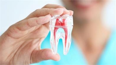
If a leaf porcelain (veneer) is planned for a tooth, it must be radiologically verified whether there is a latent (asymptomatic) infection at the root tip of that tooth. If there is a lesion at the root tip, root canal treatment should be performed first, and tissue healing should be ensured. Additionally, the root lengths of the teeth in the bone also determine the biomechanical support capacity of the restoration. Even if the tooth, which has weakened bone support and has mobility (looseness), is aesthetically aimed at, placing a heavy ceramic crown can lead to the future loss of that tooth. X-rays are the medical maps through which the physician reads the unseen.
How Are the Oral Biological Tissues’ (Tooth and Mucosa) Health Status Analyzed?
Aesthetic Dentistry clinics’ first physical contact phase by the physician is to create a biological map of the oral tissues. In this examination, each tooth surface is meticulously examined by drying it with air and water spray. It is noted whether there are fine crack lines that are not visible on enamel surfaces but become prominent with light, and whether there are V-shaped material losses related to brushing or acid erosion in the collar (neck) areas of the teeth.
The old restorations within the mouth are also tested at this stage. If there is discoloration or leakage at the edges of previously made composite fillings, it is anticipated that a new decay process may have begun beneath these fillings. In the soft tissue analysis, the color of the gums (whether healthy pink or swollen dark red), their firmness, and the presence of stippling (orange peel appearance) are examined. All these microscopic tissue records reveal which areas need improvement before starting aesthetic treatment (for example, whether dental calculus cleaning is required).
How is Functional Examination of the Temporomandibular Joint (TMJ) and Masticatory Muscles Conducted?
Satisfaction of aesthetic expectations will be short-lived if not compatible with functional masticatory mechanics. The human jaw is a dynamic, complex musculoskeletal system capable of three-dimensional movement. During the initial examination, the physician observes whether the jaw tip moves in a straight line or deviates to the right or left by asking the patient to slowly open and close their mouth. By applying slight pressure with two fingers to the joint area, they check for any displacement in the position of the disc.
The biggest contribution of this examination in aesthetic planning is the detection of bruxism (nighttime teeth grinding), which is a parafunctional habit in the patient. The chewing muscles (masseter and temporal muscles) are overly developed, showing sensitivity during palpation (manual examination), and a diagnosis of bruxism is made for patients whose chewing surfaces of their teeth have significantly worn down. This diagnosis directly affects the material of the porcelain restoration to be planned. It is a medical necessity to plan for thicker monolithic ceramics or maximized fracture-resistant zirconium instead of finely structured glass ceramics for such a patient.
How Are Facial Symmetry and Macro Aesthetic Parameters Recorded?
The perfect appearance of a dental arch within the mouth does not mean that this arch is compatible with the patient’s face. The proportion of the teeth within the facial skeleton is also evaluated within the framework of macro aesthetic rules. One of the longest and most detailed stages of the initial examination is the photographic recording session; during this session, a horizontal line (pupillary line) passing through the patient’s pupils, the tip of the nose, the curve of the lower lip, and a vertical line (midline) that passes exactly through the middle of the face are taken as references.
The doctor measures the deviation of the midpoint between the upper two teeth with respect to the facial midline in millimeters when transferring these photographs to design software on the computer. Additionally, the curve (smile arc) that the patient’s lower lip takes during smiling is recorded. The cutting edges of the new porcelain teeth to be created are planned to follow this lower lip curve in a parallel fashion. The photographic archive provides basic data regarding whether the designed teeth will have angular or rounded shapes, regardless of whether the face is square, oval, or triangular.
| Macro Aesthetic Parameters | Purpose of Evaluation in Examination |
|---|---|
| Pupillary (Pupil) Line | To determine the horizontal plane of the teeth (occlusal plane) and to identify their alignments. |
| Facial Midline | To check the alignment of the junction point of the upper two central teeth with the right-left symmetry of the face. |
| Rest Position of Lips | To measure how many millimeters the front teeth appear to be apart when the lips are relaxed (the youthful gap). |
| Smile Curve | To test how parallel the cutting edges of the upper teeth are to the smile arc of the lower lip. |
How Are Gum Levels (Pink Aesthetics) Evaluated in the Initial Examination?
The aesthetic capacity of white teeth emerges in harmony with the pink tissue that surrounds them like a frame. During the Aesthetic Dentistry examination, the depth of each tooth’s gum pocket is measured using a periodontal probe. The patient’s gum biotype (thin, medium, or thick) is identified. For individuals with a thin biotype, it is recommended to prepare much finer borders (preparations) to avoid irritating the gums with porcelain applications.
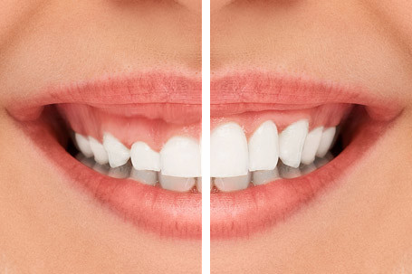
However, it is observed how much the upper lip stretches during a broad smile of the patient. If the gums appear much higher than they should be (about 2-3 mm) and the teeth look shorter/square than they are, this condition is referred to as a ‘Gummy Smile’. The doctor diagnoses this condition during the first appointment, presenting the need for gum lengthening (gingivectomy) or lip positioning procedures to the patient in a medical map format before proceeding with restorative phases.
How to Balance the Patient’s Psychosocial Expectations with Biological Compatibility?
Individuals often approach doctors with demands that are not suitable for their facial profiles, jawbones, or ages due to the influence of popular aesthetic trends in the media or their surroundings (for example, excessively large and perfectly white teeth). One of the most sensitive processes of the initial examination is balancing these abstract desires of the patient with concrete biological rules. The color or shape of a restoration to be applied to a patient should not impair that patient’s lip stretch capacity and closure dynamics.
The doctor analyzes the patient’s condition based on their own photographs. If the teeth’s lengths are extended beyond what the hospital desires to the point that it will touch the lower lip (causing a phonetic disorder), this biological limit is explained to the patient in detail. With an accurate medical guidance, expectations are revised and realistic (predictable) goals are negotiated. This psychosocial assessment is a transparent clinical approach that eliminates potential mismatches that could arise at the end of treatment from the very beginning.
How are Color Selection and Optical Assessments Determined in the First Session?
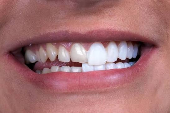
Identifying the tooth color is not just about selecting a level of whiteness; it is an optical process in which the reflection indices of light are analyzed. In the first examination, the patient’s existing teeth’s original color (the shade) is identified and recorded. Since the thickness and opacity value of the porcelain to be applied will be determined by this existing color, if thin ceramics that allow light to pass through are planned for a highly dark-colored, root-treated tooth, the underlying darkness will be reflected outward.
The hue and value of the new design to be made are selected according to the patient’s sclera and skin color references. A color that creates extreme contrast with skin tone (very white) creates an artificial focal point within the face. The doctor also considers the patient’s age group; they add to the laboratory prescription based on the initial examination data what level of transparency and blue/grey optical reflections should be present on the incisal edges of the enamel.
How is a Multidisciplinary Roadmap Created According to Avrupadent Clinic Criteria?
In corporate medical service standards, it is recognized that an aesthetic request goes beyond just porcelain applications. In Avrupadent’s process management, the first examination is concluded with the processing of the obtained raw data by all relevant specialist fields. For example, if a patient with aesthetic concerns in the anterior region has a closure disorder detected in their digital scanning data, an orthodontist may recommend a short-term transparent plate (aligner) treatment to avoid excessively cutting the teeth.
All these clinical findings are presented to the patient as a report and a sequential appointment schedule. A sequential arrangement is established, such as stabilizing the health of the gum first, renewing root treatments if necessary, followed by orthodontic alignment and finally the application of final ceramic restorations. The multidisciplinary approach adopts as a medical rule not only the creation of an aesthetic dental surface but also the solidification of the root and bone architecture that will support that surface to withstand the test of time.
What Are Frequently Asked Questions?
1. How long does the first examination take?
Since it includes stages such as taking radiological images, making photographic records, and thoroughly discussing expectations with the physician, it generally covers a comprehensive time span of between 30 and 45 minutes.
2. Is there sensitivity during the examination?
The first appointment typically involves data collection, photography, X-ray acquisition, and physical examination steps with the aid of mirrors, so it does not involve any invasive (surgical) procedures and is generally comfortable.
3. Is a procedure performed during the first appointment?
This session is primarily focused on planning and diagnosis; however, if the patient has an acute symptom or if a decision is made for detailed dental stone cleaning, which is the first step of planning, initial procedures may be applied depending on the situation.
4. Can aesthetic planning be done without X-rays?
Since understanding the root structures remaining under the gum, bone density, and potential hidden infections form the biological basis of the treatment, medical planning without X-rays (radiological imaging) is not recommended.
5. Do gum diseases affect the aesthetic process?
Taking aesthetic measurements or performing porcelain restorations while there is active gum inflammation or bleeding will disrupt biological harmony; therefore, it is a medical rule to first treat the gum diseases and achieve stabilization.
6. Is a mock-up done during the first examination?
Digital and photographic data obtained during the first examination are sent to the laboratory to prepare a three-dimensional design; thus, the physical mock-up (intraoral trial) procedure is usually applied to the patient during the next planning session.
7. Do jaw joint issues hinder design?
It is not a defect, but if there is pain, a clicking sound, or sliding in jaw movements (TME discomfort), joint stability is ensured first to prevent additional load on the porcelain pieces to be made, and the design is modified accordingly.
8. What should be taken into consideration when coming for the first examination?
Having a list of regularly used medications (especially anticoagulants), any history of chronic systemic diseases, and clearly communicating general aesthetic expectations to the doctor will enhance the efficiency of the process.
9. Are old porcelain pieces removed during the first session?
Existing prostheses are not disturbed until the treatment map is clarified and laboratory planning is completed; old porcelain pieces are only removed once the treatment phase has officially started and temporary teeth are ready.
10. Why are digital photographs taken?
Photographs are taken to measure the alignment of the teeth’s contours, lip curves, the parallelism of the pupils, and the symmetry of smiles in millimeters using computer programs (macro aesthetic analysis).
11. Does lip structure affect aesthetic planning?
The thickness of the lips, the allowance for stretching, and their resting position directly determine how much of the front teeth will be visible, serving as a fundamental reference point in extending or shortening the teeth’s shape.
12. Why are chronic diseases asked about in the first examination?
Conditions like diabetes, which can affect healing, or heart diseases that may require changes in anesthesia, are thoroughly questioned as they determine the safety of the planned treatment and the type of medical agents to be used.
13. How is teeth grinding (bruxism) assessed during an examination?
The significant increase in volume of the chewing muscles, characteristic flattening on the cutting and grinding surfaces of the teeth (wear facets), and deviations in joint movements are identified through the clinical examination by the physician.
14. How are aesthetic expectations determined?
An interactive expectation analysis is conducted by allowing the patient to freely express the areas of discomfort; this can involve reviewing previous photographs or showing dental forms from digital libraries.
15. How does the process progress after the initial examination?
The collected data undergoes a multidisciplinary consultation, a sequential appointment schedule is created for the patient, and often in the second session, a physical mock-up of the digital design is made to obtain approval for the actual procedures.