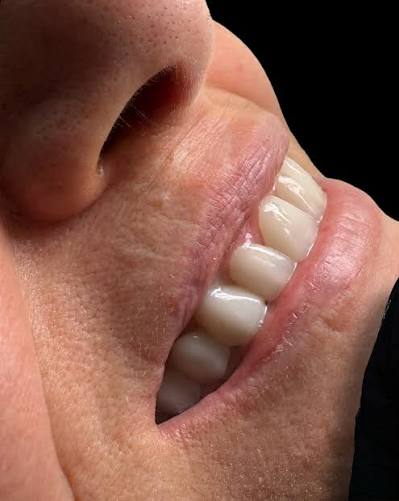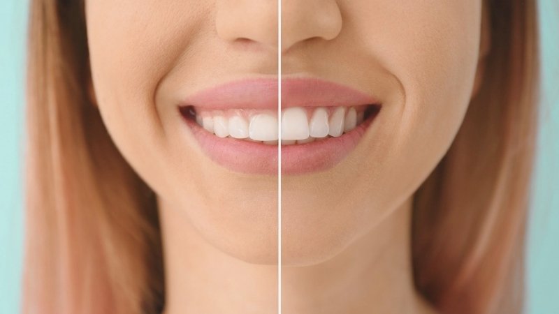
What Effects Does Gum Aesthetics Have on Smile?Gum aesthetics influences the overall character and brightness of the smile by determining how long or short the teeth appear in the mouth, whether the teeth appear to be aligned symmetrically or not, and the optical balance that is established with the curves of the lips.
The assessment of visual harmony in the mouth and jaw anatomy has moved beyond parameters such as the color or size of the tooth enamel, alongside the development process of dentistry science. In clinical approaches, the form and light transmissibility of teeth, referred to as “white aesthetics”, can only create biological harmony when combined with the “pink aesthetics” that refers to the health and symmetry of the gum tissue surrounding them. Even perfectly designed porcelain restorations can diminish aesthetic perception by losing their optical harmony with the rest of the face when placed over an asymmetric, inflamed, or overly prominent gum tissue. The discipline of Aesthetic Dentistry frames the complementary, proportional, and healthy coming together of these pink and white tissues as a fundamental medical goal.The shape, thickness, color, and amount of gum tissue varies from person to person, depending on genetic and environmental factors. The extent of upper lip mobility during speech or a broad smile determines how much of the gum tissue will be perceived from the outside. If the gum borders are wavy, irregular, or drawn back, it draws attention away from the teeth and towards this imbalance. Detailed in European dental clinic protocols, millimetric adjustments made to the gum (periodontal adjustments) bring the teeth to ideal proportions and ensure parallelism with reference points of the facial skeleton. This comprehensive guide evaluates the visual effects of gum asymmetries, the mechanics of the “Gummy Smile” (gum smile) phenomenon, the optical differences created by tissue pigmentation (coloration), and the role of pink aesthetics in planning processes based on scientific data.
How Do Asymmetries in Gum Level Alter the Dynamics of Smiling?
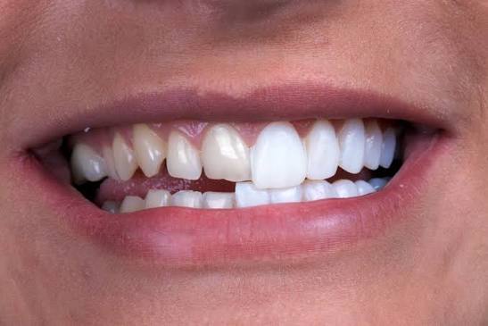
Asymmetries in gum level cause the lengths of adjacent teeth to be perceived as different (one longer, the other shorter), disrupting the parallelism between the right and left halves of the face and adding an irregular expression to the dynamics of smiling.The position of the gingival margins (the zenith points) on the tooth surface is an anatomical determinant that defines the visible crown length of the tooth in millimeters. In an ideal arrangement, the gingival zenith points of the two central incisors in the upper jaw should be located at the same horizontal plane. The gingival margin of the adjacent lateral teeth is positioned approximately one millimeter below this plane, while the margin of the canine teeth rises back to the same level as the central teeth. This slight wave-like and symmetrical curve within the natural setting reflects the dynamism and biological order of the smile. However, due to genetic dental eruption abnormalities, discrepancies, or asymmetrical jawbone development, these levels may deviate from one another.
For example, if the gingival margin of the right central incisor is positioned lower (covering more of the tooth) than the left central incisor, the right tooth will visually appear shorter and more square-shaped compared to the left. The brain instantly perceives the millimetric level difference between these two teeth and reflects it in the general smile as a feeling of contrast or disharmony. In cases where such asymmetries exist, periodontal interventions are performed to align tissue margins before planning for Aesthetic Dentistry. Using lasers or cautery devices, the remaining gingival tissue is elevated to a millimetric level, equalizing the zenith points of both teeth. Applying porcelain laminates or composite without this alignment will further accentuate the asymmetry problem instead of resolving it.
Gingival Margin (Zenith) Status Impact on Visual Perception Clinical Adjustment Approach Symmetrical and Wavy Curve A feeling of proportional tooth sizes, harmonious with the face, and a natural arrangement. No intervention is required; it can be converted directly to a prosthetic appliance. Level Difference (Asymmetric) The appearance of one tooth being short while the adjacent one appears long is a distorted expression. The matching of tissue by cutting the remaining tissue below with the help of laser or cautery (gingivectomy). Smooth (Straight Edge) Border A monoblock appearance that feels artificially unnatural and creates a sensation of excessive prosthetic teeth. The slight lowering of the level of lateral incisors through tissue shaping.
Gummy Smile: How Does It Visually Affect Tooth Length?
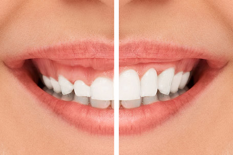
The gummy smile scenario leads to an aesthetic shift from teeth to the reddish tissue, resulting from excessive stretching of the upper lip or the gum tissue covering the teeth more than necessary, causing them to be perceived anatomically as short, square, or embedded in bone.It is a common situation, within the framework of general aesthetic ratios, for an individual’s gums to be unseen or only visible for a few millimeters when at rest or lightly smiling. However, if excessive elevation of the upper lip during a broad smile or laughter exposes the gums in a band wider than 3-4 millimeters, this clinical situation is referred to as “Gummy Smile.” This condition stems from various physiological origins, such as excessive vertical growth of the upper jaw, hypermobility of the lip muscles, or teeth remaining beneath the gum without completing the eruption process.
In cases of gummy smile, the length of the teeth is relatively short compared to their width. The ideal length of an upper incisor should be approximately 20% to 25% longer than its width. When the gum tissue covers this length, the teeth appear square, blunt, and aesthetically unpleasing. In Avrupadent procedures, a detailed periodontal mapping is created to solve this issue. If the problem arises from excessive coverage of the tooth by the gum tissue, a simple gingival contouring procedure (gingivoplasty) performed under local anesthesia will remove excess tissue, revealing the actual anatomical length of the tooth beneath the bone. If the issue stems from overactive lip muscles, medical muscle relaxant applications directed at lip positioning or related muscles are implemented to limit the visibility of the pink/red tissue.
How Does Gum Recession Affect the Aesthetics of the Root Appearance?
Gum recession leads to the exposure of yellowish root surfaces that are beyond the anatomical crown boundary of the teeth, causing the teeth to appear excessively long and resulting in the formation of dark “black triangles” between the teeth, which diminishes the perception of youthful and healthy tissue on the face.The health of the periodontal tissues functions as a barrier that tightly surrounds the tooth neck and protects the root surface from external factors. Poor brushing habits and hard brushing, having an anatomically thin tissue structure (biotype), or chronic gum infections can eventually cause this barrier to dissolve and allow the root to shift (recede). When recession occurs, the shiny white enamel layer is replaced by a yellowish, dull, and more sensitive cement (root) surface. The simultaneous visibility of these two differently colored tissues disrupts color uniformity and can lead to the perception of the tooth as stained or aged.
However, gum recession occurs not only on the surface of the tooth but also in the triangular-shaped tissue pieces (papillae) between the teeth. When the papillae dissolve, dark spaces form between the roots of the teeth. These gaps, known in dental literature as “black triangles,” cause air leakage while speaking, food accumulation, and an aged appearance in terms of aesthetics. In the restorative process of such cases, restoring the lost tissue to its previous level through surgical grafting methods or optically closing these black triangles with suitable porcelain/composite restorations is a medical priority.
Why Do Dark Colored (Pigmented) Gums Cast Shadows on the Brightness of a Smile?
Dark or bluish gums disrupt the lively pink contrast expected with the skin and lips, creating a dark frame around white porcelain teeth, which shadows the overall brightness and freshness of the smile.The clinical reference for healthy gum tissue is a light and lively color tone referred to as “gum pink” or “pale pink.” However, in some individuals, due to genetic factors, heavy tobacco use, or environmental influences, melanin pigments that give color to the skin can excessively accumulate on the gum surface. This accumulation leads to the formation of brown, black, or purplish spots (physiological pigmentation) on the gums. Regardless of how white and uniform the teeth are, the expected aesthetic contrast cannot be achieved due to the surrounding dark-colored tissue.
Aesthetic Dentistry practice implements the lightening of these dark-colored tissues through a procedure known as “depigmentation” (peeling). Nowadays, using laser technologies, the uppermost thin and pigmented epithelial layer of the gum is vaporized under local anesthesia. The new tissue that emerges heals in a healthy and light pink manner, without any melanin accumulation. The lightening of the framework allows the surface (teeth) to shine in a much more fresh, clear, and attractive way, representing an important supportive medical procedure.
How is Pink and White Aesthetic Harmony Established in a Biological Framework?
Pink and white aesthetic harmony is established by blending the ratios of the width and height of the teeth (white aesthetic) with the height and curvature of the gum line (pink aesthetic) according to the principles of the golden ratio, and planning it in a way that respects chewing function and biological width.In dentistry, aesthetics is the science of reflecting the mathematical ratios inherent in nature (golden ratio) onto tissues. White aesthetics refers to the effects of porcelain or composite materials on the shape, color, and light transmissibility of the teeth; while pink aesthetics encompasses the symmetry and health of the living tissues surrounding these materials. These two fields cannot be thought of independently from each other. If a perfectly processed glass ceramic (e-max) laminate is placed on an inflamed, bleeding gum, a poor clinical outcome is achieved.
Establishing this balance in the biological substrate depends on preserving the anatomical rule known as “biological width.” Biological width is a natural defense zone that is approximately 2-3 millimeters thick, ranging from the gum line to the bony tissue. If the margin of the porcelain restoration to be performed is placed too deeply, violating this area, the body perceives this restoration as a foreign object, leading to chronic inflammation in that region. In the management processes of Avrupadent, restoration planning carefully approaches these biological distances, maintaining the health of the gum tissue and ensuring that the aesthetic contour remains intact for years.
What Technological Methods are Used in Gum Shaping Procedures?
Gum shaping procedures are performed using diode laser systems that simultaneously provide bleeding control and accelerate tissue healing, electrocautery devices, or precise scalpels, employing minimally invasive (tissue-preserving) methods in clinical settings.In previous years, when gum leveling procedures (gingivectomy) were performed with classical surgical knives (scalpels), the bleeding and tissue swelling that occurred after the procedure delayed the acquisition of prosthetic measurements for days. The integration of modern technologies into clinics has transformed this process into a highly comfortable dimension for both patients and practitioners. Electrocautery devices and especially soft tissue lasers (diode lasers) have become primary tools in reconstructing gum aesthetics.
Laser waves vaporize the gum tissue at a millimeter level while simultaneously sealing the capillaries (coagulation). This way, there is almost no bleeding during the operation. The absence of bleeding allows the surgeon to align asymmetrical tissues with a clearer line of sight. Additionally, the laser’s ability to reduce bacterial load in the tissue supports the healing process (resolution) after the procedure, helping the patient to quickly return to normal life. This technological advancement acts as a medical bridge that allows the measurement phase required for porcelain restorations to be completed in much shorter periods.
How Does the Gum Structure (Biotype) Guide Restoration Processes?
The gum biotype; in thin and transparent tissues requires only the selection of metal-free restorations to prevent the reflection of the material beneath the porcelain, while in thick and fibrotic tissues, it allows for easier planning of the preparations (steps) due to being more tolerant to mechanical pressures, thus guiding the processes.Individuals’ gum thicknesses vary, just like skin types, and these differences are classified in clinical literature as “periodontal biotype.” Individuals with a thick biotype have gums that are dense, fibrotic, and quite resistant to external mechanical effects. Adjusting margins is easier when performing tooth cutting in such tissue, and the risk of recession in the gum is lower. Moreover, this thick tissue does not reflect the color of the restoration placed underneath or any minor mismatches outside (it has a high masking ability).
However, for patients with a thin biotype, the gum tissue is as delicate as thin paper. This tissue is extremely sensitive to trauma and will show deformation under even the slightest pressure. When porcelain with a metal framework is applied to a patient with thin tissue, the underlying metal’s gray color is instantaneously noticeable as a purplish line due to the transparency of the gum. Because of this biological reality, all procedures planned for patients with a thin biotype must be shaped using high-light-transmitting glass ceramics (e-max) or biocompatible materials like zirconium; this is a fundamental medical protocol that must be adhered to according to Europadent standards.
Gum Biotype Anatomical and Physiological Characteristics Restorative Planning Criteria Thick and Flat Biotype Tissue is thick, richly vascularized, and quite resistant to deformation. Easily tolerates mechanical procedures, and does not reflect the color of the underlying material. Thin and Ruffled Biotype Tissue is transparent, delicate, the triangular papillae are long, and sensitive to trauma. Only metal-free, biocompatible ceramics should be chosen, and procedures must be performed with great caution.
How Do Inflammation and Swelling in the Gums Affect Aesthetic Planning?
Inflammation and swelling can mask the actual anatomical position of the gum borders, leading to incorrect measurements being taken; therefore, before aesthetic procedures are initiated, these conditions must be treated to ensure that the tissue reaches a healthy, tight, and non-bleeding form, thus directly shaping the planning.The inadequacy of oral care, plaque accumulation, or genetic predispositions can lead to gum diseases (gingivitis or periodontitis) characterized by redness, swelling, and bleeding at the slightest touch. Regardless of how significant the size or color disorder in a patient’s teeth may be, if there is active inflammation in the gums, the procedures of Aesthetic Dentistry are unequivocally postponed due to a medical rule. No prosthesis constructed on an unhealthy foundation can have a predictable future.
Swollen gums appear much larger than their actual size and are in a shifted position. When this is the case, tooth trimming is performed, and if measurements are taken, the porcelains that will be produced in the laboratory will be prepared according to this swollen condition. In the following weeks, as the patient’s gums heal and the swelling subsides (returns to its normal level), the edges of the porcelain made will remain outside and create visible aesthetic gaps between the tooth and the crown. Moreover, in a bleeding environment, the adhesion strength of the medical resin cements (adhesives) to the tooth is weakened, increasing the risk of crown displacement. Therefore, periodontal treatment (removal of tartar and smoothing of the root surface) should first be applied to ensure tissue stabilization, followed by moving on to the second phase of aesthetic planning.
What are Frequently Asked Questions?
1. Does gum asymmetry directly affect the smile?
Yes, having gum levels different between adjacent teeth causes one tooth to look longer than the other, disrupting the overall symmetry of the smile and creating a distorted perception of beauty.
2. Does gum shaping elongate the tooth length?
This procedure does not extend the natural length of the tooth within the bone; it merely elevates the excess red tissue above the tooth, revealing the visible (clinical) crown length in the mouth.
3. In the case of a gummy smile, is only the gum treated?
It varies depending on the source of the case; if the issue is excess gum, tissue is only shaped with a laser, but if the problem arises from excessive muscle contraction of the lip, medical applications like botulinum toxin are also included in the treatment.
4. In which situations are pink aesthetic procedures planned?
Situations include when more than 3 mm of gum is visible while smiling (gummy smile), asymmetry in gum margins, presence of bruising/staining in the tissues, and cases where tooth lengths are perceived as very short.
5. Can bruising in the gums be addressed with aesthetic procedures?
Yes, purple and brown discolorations due to physiological melanin pigmentation or smoking can be reverted to a healthy light pink color through a depigmentation (peeling) procedure using laser technology.
6. How does gum recession affect restorative planning?
The root surfaces exposed due to recession change the boundary of the porcelain finish. To maintain aesthetic appearance, the receded tissue must first be replaced with periodontal grafts, or the porcelain must be designed in an anatomical form to cover this exposure.
7. What color and form should healthy gums have?
Clinically, healthy gums should have a pale “rosewood” or a light pink color, with a matte consistency that tightly envelops the tooth, and they should not bleed when brushing. They should also have a slightly stippled texture that resembles orange peel.
8. Is gum treatment necessary before porcelain veneers?
If there is inflammation, bleeding, or accumulation of tartar in the tissues, gum treatment must be performed first to stabilize the biological foundation and maintain the fit and quality of the porcelain on the tooth.
9. What is the purpose of laser gum procedures?
Lasers can shape tissue while simultaneously cauterizing blood vessels to control bleeding, eliminate bacteria from the area, and provide a much more comfortable healing process after surgery compared to traditional methods.
10. Why do black spaces (black triangles) form between teeth?
Black triangles form as a result of the melting (recession) of triangular gum papillae between teeth due to gum infections or traumatic brushing, leading to a dark, aged appearance during smiling.
11. How does gum biotype influence aesthetic outcomes?
In transparent tissues with a specific biotype, zirconium or full ceramics that allow light to pass through should be chosen to avoid reflecting the color of the underlying structure; precise border preparations must be made suitable for tissue thickness.
12. What is the relationship between the gum line and gum level?
The line formed by the upper lip while smiling should be parallel and proportional to the borders of the gums. Asymmetrical positioning that violates the curvature of the lips weakens the sense of aesthetic unity and harmony.
13. Can aesthetic porcelain be applied over inflamed gums?
According to medical principles, applying prostheses over swollen and bleeding tissues is not medically approved, as it may lead to porcelain edges being exposed when the gum level decreases in the future and cause leakage.
14. How is pink aesthetic analysis conducted in Avrupadent procedures?
Using the patient’s digital intraoral scans and high-resolution photographs, gum levels are measured millimetrically through software, and the biological distance to the bone limits is supported by radiological findings.
15. How is tissue adaptation monitored after gum surgeries?
After shaping procedures, a few days of healing period is expected for the tissue to adapt to its new anatomy, after which temporary teeth are placed, and the biological adaptation of the gums to the porcelain form is monitored through routine clinical appointments.
The assessment of visual harmony in the mouth and jaw anatomy has moved beyond parameters such as the color or size of the tooth enamel, alongside the development process of dentistry science. In clinical approaches, the form and light transmissibility of teeth, referred to as “white aesthetics”, can only create biological harmony when combined with the “pink aesthetics” that refers to the health and symmetry of the gum tissue surrounding them. Even perfectly designed porcelain restorations can diminish aesthetic perception by losing their optical harmony with the rest of the face when placed over an asymmetric, inflamed, or overly prominent gum tissue. The discipline of Aesthetic Dentistry frames the complementary, proportional, and healthy coming together of these pink and white tissues as a fundamental medical goal.
The shape, thickness, color, and amount of gum tissue varies from person to person, depending on genetic and environmental factors. The extent of upper lip mobility during speech or a broad smile determines how much of the gum tissue will be perceived from the outside. If the gum borders are wavy, irregular, or drawn back, it draws attention away from the teeth and towards this imbalance. Detailed in European dental clinic protocols, millimetric adjustments made to the gum (periodontal adjustments) bring the teeth to ideal proportions and ensure parallelism with reference points of the facial skeleton. This comprehensive guide evaluates the visual effects of gum asymmetries, the mechanics of the “Gummy Smile” (gum smile) phenomenon, the optical differences created by tissue pigmentation (coloration), and the role of pink aesthetics in planning processes based on scientific data.
How Do Asymmetries in Gum Level Alter the Dynamics of Smiling?
The position of the gingival margins (the zenith points) on the tooth surface is an anatomical determinant that defines the visible crown length of the tooth in millimeters. In an ideal arrangement, the gingival zenith points of the two central incisors in the upper jaw should be located at the same horizontal plane. The gingival margin of the adjacent lateral teeth is positioned approximately one millimeter below this plane, while the margin of the canine teeth rises back to the same level as the central teeth. This slight wave-like and symmetrical curve within the natural setting reflects the dynamism and biological order of the smile. However, due to genetic dental eruption abnormalities, discrepancies, or asymmetrical jawbone development, these levels may deviate from one another.
For example, if the gingival margin of the right central incisor is positioned lower (covering more of the tooth) than the left central incisor, the right tooth will visually appear shorter and more square-shaped compared to the left. The brain instantly perceives the millimetric level difference between these two teeth and reflects it in the general smile as a feeling of contrast or disharmony. In cases where such asymmetries exist, periodontal interventions are performed to align tissue margins before planning for Aesthetic Dentistry. Using lasers or cautery devices, the remaining gingival tissue is elevated to a millimetric level, equalizing the zenith points of both teeth. Applying porcelain laminates or composite without this alignment will further accentuate the asymmetry problem instead of resolving it.
| Gingival Margin (Zenith) Status | Impact on Visual Perception | Clinical Adjustment Approach |
|---|---|---|
| Symmetrical and Wavy Curve | A feeling of proportional tooth sizes, harmonious with the face, and a natural arrangement. | No intervention is required; it can be converted directly to a prosthetic appliance. |
| Level Difference (Asymmetric) | The appearance of one tooth being short while the adjacent one appears long is a distorted expression. | The matching of tissue by cutting the remaining tissue below with the help of laser or cautery (gingivectomy). |
| Smooth (Straight Edge) Border | A monoblock appearance that feels artificially unnatural and creates a sensation of excessive prosthetic teeth. | The slight lowering of the level of lateral incisors through tissue shaping. |
Gummy Smile: How Does It Visually Affect Tooth Length?
It is a common situation, within the framework of general aesthetic ratios, for an individual’s gums to be unseen or only visible for a few millimeters when at rest or lightly smiling. However, if excessive elevation of the upper lip during a broad smile or laughter exposes the gums in a band wider than 3-4 millimeters, this clinical situation is referred to as “Gummy Smile.” This condition stems from various physiological origins, such as excessive vertical growth of the upper jaw, hypermobility of the lip muscles, or teeth remaining beneath the gum without completing the eruption process.
In cases of gummy smile, the length of the teeth is relatively short compared to their width. The ideal length of an upper incisor should be approximately 20% to 25% longer than its width. When the gum tissue covers this length, the teeth appear square, blunt, and aesthetically unpleasing. In Avrupadent procedures, a detailed periodontal mapping is created to solve this issue. If the problem arises from excessive coverage of the tooth by the gum tissue, a simple gingival contouring procedure (gingivoplasty) performed under local anesthesia will remove excess tissue, revealing the actual anatomical length of the tooth beneath the bone. If the issue stems from overactive lip muscles, medical muscle relaxant applications directed at lip positioning or related muscles are implemented to limit the visibility of the pink/red tissue.
How Does Gum Recession Affect the Aesthetics of the Root Appearance?
The health of the periodontal tissues functions as a barrier that tightly surrounds the tooth neck and protects the root surface from external factors. Poor brushing habits and hard brushing, having an anatomically thin tissue structure (biotype), or chronic gum infections can eventually cause this barrier to dissolve and allow the root to shift (recede). When recession occurs, the shiny white enamel layer is replaced by a yellowish, dull, and more sensitive cement (root) surface. The simultaneous visibility of these two differently colored tissues disrupts color uniformity and can lead to the perception of the tooth as stained or aged.
However, gum recession occurs not only on the surface of the tooth but also in the triangular-shaped tissue pieces (papillae) between the teeth. When the papillae dissolve, dark spaces form between the roots of the teeth. These gaps, known in dental literature as “black triangles,” cause air leakage while speaking, food accumulation, and an aged appearance in terms of aesthetics. In the restorative process of such cases, restoring the lost tissue to its previous level through surgical grafting methods or optically closing these black triangles with suitable porcelain/composite restorations is a medical priority.
Why Do Dark Colored (Pigmented) Gums Cast Shadows on the Brightness of a Smile?
The clinical reference for healthy gum tissue is a light and lively color tone referred to as “gum pink” or “pale pink.” However, in some individuals, due to genetic factors, heavy tobacco use, or environmental influences, melanin pigments that give color to the skin can excessively accumulate on the gum surface. This accumulation leads to the formation of brown, black, or purplish spots (physiological pigmentation) on the gums. Regardless of how white and uniform the teeth are, the expected aesthetic contrast cannot be achieved due to the surrounding dark-colored tissue.
Aesthetic Dentistry practice implements the lightening of these dark-colored tissues through a procedure known as “depigmentation” (peeling). Nowadays, using laser technologies, the uppermost thin and pigmented epithelial layer of the gum is vaporized under local anesthesia. The new tissue that emerges heals in a healthy and light pink manner, without any melanin accumulation. The lightening of the framework allows the surface (teeth) to shine in a much more fresh, clear, and attractive way, representing an important supportive medical procedure.
How is Pink and White Aesthetic Harmony Established in a Biological Framework?
In dentistry, aesthetics is the science of reflecting the mathematical ratios inherent in nature (golden ratio) onto tissues. White aesthetics refers to the effects of porcelain or composite materials on the shape, color, and light transmissibility of the teeth; while pink aesthetics encompasses the symmetry and health of the living tissues surrounding these materials. These two fields cannot be thought of independently from each other. If a perfectly processed glass ceramic (e-max) laminate is placed on an inflamed, bleeding gum, a poor clinical outcome is achieved.
Establishing this balance in the biological substrate depends on preserving the anatomical rule known as “biological width.” Biological width is a natural defense zone that is approximately 2-3 millimeters thick, ranging from the gum line to the bony tissue. If the margin of the porcelain restoration to be performed is placed too deeply, violating this area, the body perceives this restoration as a foreign object, leading to chronic inflammation in that region. In the management processes of Avrupadent, restoration planning carefully approaches these biological distances, maintaining the health of the gum tissue and ensuring that the aesthetic contour remains intact for years.
What Technological Methods are Used in Gum Shaping Procedures?
In previous years, when gum leveling procedures (gingivectomy) were performed with classical surgical knives (scalpels), the bleeding and tissue swelling that occurred after the procedure delayed the acquisition of prosthetic measurements for days. The integration of modern technologies into clinics has transformed this process into a highly comfortable dimension for both patients and practitioners. Electrocautery devices and especially soft tissue lasers (diode lasers) have become primary tools in reconstructing gum aesthetics.
Laser waves vaporize the gum tissue at a millimeter level while simultaneously sealing the capillaries (coagulation). This way, there is almost no bleeding during the operation. The absence of bleeding allows the surgeon to align asymmetrical tissues with a clearer line of sight. Additionally, the laser’s ability to reduce bacterial load in the tissue supports the healing process (resolution) after the procedure, helping the patient to quickly return to normal life. This technological advancement acts as a medical bridge that allows the measurement phase required for porcelain restorations to be completed in much shorter periods.
How Does the Gum Structure (Biotype) Guide Restoration Processes?
Individuals’ gum thicknesses vary, just like skin types, and these differences are classified in clinical literature as “periodontal biotype.” Individuals with a thick biotype have gums that are dense, fibrotic, and quite resistant to external mechanical effects. Adjusting margins is easier when performing tooth cutting in such tissue, and the risk of recession in the gum is lower. Moreover, this thick tissue does not reflect the color of the restoration placed underneath or any minor mismatches outside (it has a high masking ability).
However, for patients with a thin biotype, the gum tissue is as delicate as thin paper. This tissue is extremely sensitive to trauma and will show deformation under even the slightest pressure. When porcelain with a metal framework is applied to a patient with thin tissue, the underlying metal’s gray color is instantaneously noticeable as a purplish line due to the transparency of the gum. Because of this biological reality, all procedures planned for patients with a thin biotype must be shaped using high-light-transmitting glass ceramics (e-max) or biocompatible materials like zirconium; this is a fundamental medical protocol that must be adhered to according to Europadent standards.
| Gum Biotype | Anatomical and Physiological Characteristics | Restorative Planning Criteria |
|---|---|---|
| Thick and Flat Biotype | Tissue is thick, richly vascularized, and quite resistant to deformation. | Easily tolerates mechanical procedures, and does not reflect the color of the underlying material. |
| Thin and Ruffled Biotype | Tissue is transparent, delicate, the triangular papillae are long, and sensitive to trauma. | Only metal-free, biocompatible ceramics should be chosen, and procedures must be performed with great caution. |
How Do Inflammation and Swelling in the Gums Affect Aesthetic Planning?
The inadequacy of oral care, plaque accumulation, or genetic predispositions can lead to gum diseases (gingivitis or periodontitis) characterized by redness, swelling, and bleeding at the slightest touch. Regardless of how significant the size or color disorder in a patient’s teeth may be, if there is active inflammation in the gums, the procedures of Aesthetic Dentistry are unequivocally postponed due to a medical rule. No prosthesis constructed on an unhealthy foundation can have a predictable future.
Swollen gums appear much larger than their actual size and are in a shifted position. When this is the case, tooth trimming is performed, and if measurements are taken, the porcelains that will be produced in the laboratory will be prepared according to this swollen condition. In the following weeks, as the patient’s gums heal and the swelling subsides (returns to its normal level), the edges of the porcelain made will remain outside and create visible aesthetic gaps between the tooth and the crown. Moreover, in a bleeding environment, the adhesion strength of the medical resin cements (adhesives) to the tooth is weakened, increasing the risk of crown displacement. Therefore, periodontal treatment (removal of tartar and smoothing of the root surface) should first be applied to ensure tissue stabilization, followed by moving on to the second phase of aesthetic planning.
What are Frequently Asked Questions?
1. Does gum asymmetry directly affect the smile?
Yes, having gum levels different between adjacent teeth causes one tooth to look longer than the other, disrupting the overall symmetry of the smile and creating a distorted perception of beauty.
2. Does gum shaping elongate the tooth length?
This procedure does not extend the natural length of the tooth within the bone; it merely elevates the excess red tissue above the tooth, revealing the visible (clinical) crown length in the mouth.
3. In the case of a gummy smile, is only the gum treated?
It varies depending on the source of the case; if the issue is excess gum, tissue is only shaped with a laser, but if the problem arises from excessive muscle contraction of the lip, medical applications like botulinum toxin are also included in the treatment.
4. In which situations are pink aesthetic procedures planned?
Situations include when more than 3 mm of gum is visible while smiling (gummy smile), asymmetry in gum margins, presence of bruising/staining in the tissues, and cases where tooth lengths are perceived as very short.
5. Can bruising in the gums be addressed with aesthetic procedures?
Yes, purple and brown discolorations due to physiological melanin pigmentation or smoking can be reverted to a healthy light pink color through a depigmentation (peeling) procedure using laser technology.
6. How does gum recession affect restorative planning?
The root surfaces exposed due to recession change the boundary of the porcelain finish. To maintain aesthetic appearance, the receded tissue must first be replaced with periodontal grafts, or the porcelain must be designed in an anatomical form to cover this exposure.
7. What color and form should healthy gums have?
Clinically, healthy gums should have a pale “rosewood” or a light pink color, with a matte consistency that tightly envelops the tooth, and they should not bleed when brushing. They should also have a slightly stippled texture that resembles orange peel.
8. Is gum treatment necessary before porcelain veneers?
If there is inflammation, bleeding, or accumulation of tartar in the tissues, gum treatment must be performed first to stabilize the biological foundation and maintain the fit and quality of the porcelain on the tooth.
9. What is the purpose of laser gum procedures?
Lasers can shape tissue while simultaneously cauterizing blood vessels to control bleeding, eliminate bacteria from the area, and provide a much more comfortable healing process after surgery compared to traditional methods.
10. Why do black spaces (black triangles) form between teeth?
Black triangles form as a result of the melting (recession) of triangular gum papillae between teeth due to gum infections or traumatic brushing, leading to a dark, aged appearance during smiling.
11. How does gum biotype influence aesthetic outcomes?
In transparent tissues with a specific biotype, zirconium or full ceramics that allow light to pass through should be chosen to avoid reflecting the color of the underlying structure; precise border preparations must be made suitable for tissue thickness.
12. What is the relationship between the gum line and gum level?
The line formed by the upper lip while smiling should be parallel and proportional to the borders of the gums. Asymmetrical positioning that violates the curvature of the lips weakens the sense of aesthetic unity and harmony.
13. Can aesthetic porcelain be applied over inflamed gums?
According to medical principles, applying prostheses over swollen and bleeding tissues is not medically approved, as it may lead to porcelain edges being exposed when the gum level decreases in the future and cause leakage.
14. How is pink aesthetic analysis conducted in Avrupadent procedures?
Using the patient’s digital intraoral scans and high-resolution photographs, gum levels are measured millimetrically through software, and the biological distance to the bone limits is supported by radiological findings.
15. How is tissue adaptation monitored after gum surgeries?
After shaping procedures, a few days of healing period is expected for the tissue to adapt to its new anatomy, after which temporary teeth are placed, and the biological adaptation of the gums to the porcelain form is monitored through routine clinical appointments.