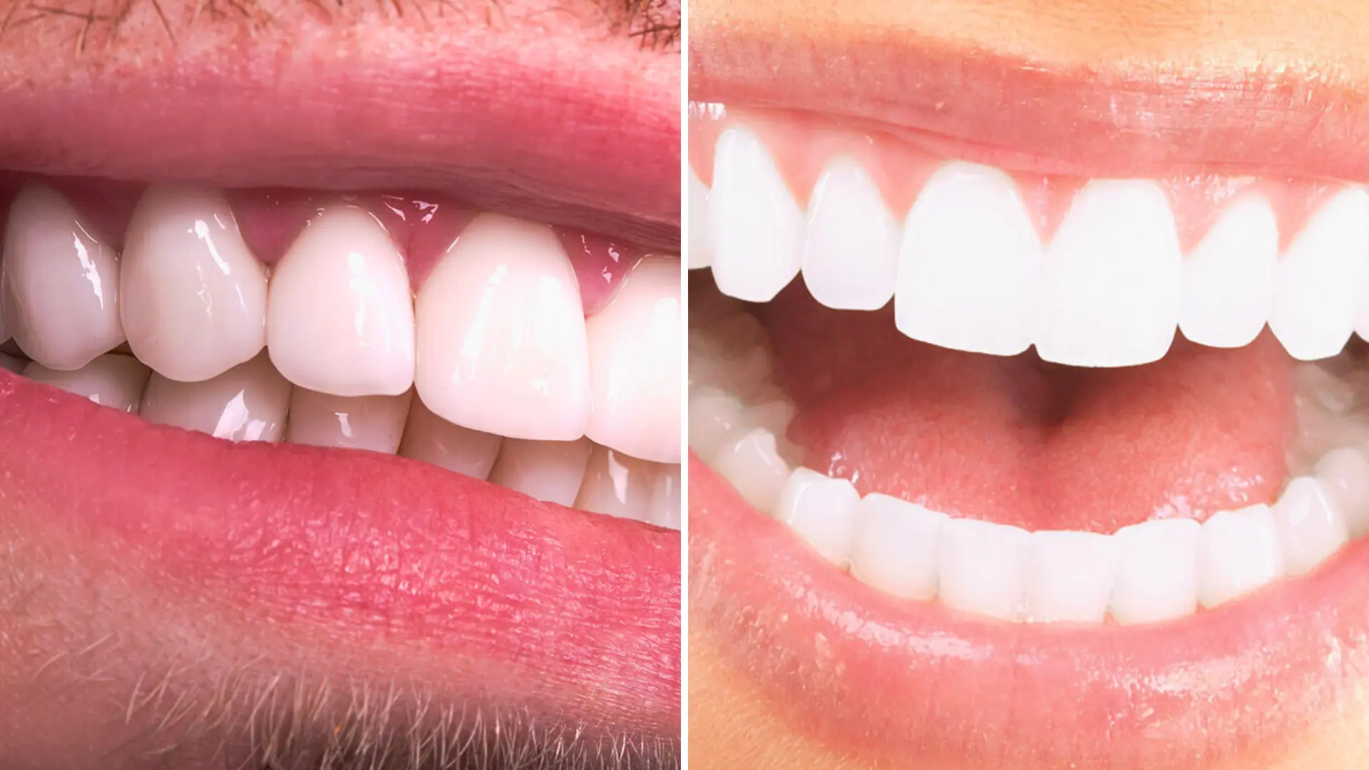
What Are the Differences Between Laminate and Zirconium?
The process of restoring morphological asymmetries in the oral and dental structure, tissue losses, and discoloration problems through medical protocols is shaped around innovative solutions offered by biomaterials science. Current clinical disciplines have shifted away from the incompatibility of metal alloys with tissues, focusing on high biocompatibility rates of crystalized elements and glass ceramics. In the planning of Aesthetic Dentistry, the concepts that doctors and patients encounter most frequently, laminate (leaf porcelain) and zirconium, although they seem to serve the same visual improvement goal, are actually two distinct medical approaches that differ entirely in terms of structural properties, application methods on teeth, and light scattering indices.
The decision regarding which material will restore a tooth depends on a multidisciplinary analysis based on the severity of the occlusal (biting) forces applied to that tooth, the extent of existing substance loss in the tooth, and the desired optical depth. Since both applications do not have a metal substructure, they do not cause corrosion (oxidation) or gray reflections at the gum line; however, the volumes of the materials covering the tissue differ. In clinical evaluations conducted in Avrupadent’s process management, the patient’s expectations are filtered through skeletal limits to determine which material is more suitable for the biological structure. In this comprehensive medical guide, the chemical compositions of laminate and zirconium materials, intervention limits on dental tissue, light transmissivity (translucency) properties, and interactions with jaw mechanics are objectively addressed.
What Are the Basic Definitions of Laminate and Zirconium Applications?
In medical literature, lamina restorations, also known as “veneer,” can be likened to a contact lens due to their structure. They are produced in a laboratory environment from special lithium disilicate or feldspathic glass ceramics and are applied only to the visible front surface without touching the tongue/palate side. Although their thin structures can cause the material to be fragile on its own, when integrated with resin cements (medical adhesives), they behave like a single piece with the enamel tissue of the tooth, gaining high functional tolerance.
Zirconium (zirconium dioxide) is the adapted form of a naturally occurring white and extremely hard element for the medical field. Zirconium applications operate under the logic of “crown” (coating); in other words, it encases the entire surfaces of the tooth, fully enclosing it. These white and resilient zirconium blocks have replaced the gray skeletons under classic metal-supported porcelains. Zirconium can be processed with porcelain powder or produced as monolithic (single piece), serving as a supportive column that protects the tooth against mechanical pressures.
How Does the Amount of Intervention on Tooth Tissue Differ Between the Two Methods?
The principles of preventive dentistry are primarily based on limiting the unnecessary sacrifice of the natural and healthy dental tissue of the patient. Laminate applications are a direct reflection of this preventive philosophy. A microscopic level enamel preparation is made only on the surface facing the lips, without disturbing the structural integrity of the tooth. In fact, in some suitable anatomical cases (when the teeth are smaller or positioned further back than normal), it is possible to apply ‘prepless’ laminates without any tooth shaving.
In contrast, zirconium crowns require a certain thickness to be able to cover the tooth from all sides and withstand chewing forces. To form this thickness, a tissue removal process is applied circumferentially from the front, back, sides, and chewing surfaces of the tooth. The reduction procedure performed for zirconium is broader in scope compared to laminates; however, in cases where the tooth is already largely decayed, broken, or weakened, this intervention acts as protective armor, helping to absorb destructive forces from the outside instead of weakening the tooth.
In Which Anatomical and Clinical Situations Should Which Material Be Preferred?
Aesthetic Dentistry analyzes the hospital’s functional requirements directly shaping material selection. For patients with gaps (diastema) in the front teeth, small cracks related to age on the enamel surface, or hereditary stains, laminates are the ideal solution. Since there is no approach to the nerve layer of the tooth (pulpa), the likelihood of sensitivity after the procedure is quite low, and due to its translucency, it creates a natural enamel illusion.
However, if there are large fillings in the teeth, if fractures due to trauma cover a large part of the tooth, or if the tooth has lost vitality due to root canal treatment and has become fragile, thin laminates are insufficient to mechanically support the tooth. In such resistance-requiring scenarios, zirconium crowns come into play. Additionally, when it comes to closing gaps where multiple adjacent teeth are missing, it becomes essential to use bridge (fixed) systems, and since the material must have high flexibility and break resistance, zirconium blocks become an indispensable choice as a supportive skeleton.
| Clinical Case Profile | Recommended Medical Approach | Reason for Planning |
|---|---|---|
| Small Gaps in Front Teeth (Diastema) | Porcelain Laminate (Veneer) | Optically closing gaps by minimally shaping the tooth structure. |
| Root Treated and Large Filled Tooth | Zirconium Crown (Cover) | Providing structural protection against mechanical forces by wrapping the fragile tooth completely (360 degrees). |
| Mild Crookedness and Color Disorder | Porcelain Laminate (Veneer) | Creating alignment and color control with thin ceramics in undesirable orthodontic cases. |
| Multiple Tooth Loss (Bridge Requirement) | Zirconium-Supported Systems | Providing high resistance to loads that the body teeth will carry. |
What Differences Can Be Observed in Terms of Light Transmittance and Optical Properties?
The factor that determines the degree of naturalness of biomaterials inside the mouth is how they interact and break down light. Natural tooth enamel not only reflects the light that falls on it but also absorbs it and transmits it to the dentin layer (translucency). Laminates produced from lithium disilicate or feldspathic powders replicate this light transmittance thanks to their glass-like structures. The bluish transparency felt particularly in the cutting edges of the tooth is achieved at quite high standards with these materials.
Zirconium dioxide, due to its dense crystalline structure, has a whiter and more opaque character (transmitting less light) compared to glass ceramics. This opacity can be an advantage rather than a disadvantage in some clinical scenarios. If the natural color of the tooth to be restored (the root color) has turned black or dark gray due to root canal treatment or old amalgam fillings, laminates that transmit a lot of light will reflect this dark color outward. Zirconium, thanks to its masking capacity, allows the underlying dark tissue to be captured, offering a clean, bright, and uniform whiteness outward.
What are the tolerances against biomechanical resistance and bending forces?
During chewing, serious vertical and horizontal pressures are exerted on the teeth by the jaw and chin mechanism. Zirconium is found in a hardness scale comparable to that of metals in structure. Thanks to its properties in the crystal phase, it exhibits an unparalleled mechanical behavior that slows down the progression of micro-cracks that can form on its surface by self-blocking (phase transformation toughness). Therefore, the use of zirconium is strongly supported medically in areas where molar teeth are located and chewing activities occur most intensely.
Laminates are very delicate and thin ceramic sheets while being handled by a technician in the laboratory; they can easily break. However, in a clinical setting, after the tooth surface is micro-porous enough to open pores, and after using “adhesive” (chemical) bonding resin cements, the situation changes. The porcelain fuses with the tooth enamel as a single block, creating a biochemical locking mechanism. Thanks to this locking, the laminate becomes as resistant to normal thermal stress as natural enamel. Nevertheless, they are more sensitive to abnormal mechanical trauma, such as breaking hard-shelled foods with teeth, compared to zirconium.
What are the time differences between treatment processes and laboratory procedures?
In modern clinics, Aesthetic Dentistry applications are completed in much shorter times compared to previous years with the introduction of digital workflows (CAD/CAM systems). In veneer treatment, a very light abrasion is performed on the front surface of the teeth in the first session, digital measurements are taken with intraoral scanners, and temporary crowns are placed on the patient. After laboratory production, the porcelain veneers are usually permanently bonded to the enamel in the second or third session. This entire process can be completed in 5 to 10 days, depending on the progress of the trial.
In the zirconium process, reducing the teeth by 360 degrees (preparation) requires more clinical time and attention. After measurements are taken, zirconium infrastructure is milled in the laboratory, and a “framework trial” session is frequently organized to test the compatibility of this infrastructure with the gum and occlusion (bite). If everything meets medical standards, the porcelain powders are layered on top to achieve the final form. These additional trials in the zirconium process may slightly extend the total time spent in the clinic compared to veneers; however, they are necessary steps that should not be overlooked to ensure final compatibility.
How Do Occlusal Disorders Affect Material Selection?
In dentistry, functional examination is the supporting column of aesthetic planning. The relationship of contact between the upper and lower jaw is called occlusion. If the front teeth close end-to-end (edge-to-edge) with the lower teeth or if the teeth hit each other very hard while moving the lower jaw forward, backward, or sideways, this creates a destructive stress area on restorations. Thin porcelain veneers may not withstand these vertical and horizontal intense crushing forces for long and can either fail or detach from the surface they were bonded to.
Additionally, individuals with sleep apnea or stress-related bruxism (nighttime teeth grinding and clenching) exert an excessive force on the chewing muscles. To absorb the pressure created by these muscles, the application of zirconium restorations, which have bite resistance, becomes a medical necessity. Even after applying zirconium crowns, the physician uses medical night splints during sleep to protect the joint and the crowns, thereby supporting the biological lifespan of the process.
How is Pink Aesthetics (Gum Harmony) Achieved in Both Cases?
The true appearance of restorations within the mouth is measured not only by the shape of the tooth but also by the symmetry and health of the gum tissue surrounding that tooth. When old-type metal-supported porcelain crowns were used, the underlying chrome-cobalt alloys would react over time with the gum tissue, causing oxidation and the formation of a dark, purplish frame in the neck area. The zirconium and laminate materials used today have a completely white and light-permeable chemistry, which does not cause any toxic or allergic discoloration in the gum tissue.
In both methods, the edge boundaries of the restorations are adjusted at a microscopic level to be in line with the gum or remain very slightly within the gum pocket. Since the tooth preparation for laminate restorations is quite minimal, the contact established with the gum is much more superficial; this reduces the likelihood of tissue inflammation. For zirconium crowns, achieving accurate alignment of the margin and ensuring the porcelain edge does not press against the gum (preservation of biological width) is directly dependent on the craftsmanship of the dentist and the quality of the digital scanning.
What Medical Criteria Are Used for Selection in Avrupadent Clinics?
In medical applications, each hospital’s physiological structure and visual expectations are unique variables. Instead of presenting ready-made templates to the patient in European treatment processes, a multidisciplinary examination concept is applied. If only asymmetry and discoloration are present in the patient’s anterior teeth area and the enamel is in a healthy state, the process is automatically designed in favor of laminate treatment with the principle of preserving live tissue.
However, if deep caries have progressed to the dentin layer, structural weaknesses from root canal treatments, or missing tooth gaps are detected as a result of radiological evaluations; planning will be directed towards zirconium systems to strengthen the supporting infrastructure. Based on the measurements obtained in digital smile design programs, the optical reflection values, thickness, and compatibility with the chewing muscles of the material to be applied to the patient are determined through collective consultation. This analytical and measurable approach elevates the biological compatibility of the treatment to high standards, along with its aesthetic benefits.
What are the Frequently Asked Questions?
1. Can laminate and zirconium applications be combined in the same session?
Depending on the functional needs inside the mouth, both materials can be used on the same patient; for example, while an aesthetic laminate is applied to the anterior teeth, it is possible to apply zirconium in the same session to the posterior teeth where significant substance loss has occurred.
2. Do both restorations change color with coffee or tea consumption?
No, both lithium disilicate glass ceramics and zirconium blocks are glazed in the laboratory at high temperatures, making their surfaces non-porous; they do not absorb organic pigments from foods.
3. Which option should be included in the plan for those with bruxism (tooth grinding)?
Due to the excessive force applied by the muscles in bruxism (tooth grinding), zirconium crowns, which have high structural hardness and fracture resistance, should be included in the plan instead of fragile thin porcelain.
4. Can leaf porcelain be restored if it gets damaged?
If the porcelain leaf breaks without undergoing any deformation, it can be reintegrated to the tooth by a dentist using chemical surface cleaning and special adhesive resins.
5. Do cavities occur under zirconium crowns?
If a compatible step is performed and the zirconium is accurately constructed with dental cements, the tooth will not decay under it. However, if the patient neglects daily flossing and interproximal cleaning, it is possible for bacteria to seep through the gum line (secondary decay).
6. Which material requires less intervention for gaps between the front teeth?
The method of laminates, which requires minimal intervention in dental tissue to close gaps between the front teeth (diastema), and sometimes involves no grinding at all (prepless), is the most conservative medical procedure.
7. Which method is more compatible with the structure of thin gum tissue?
Both materials are biocompatible since they are metal-free; however, laminates cause less tooth reduction and can maintain the boundary slightly above the gum tissue level (supragingival), therefore being gentler on thin gums.
8. Can laminates be applied to the back (chewing) areas of teeth?
The intense vertical pressures applied in the rear molar area, where the chewing force is concentrated and the masticatory process occurs, can break thin glass structures. Therefore, in those areas, thicker ceramics or zirconium are generally planned.
9. How do zirconium-based crowns reflect light?
Current zirconium blocks have a semi-transparent structure; they are not as opaque as metal but also do not transmit light as effectively as lithium disilicate laminates. They absorb light to a certain degree and serve to prevent the underlying dark color from reflecting outward.
10. Which requires more abrasion from the dental tissue?
In zirconium application, an average of 1 – 1.5 mm of abrasion is done from every surface of the tooth to allow the material to surround it completely, while in laminates, only 0.3 – 0.7 mm of abrasion is done from the front surface, meaning that zirconium requires more intervention.
11. Can restorations be repaired if they break?
Large porcelain fractures that occur in the oral environment are generally not repaired; a new one is produced in the laboratory by taking new impressions. Very small and surface-level remnants can rarely be temporarily masked with composite filling materials.
12. Is there a difference between the daily maintenance routines of both applications?
There is no difference in terms of routine brushing habits. In both applications, dental floss and interproximal brushes should be used to ensure no bacterial plaque remains, especially at the gum junctions.
13. Which material masks in teeth that have undergone root canal treatment and have become discolored?
After canal treatment, discolored teeth may reflect the underlying dark color; for this reason, zirconium bases that have high opacity and coverage are clinically included in the plan to mask the underlying color.
Is the age factor decisive in the choice of laminate or zirconium?
Rather than chronological age, the condition of the enamel on the teeth and the amount of wear are decisive. However, since the surface area where laminates will be constructed decreases in older age groups where the enamel structure is very thin, it may be necessary to turn to zirconium.
What steps are taken when there is indecision between two materials during Avrupadent processes?
Based on clinical examination findings, three-dimensional digital jaw scans, and functional bite analysis, a roadmap based on objective medical facts is provided by evaluating the patient’s existing tooth loss rate in digital programs.