In which situations can composite bonding procedures be preferred?
The primary focus of restorative procedures within dental practice is to shape the patient’s biological tissue at a minimal level, restoring the morphology that is lost or congenitally missing to an appearance and function that is true to its original form. The structure of the front teeth, which plays an important role in individuals’ social interactions, can be exposed to trauma, genetic placement disorders, or abrasions. Compensating for such deformities in a clinical setting relies on the micro-mechanical adhesion principles provided by the disciplines of Aesthetic Dentistry. Composite resin materials, which do not require laboratory production stages necessary for porcelain applications, and are often shaped in the same session through the direct work of the dentist, have a wide range of applications today.
In clinical evaluations conducted according to European standards, each individual’s enamel thickness, light transmittance index (translucency), and jaw closure movements exhibit unique characteristics. Ensuring that the material applied does not remain foreign to this biological system is only possible by integrating the nanoparticles within the material using a layering technique (stratification) that is compatible with the tooth. Distributing color pigments into the correct layers, obscuring the optical borders with adjacent teeth, and achieving a durability level of the surface polish that can withstand for a long time are among the fundamental stages of this application. In this detailed medical guide we have prepared, the chemical nature of composite bonding applications, its role in repairing spaces and fractures between teeth, its clinical function in eliminating asymmetries, and the daily maintenance processes after the procedure are examined based on objective data.
What is Composite Bonding Application and How is Its Basic Function Explained?
Composite bonding, also known in the medical literature as adhesive restoration systems, refers to the process of bonding composite resins, which are formed by the combination of inorganic filler particles (such as silica, barium glass, etc.) with an organic polymer matrix (such as Bis-GMA, UDMA), to enamel and dentin tissues through chemical and micro-mechanical means. Unlike traditional amalgam fillings, composite materials integrate as if they were a part of the tooth itself by using adhesive agents (bonds) that bind to acid-etched tissues instead of physically pressing into a cavity formed in the tooth. This integration results in a structure that hardens and locks onto the tooth through the activation of photoinitiators (camphorquinone) within the material by blue spectrum halogen or LED light sources.
The fundamental function of this process is not only to fill the missing volume but also to mimic the biomechanical strain coefficient of the tooth (elastic modulus). When biting pressure is applied to a tooth, it shows a certain degree of elastic deformation. Since the physical properties of composite resins offer values very close to the tooth’s dentin layer, they deform under pressure and transmit force in a homogeneous manner. Additionally, in terms of optical functions, composites behave like natural tooth enamel by breaking (opalescence) and reflecting (fluorescence) light due to the different particle sizes within them, which prevents the material from appearing as an inanimate object in the mouth. Both form restoration and biomimetic optical restoration are among the main objectives of bonding applications.
How Does Composite Bonding Play a Role in Closing Gaps Between Teeth (Diastema)?
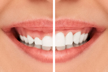
The genetic mismatch between the width of the jaw bone and the size of the teeth during human anatomical development leads to gaps between teeth that are expected to be next to each other. These gaps, known as diastema, create visual asymmetry, especially when located between the upper central incisors, and can disrupt the flow of air, causing phonetic interruptions in the pronunciation of sounds like ‘S’ and ‘F’. In cases where orthodontic wire or plate treatments are not preferred, the composite bonding method is an effective restorative tool that closes these gaps while preserving the tooth’s own tissue.
During the diastema closure process, the dentist does not remove any tissue from the front or back surface of the teeth. Special acid etching procedures are applied to the surfaces of the teeth facing each other (mesial or distal) to create micro-level retention areas. Afterwards, transparent bands (matrices) are used to add composite material in millimetric ratios to the sides of the tooth. While doing this addition, care is taken to ensure that the gum papilla (the triangular tissue between two teeth) is not crushed and that a proper emergence profile is created to maintain its healthy anatomical form. Thanks to the added material, the gaps are closed, but in order to maintain the balance of the tooth’s width-height (golden ratio), an equal volume of material is added to both teeth, which is a fundamental medical measure considered in establishing symmetry.
Why Does This Method Come to Fore in the Repair Process of Broken or Cracked Teeth?
Mechanical traumas, falls, sports injuries, or uncontrolled impacts against a hard object can cause micro-cracks (crack lines) or noticeable tissue ruptures in the enamel, which is the outer protective shield of the teeth. Particularly, these cracks occurring at the incisal edges of the anterior teeth expose the underlying dentin tissue, which is more vulnerable and susceptible to external factors, leading to sensitivity in the patient and aesthetic disturbance. Composite bonding is the only direct restoration method employed to replace the lost part of the tooth while sitting in the dentist’s chair.
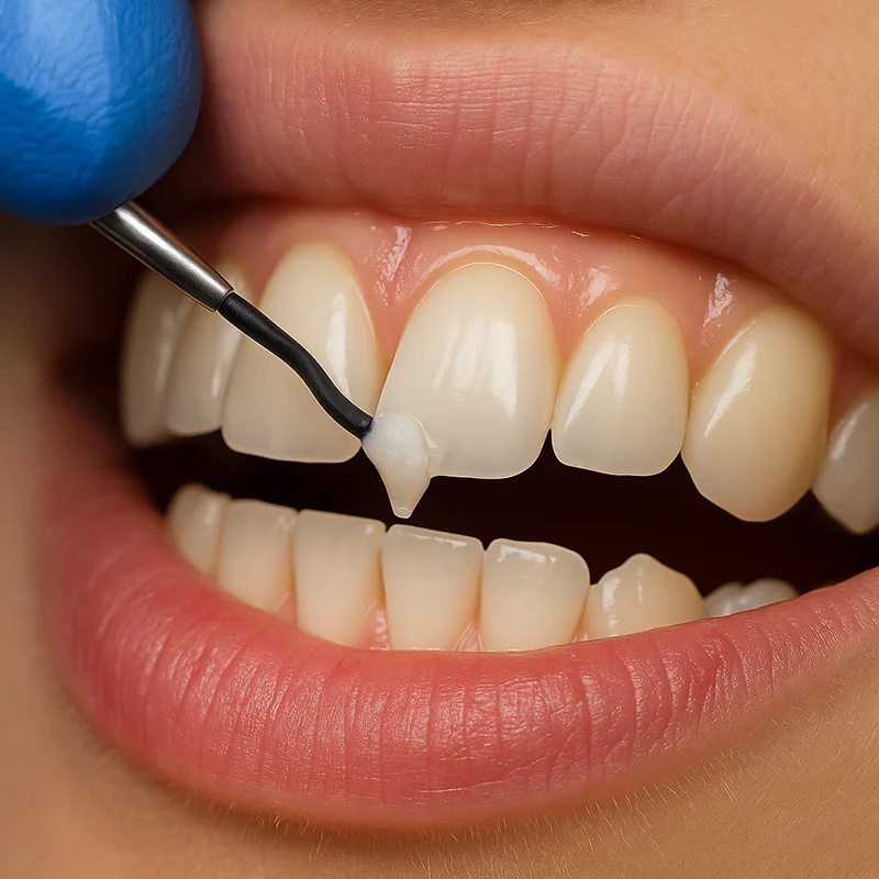
During the repair process, the sharp edges of the broken margin are lightly tapered to conceal the composite’s visible bonding line (margin) (bevel application). This tapering supports the invisible blending of the added material with the outer surface. In the layering technique applied to the broken area, the internal dentin layer is initially mimicked with opaque (less light-transmitting) composites, followed by the placement of more translucent enamel composites on the outer surface. Thus, when light strikes the surface of the tooth, a boundary line (demarcation line) between the repaired area and the natural tooth structure cannot be discerned; the material reaches the original optical depth of the tooth, ensuring organic integrity.
| Anatomical Issue | Application Function of Composite Bonding |
|---|---|
| Incisal Edge Fractures (Trauma) | Layered restoration of the broken area by creating a bevel (taper) and gaining volume through adhesive methods. |
| Enamel Cracks (Crack Lines) | Cleaning the surface of the crack line and sealing it with liquid resins that penetrate into it, preventing discolorations. |
| Abrasion Losses | Filling the abraded areas (cervical areas) with composites matching the tooth color to limit sensitivity. |
In Which Situations is Composite Bonding Applied to Mask Color Changes?
The discolorations in teeth fall into two main categories: External (extrinsic) discolorations are usually removed through surface cleaning procedures; internal (intrinsic) discolorations are permanent pigmentation that has occurred during the structural development of enamel and dentin tissues. Teeth that have experienced high doses of fluorine exposure (fluorosis) during childhood, certain antibiotics used (tetracycline derivatives), or teeth that have lost vitality after root canal treatment, exhibit dark brown, gray, or opaque white spots within their internal structure. These types of intense internal stains may not respond adequately to standard teeth whitening (bleaching) procedures.
Aesthetic Dentistry uses the masking capability of composite materials to cover these stains that cannot be lightened by whitening. Highly opaque resin components (opaquer) are placed in a very thin layer on the front surface of the tooth to block the reflection of the underlying dark or defective color. More transparent composite layers applied over this masking layer add vitality and brightness to the tooth. Unlike porcelain applications, disguising the stain without abrading the front surface of the tooth (or doing so at a microscopic level) is an important medical approach that supports the preservation of the vitality of dental tissue.
How Does Application Provide Clinical Contribution in Correcting Tooth Size Discrepancies?
The aesthetic harmony in a smile’s dynamics is not only dependent on the color of the teeth but also on the symmetrical relationship between the heights of adjacent teeth. Conditions such as teeth grinding (bruxism), acid erosion caused by acidic food consumption, or developmental conditions where adjacent teeth (laterals) are small and tapered like wedges (microdontia) can lead to an inconsistency in the appearance of the teeth. When the upper incisors are worn down, this results in a shortened vertical dimension of the face and an aged expression. If the teeth that are expected to follow the curvature of the upper lip during a smile are lined up unevenly or asymmetrically, it diminishes the perception of harmony on the face.
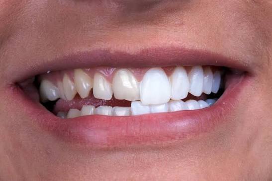
In cases of tooth length discrepancies, composite bonding is performed by adding material to the incisal edges of shortened or misshapen teeth, restoring the tooth to its original length. The dentist extends the teeth millimetrically based on the patient’s lip stretch allowance and the curvature of the lower lip (smile arc). The most critical aspect to consider during this lengthening process is that the added composite section does not come into hard contact with the lower teeth when the patient moves their lower jaw forward and sideways (eccentric movements). The occlusion dynamics are carefully adjusted so that the new form is not only visually satisfying but also functions without creating a mechanical barrier within the chewing system.
How Are Anterior Region Asymmetries Regulated with Composite Bonding in Smile Design?
The facial skeleton and smile structure contain minimal differences between the right and left sides as a natural necessity. However, when these differences reach the extent of the axial rotations of the teeth, one tooth being positioned more posteriorly than another, or the angles being different, an eye-catching picture of asymmetry emerges. In dentistry, the macro aesthetic rules require the vertical midline of the face to pass through the exact junction of the teeth and the horizontal plane to be parallel to the pupils.
In cases of slight misalignments or positioning of teeth that are set back, short-session composite additions are used as an alternative to the months-long orthodontic (braces/trays) treatment processes. Composite is placed on the front surface of the backward tooth in an appropriate volume, making the tooth optically appear more forward and aligning it with the adjacent tooth. At the same time, the lateral edges of the teeth (incisal embrasures) are shaped according to the facial form; while straight edges are created for individuals with a square face, in an oval facial structure, the corners of the composite are rounded to add an organic softness to the face. These optical illusions and volumetric adjustments provide significant medical support in masking asymmetries.
How is Dental Tissue Processed During Application According to Europadent Standards?
Clinical evaluations conducted in European dental management show that restorative procedures are carried out according to a specific protocol chain. The composite bonding procedure consists of a series of consecutive steps, typically completed in a single appointment, following the dentist’s accurate clinical analysis. Firstly, the color selection must be made under natural daylight before the tooth is dry; because the dehydrated (dry) tooth enamel’s light scattering property temporarily changes and appears whiter. Once the correct color (hue, chroma, value) is determined, the surfaces of the teeth to be treated are completely isolated from all moisture and saliva. This isolation, usually achieved with rubber sheets called “rubber dam,” is a fundamental clinical requirement that directly determines the quality of chemical bonding.
After isolation, phosphoric acid is applied to the tooth surface for an average of 15-20 seconds. This acid microscopically dissolves the enamel prisms, creating a rough, matte surface like a sponge. Once the acid is rinsed off and dried, the adhesive agent (bond) in a thin liquid form, which will penetrate the dentin and enamel tissues, is applied to the tooth and cured with light. Then, composite resins in the predetermined colors are layered onto the tooth using special hand instruments (spatulas) or fine brushes. The dentist shapes the tooth like an anatomical sculpture, working the surface details and developmental grooves into the material. The final and most decisive stage of the procedure is the polishing stage. Using special abrasives, rubber cups, and diamond-containing polishing pastes, the composite surface is polished until it reaches the smoothness of natural enamel. Properly performed polishing increases light reflection and reduces potential future food staining.
What Details Does the Daily Oral Care Routine After Application Rely On?
The ability of applied composite restorations to maintain their optical and mechanical properties over many years depends on the hygiene and usage habits in the home environment supporting the success achieved in the clinic. Since composite materials contain an organic polymer content, they are somewhat more permeable to external pigments compared to porcelain structures. Therefore, rinsing the mouth with water or brushing the teeth after consuming highly pigmented substances such as tea, coffee, red wine, or tobacco products is crucial to prevent superficial staining.
There are also limitations that need to be observed from a mechanical usage perspective. Although composite additions are solid structures produced with high technology, they should not be subjected to abnormal forces such as grinding hard-shelled foods like hazelnuts or walnuts with anterior teeth, or using teeth to open packages, or biting nails. Such excessive stress loads can lead to chipping at the edges of the material. Furthermore, in the brushing routine, it is recommended to prefer soft and non-abrasive fluoride toothpaste instead of excessively coarse (aggressive abrasive) whitening toothpastes that could scratch the composite surface microscopically. Regular visits to the dentist to renew the polish of the composite surface with professional tools are the most supportive step in extending the aesthetic lifespan of the application.
- Brushing Technique: Regular cleaning should be done using toothbrushes with medium or soft bristles and low abrasive toothpaste.
- Interdental Cleaning: Daily use of dental floss or an interdental brush should not be neglected to maintain the health of the gum tissue and the integrity of the composite.
- Para-functional Habits: Individuals with a history of nighttime teeth grinding (bruxism) should make an effort to use a night guard to protect their restorations from mechanical pressure.
- Stain Control: The consumption of dark-colored liquids should be kept under control, and clinical polishing should be performed at regular intervals (6-12 months).
What are Frequently Asked Questions?
1. How long does the composite bonding procedure take?
Depending on the number of teeth to be treated and the details of the modifications to be made, the duration can vary; however, the treatment of a single tooth generally takes between 30 to 45 minutes, and most cases are completed clinically in a single session.
2. Is anesthesia needed during the procedure?
When there is no deep excavation into the live tissue (dentin) or nerve layer (only surface addition), local anesthesia or numbing is not necessary, making this a process that can proceed quite comfortably for the patient.
3. Does the color of the applied composite material change over time?
Due to the polymer structure in its content, surface staining may occur over the years as a result of heavy consumption of tea, coffee, and tobacco; however, these stains can be easily removed and restored to their original brightness with a brief polishing procedure performed by the dentist.
4. Does closing the gaps between teeth affect speech?
Since the shapes of the teeth in diastema closure procedures are designed to be compatible with the contact limits of the lips and tongue (phonetic distances), issues such as slurring or pronunciation problems in the patient’s speech are avoided.
5. Can foods like apples be bitten with teeth that have undergone composite bonding?
Consuming natural foods (like apples and pears) with their normal biting force does not cause any problems; however, it is crucial to avoid abnormal forces such as biting into ice or hard-shelled nuts with the front teeth.
6. Will there be sensitivity in the teeth after the procedure?
Because it is a method that preserves a large amount of tooth structure, the development of permanent sensitivity to heat and cold after the procedure cannot be predicted; sometimes, experiencing mild sensations for a few days during the adaptation period is physiological.
7. How does composite material adhere to the tooth?
Special acids applied to the tooth enamel create microscopic pores on the surface; medical adhesives in liquid form penetrate these pores, cure under blue light, and ensure that the composite adheres to the tooth with a chemical lock.
8. Can composite bonding be applied to every tooth?
Composite bonding can be applied to the front or back teeth if the enamel tissue is sufficient, the loss of substance in the tooth remains within reasonable limits, and there are no excessive severe biting forces.
9. What is the difference between porcelain laminates and composite bonding?
Porcelain laminates are glass ceramics that are prepared in a laboratory environment, require multiple sessions, and have a higher resistance to staining. Composite bonding, on the other hand, is a more practical and protective option that the dentist applies in a single session in the clinic, without touching the tooth itself.
10. Do composite restorations wear down over time?
Depending on the brushing technique, the abrasive content of the paste used, and the patient’s chewing forces, there may be micro-level wear over very long periods; however, these areas can be easily repaired in the clinic with small composite additions.
11. Can composite bonding be applied to individuals who grind their teeth at night?
In patients with bruxism (teeth grinding), the composites applied may have a risk of breaking or chipping due to abnormal pressure during sleep; using a protective night guard after treatment is necessary for the longevity of the application in these patients.
12. Is it necessary to clean tooth calculus before application?
Creating a healthy boundary of the gum tissue and cleaning the tooth calculus before the procedure is a fundamental rule to stabilize the oral flora and prevent any fluids from the gum tissue from disrupting the adhesion of the composite to the tooth.
13. Does removing composite bonding damage the tooth?
In cases where there is no structural abrasion on the tooth during the procedure (in prepless techniques), the composite material can be safely returned to the tooth’s original natural state by the dentist using special polishing tools.
14. How often should routine dental check-ups be done after treatment?
It is recommended to have routine check-ups every 6 months, as is standard in general dentistry, to assess the edge fit of restorations, the surface gloss, and the health of the gums.
15. How is color matching ensured within Avrupadent?
In clinical procedures, the color codes of the patient’s natural tooth enamel and dentin layers are determined using special scales; the opacity and translucency values of the composite material are layered according to the different areas of the tooth, creating an organic harmony.