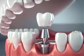
Is it possible to perform an implant years after tooth loss?
Implant treatment years after tooth loss is a medical procedure that can be applied if the individual’s jawbone volume and overall health condition are suitable. Although it is not a direct obstacle for treatment to have a long time pass since the tooth loss, the condition of the bone tissue in the area must be examined through radiological methods to create a personalized treatment plan. In necessary cases, additional surgical procedures aimed at increasing the bone quantity can make the area suitable for treatment.
The gaps left by teeth extracted a long time ago may undergo some anatomical changes over time. Among these changes are the resorption (melting) that occurs in the jawbone and the compensation of neighboring teeth into the gap. However, thanks to modern dental practices, even if many years have passed, the relevant areas are assessed based on detailed scans, and treatment options are determined. Not only the area where the missing tooth is located but also the dynamic structures of the patient’s entire mouth and systemic diseases are fundamental parts of this planning.
How is the treatment for a tooth that has been missing for many years evaluated?
The treatment to be applied for a tooth that has been missing for many years begins with the evaluation of the jawbone’s width, height, and density using three-dimensional dental tomography (CBCT). Based on the obtained data, the distances to nerve pathways and sinus cavities are measured to determine the size and angle of the titanium material that will be placed in the relevant area. This preliminary assessment is the most crucial stage of the process.
In the clinical examination performed, not only the bone structure is assessed, but also the thickness of the gum and the condition of surrounding tissues are examined. Years passing since tooth loss can lead to certain structural losses in surrounding tissues. Therefore, the assessment process requires a detailed analysis of the biomechanical capacity of the area rather than a standard examination. Based on the results of the analysis obtained, the patient is only informed and the steps of the process are explained with medical justifications.
What Anatomical Changes Occur in the Jawbone Years After Tooth Extraction?
In the years following tooth extraction, due to the inability of the chewing force to be transmitted to that area, cellular activity in the jawbone decreases, and horizontal and vertical bone resorption occurs. Additionally, anatomical changes such as sagging towards the base of sinus cavities, as well as bone resorption in the posterior regions of the upper jaw, and the nerve canal approaching the bone surface in the lower jaw are observed.
Bone tissue tends to maintain its volume due to the functional loads imposed on it. When a tooth is lost, this load is eliminated, and the body begins to gradually resorb the bone tissue in that area. This is a biological adaptation process. Understanding the time-dependent development of the related changes is important for the quality of the assessment to be made. The table below summarizes the possible anatomical changes that can occur during the process after tooth loss based on stages for informational purposes.
| Process / Time Frame | Expected Biological Changes | Anatomical Outcomes |
|---|---|---|
| First 6 Months | Improvement of jaw deficiency and onset of bone formation. | The period during which the fastest bone volume loss occurs and the tissue is remodeled. |
| Between 1 – 3 Years | Narrowing in the crown (bone peak) width and slight decreases in the vertical direction. | The adjacent teeth starting to tilt towards the deficiency. |
| 5 Years and Over | Severe bone loss, sinus sagging, and narrowing in the nerve distance. | Asymmetric overloads caused by the shift of chewing forces to other areas. |
How is the treatment process planned for individuals with observed jawbone loss in İzmir?
Why might a bone graft (bone powder) be necessary in treatments performed years later?
A bone graft may be necessary to support, expand, and make suitable a jawbone that has lost its volume and density as a result of long-term toothlessness, either horizontally or vertically. It is a medical requirement that healthy bone tissue of a certain thickness surrounds the titanium roots so they can be completely stabilized within the bone and withstand functional chewing forces.
The graft materials used are special materials that are biocompatible with the human body and promote bone formation (osteogenesis). The grafts applied during this procedure gradually become enveloped by the body’s own bone cells, taking on a form similar to original bone tissue. In the upper jaw, particularly in the area where molars are located, the procedure of pushing up the sinus membrane and filling it with bone powder from below (sinus lifting) is among the frequently applied methods. The primary purpose of these procedures is to create a healthy foundation.
What is Sinus Droop and How Does It Affect Upper Jaw Procedures?
Sinus droop is the condition where the bone in the area left empty after the loss of teeth in the back region of the upper jaw melts, thereby narrowing the bone volume as the maxillary sinuses, which have air spaces, expand downward. This anatomical change may necessitate planning an additional surgical procedure called ‘sinus floor elevation’ because it reduces the bone height needed for interventions on the upper jaw.
The sinus floor elevation procedure can be performed using two different methods, open and closed. The choice of method depends on the amount of existing bone. The procedure is carried out after numbing the relevant area with local anesthesia. Supporting anatomical structures is implemented not only for the structural integrity of the treatment but also as a safety protocol to prevent damage to the airways or adjacent tissues.
What Should the General Health Criteria Be for Older Adults and Long-Term Tooth Loss?
When planning treatment for older adults or long-term tooth loss, it is fundamental that the individual’s systemic diseases such as diabetes, hypertension, osteoporosis, and heart disorders are under control. The use of medications that affect blood coagulation values and bone metabolism is evaluated; procedures can be applied to individuals who are regularly monitored for these diseases and have received doctor approval.
It is very important to share the detailed medical history with the dentist. In particular, patients using bone-density-regulating medications or blood thinners must consult their own doctors. Age alone does not constitute an obstacle; the crucial factor is biological age and overall systemic health condition. Coordinating with general medical disciplines prior to surgical interventions in the field of oral and dental health is a standard procedure to minimize the risk of complications.
- Controlled Diabetes: Regular fluctuations in blood sugar levels are important for tissue healing.
- Bone Metabolism: The conditions of those using osteoporosis medications need to be evaluated through consultation between doctors.
- Cardiovascular Health: Blood pressure and coagulation times affect local anesthesia and surgical procedure durations.
- Radiotherapy/Chemotherapy History: Since radiotherapy treatments applied to the head and neck region can affect bone quality, special evaluation is required.
How is the Jaw Surgery and Treatment Process Carried Out Within Avrupadent?
In the clinics of Avrupadent, maxillofacial surgery processes are carried out within the framework of a multidisciplinary approach that includes obtaining the patient’s medical history, reviewing radiological examinations, and conducting a detailed intraoral examination. Based on the findings detected, a specific protocol is established for the patient, with each intervention performed under sterile clinical conditions, according to the principles of Implantology science, and implemented after obtaining the informed consent of the hospital.
The clinical procedure starts with the local anesthesia of the relevant area and progresses with the preparation of sites at the planned positions and openings and the placement of materials. After the procedure, the patient is given detailed written and verbal instructions on nutrition and hygiene rules to follow during the healing period. Regular use of prescribed medications and participation in the specified follow-up sessions are the patient’s responsibility to ensure the process progresses as expected.
What to Expect in the Healing Process of the Procedure Performed After Years?
The healing process after the procedure encompasses an average time frame of 2 to 6 months for the integration of bone with the titanium surface (osseointegration), depending on the body’s biological structure. In cases where additional bone grafting procedures are performed, this period may extend somewhat. It is normal to observe slight swelling or sensitivity in the area during the first few days as a tissue reaction.
The first week of the healing process involves the soft tissue recovery of the surgical area. During this period, it is necessary to adhere to the dietary restrictions recommended by the dentist and to avoid extremely hot/cold foods. The main healing inside the bone occurs at the cellular level and cannot be felt by the patient. The completion of the healing process will be measured through radiological controls and clinical stability tests performed by the doctor.
| Recovery Phase | Expected Condition | Important Notes for the Patient |
|---|---|---|
| First 24 – 48 Hours | Mild discomfort (swelling) related to the surgery site and tissue sensitivity. | Application of ice compress, taking the prescribed medications on time, avoiding clenching and moving around excessively. |
| First Week | Significant healing of soft tissue (gum) and sutures coming together. | Avoidance of hard and grainy foods, protection of the area from trauma while brushing. |
| 2 – 6 Months | Continuation of the osseointegration (bone bonding) process at the cellular level. | Regular check-ups with the doctor should not be neglected, maintain high general oral hygiene. |
What Issues Can Be Observed If Problems Are Not Addressed in Areas Left Without Teeth for a Long Time?
If dental areas are left empty for a long time, anatomical deformities can be observed such as neighboring teeth tilting into those spaces, the opposing tooth elongating into the empty space, and the surrounding jawbone gradually resorbing. Additionally, this situation can lead to uneven distribution of chewing forces on the jaw, paving the way for temporomandibular joint (TMJ) discomfort and digestive issues.
The dental system operates within a biomechanical integrity where all components support each other. The absence of a link in the system increases the load on the other teeth. Developing a one-sided chewing habit when eating can lead to wear on the teeth on the used side and asymmetrical development of the facial muscles. For informational purposes, the structural conditions that are frequently encountered in long-term tooth loss situations are listed below:
- Loss of Function: Inability to perform the chewing process can lead to insufficient mastication of food.
- Aesthetic Changes: Changes in the facial profile can occur due to the collapse of the cheek and lip tissues, especially in cases of loss in the anterior region or multiple posterior regions.
- Phonetic (Speech) Problems: Tooth deficiencies may cause difficulties in the pronunciation of certain letters.
- Sensitivity in Neighboring Teeth: Excessive sensitivity can be observed due to the exposure of the root surfaces of tilted or elongated teeth.
How Do Current Imaging Technologies Contribute to Treatment Planning in Implantology?
Current imaging technologies such as 3D Volumetric Tomography and digital intraoral scanners aid in accurately detecting bone thickness, nerve pathways, and vascular routes with millimeter measurements. The digital data obtained allows for simulation of the treatment in a computer environment, providing a safe guide that minimizes surgical risks in Implantology applications.
While traditional two-dimensional X-ray (panoramic) films provide limited information about bone structure in some cases, three-dimensional systems allow examination of the tissue from every angle. Using surgical guides allows for planned openings and depths to be transferred directly from digital media to the patient’s mouth. These devices assist in managing the duration of the operation while supporting a respectful approach to surrounding tissues.
How Should Oral and Dental Hygiene Be Maintained After Treatment is Completed?
After the treatment process is completed and the upper structure prostheses are placed, oral hygiene should be maintained by brushing with fluoride toothpaste at least twice a day, using an interdental toothbrush, and dental floss. The use of oral rinses is a supportive measure to prevent food accumulation in the surrounding tissues, and a professional clinical examination should be performed at least once every 6 months.
Although the titanium material itself does not decay, the surrounding gum tissue (peri-implant tissue) is susceptible to bacterial plaque accumulation. Inadequate oral hygiene may lead to infections known as peri-implantitis, which can cause bone resorption, similar to gum inflammation seen in natural teeth. When long-term use is aimed, the patient’s personal oral care is one of the structural elements of the medical procedures performed.
What Should Individuals Wanting to Get Implants Years Later Know Before Their First Check-Up?
Individuals wishing to receive treatment after years should inform their physicians of their medical history, current blood tests, lists of medications they regularly use, and any dental X-rays from previous years in a complete manner during their first check-up. This information is essential for the doctor to healthily analyze the area’s bone dynamics, the rate of bone resorption, and the biological responses the patient can provide to surgical procedures.
Each person’s jaw and chin anatomy is unique like a fingerprint. Therefore, the experiences of those around should not be considered a reference for the hospital’s own situation. The roadmap that will emerge from the assessment by European dentists will be completely based on the individual’s current radiological and clinical findings. Proper information and a transparent evaluation process are essential for taking healthy steps.