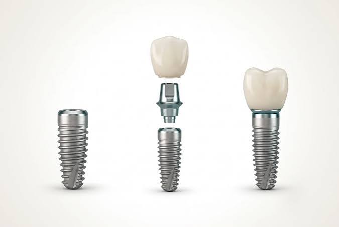
How is Bone Insufficiency Evaluated in Implant Treatment in Izmir?
Bone insufficiency in implant treatment in Izmir is evaluated by measuring the width, height, and density of the jawbone in millimeters using three-dimensional dental tomography (CBCT). This radiological analysis is combined with the patient’s medical history and systemic health condition to provide objective medical data about whether the bone can support titanium material or not. Identifying the anatomical boundaries in the relevant area (nerve canals, sinus cavities) and creating the structural map of the bone constitutes the most fundamental phase of the surgical treatment protocol to be applied.
The procedure of placing medical artificial roots into the jawbone to compensate for tooth deficiencies is entirely based on the cellular foundation provided by human anatomy. The more robust the foundation of a structure is, the longer the stability of the structure built on it lasts; in dental practice, this “foundation” is the patient’s own living jawbone (alveolar crest). However, prolonged edentulism, physiological aging, traumas, or periodontal diseases can lead to the volumetric resorption of this foundation. The true expertise of the physician is not merely to place a material in every empty space but to analyze scientifically whether there is healthy bone tissue sufficient to surround that material. In current medical protocols practiced in the Izmir location, individuals with insufficient bone volume are not seen as untreatable; rather, medical plans are implemented to reconstruct the area according to the degree of this insufficiency (augmentation).
What Technologies are Used for Jawbone Analysis in Izmir Avrupadent Clinics?
The jawbone analysis at İzmir Avrupadent clinics is performed with millimetric precision using three-dimensional dental volumetric tomography (CBCT) devices and digital intraoral scanners. Thanks to these technologies, not only the outer surface of the bone but also the density of the trabecular (spongy) structures within is examined, allowing for transparent surgical planning in a digital environment.
In a clinical examination, the dentist can only observe the patient’s gum from the outside or estimate the bone thickness by touch. However, the gum tissue (mucosa) often hides the underlying thin or resorbed bone structure like a thick cover. In the Avrupadent procedure, radiological imaging devices are at the heart of the diagnostic process, in line with health regulations standards of 2026. Digital devices display tissue depths that are unreachable by the human eye or traditional methods in cross-sectional views. With these collected digital data, the physician can visually explain the patient’s existing bone profile and present the dimensions of bone insufficiency with objective evidence.
Why Is Three-Dimensional Dental Tomography (CBCT) Essential for Bone Assessment?
Three-dimensional dental tomography (CBCT) is essential for bone assessment as it presents the bone thickness (width) and the actual positions of anatomical structures (nervous pathways, air cavities) with depth perception, which traditional two-dimensional X-rays cannot show. These devices document bone insufficiency in millimetric detail before surgical operations, defining the medical safety limits.
Panoramic X-rays, commonly used in dentistry, show the entire jaw as if it were on a flat piece of paper in two dimensions. While the vertical length of the bone can be seen from above and below, the thickness from front to back can never be known. However, the jawbone generally loses width rather than height (it thins like a knife blade). The tomography device scans the jaw in horizontal, vertical, and cross-sectional slices and transfers the data to the computer. The titanium root is digitally placed within this virtual bone model to test whether it remains exposed outside. If the bone thickness is below 1-2 millimeters, the necessity of using bone powder (graft) is definitively determined.
| Evaluation Criteria | Panoramic X-ray (2D) | Dental Tomography / CBCT (3D) |
|---|---|---|
| Bone Width (Thickness) | Cannot be seen. Only height can be estimated. | Measured in millimeters, horizontal resorption can be clearly detected. |
| Position of Anatomical Structures | Tissues overlap (superimpose), depth can be misleading. | The nerve canal and sinus membrane can be tracked in three-dimensional space with precise positioning. |
| Bone Density | Presents insufficient data about bone quality (hardness). | The trabecular (spongy) tissue of the bone is analyzed for hardness using Hounsfield units. |
What Biological Reasons Lead to Jawbone Resorption?
Bone resorption in the jaw; arises due to the absence of stimulation of bone cells (osteoblasts) following tooth extraction, due to the elimination of chewing pressure in the region, advancing age, prolonged use of removable prostheses (denture), and biological reasons such as destructive inflammation in the bone tissue caused by periodontal diseases (periodontitis).
Tissues in the human body function on the “use it or lose it” principle. A healthy tooth sends vibrations (mechanical stress) to the jawbone through the periodontal fibers around its root during each chewing motion. This vibration stimulates bone-forming cells (osteoblasts) and helps maintain bone volume. This stimulation is halted the moment the tooth is extracted. The body then begins to resorb the bone cells in that now non-functional area. This resorption process, which starts in the first 6 months, deepens both horizontally and vertically over the years. Additionally, the prolonged placement of a removable (clip-on) denture in the area of the missing tooth accelerates bone resorption by creating a disproportionate pressure and compression on the bone through the soft tissue.
What are the Anatomical Differences Between the Upper Jaw and Lower Jaw Bone Structures?
The upper jawbone (maxilla) is spongy, softer, and contains air spaces (sinuses), while the lower jawbone (mandible) is much denser and harder (cortical) because it is attached to movable muscles, and it has a central nerve canal passing through it. This difference in density is a decisive anatomical factor in evaluations of bone deficiency and cellular healing processes.
Dental surgery in jaw surgery works in two different realms. The lower jaw is a dynamic bone where chewing muscles generate high forces. To withstand these forces, the evolutionary dental shell (cortical layer) is very hard and thick. The bone insufficiency experienced in the lower jaw is usually vertical, and the distance to the inferior alveolar nerve below is the greatest risk factor. The upper jaw, however, is a static bone fixed to the skull. Its structure resembles a sponge (trabeculae). When teeth are lost in the back regions of the upper jaw, not only does the bone resorb; at the same time, the sinuses, which are air cavities, sag downward, narrowing the bone volume in a bi-directional manner. These differences necessitate that the surgical technique be specifically chosen according to the region.
What Should Be Considered to Preserve Bone Volume After Tooth Extraction?
To preserve bone volume after tooth extraction, attention should be paid to performing the extraction procedure in a non-traumatic manner without damaging the bone walls, ensuring the extraction site (socket) is free from infections, and if anatomical conditions permit, placing medical titanium material in the same session as the tooth extraction (immediate protocol) or filling the area with grafts for healing purposes.
When a tooth extraction decision is made, the doctor plans not only to remove that tooth but also to protect the socket that will remain in that area. If excessive pressure is applied to the bone during extraction with forceps or elevators, and if the thin bony wall holding the tooth breaks, it creates a residual cavity in that area. In current medical approaches, the extraction socket is cleaned carefully. If a future treatment is planned for that area, medical sponges or bone powders that support blood clotting are placed in the cavity, and the gum is stitched up. This protective protocol (socket preservation) is applied with the aim of keeping the ideal bone foundation ready for the main surgical intervention to be performed months later.
Why Are Bone Width and Height Critical Medical Constraints in Implantology Applications?
Implantology applications require a minimum of 1-2 millimeters of healthy bone wall to fully surround the titanium material to be placed in the jaw, making bone width and height a critical medical constraint. Without sufficient bone volume, exposure of the outer surface of the material can lead to tissue infections (peri-implantitis) and pave the way for failure of the medical procedure in the long term.
According to biological principles, when a medical titanium root is placed in the jawbone, it must transmit the chewing forces (functional load) equally to the surrounding bone. It is expected that the bone area where a structural body with an average diameter varying between 3.5 to 4.5 mm will be placed has a minimum width of 6-7 mm horizontally. If the bone is very thin and the grooves of the material are left exposed during the surgical procedure (dehiscence or fenestration), the gum cannot cover the exposed metal. This situation prepares the ground for bacteria to seep directly into the bone. Therefore, bone thickness is an unbendable anatomical rule.
What Medical Alternatives Are Considered When Bone Volume is Insufficient?
When bone volume is insufficient, medical alternatives such as adding biocompatible bone powder (graft) in the area, expanding the tissue through bone splitting techniques, elevating the sinus floor, or using the patient’s existing healthy bone areas through open placement (e.g., All-on concepts) are presented to the patient based on clinical and radiological evaluations.
In the modern dental protocols implemented in Izmir, bone insufficiency is not considered an insurmountable condition. If the patient’s systemic health permits, the physician may opt for reconstructing the missing anatomy (augmentation). If there is a horizontal constriction, the bone is stretched in half, and material is placed in between. If there is a vertical insufficiency, the area is elevated using special block grafts. However, if the patient is of advanced age or unable to withstand the trauma that supplementary bone surgeries may cause due to systemic diseases, the physician will adjust the process based on existing bone using open surgical concepts (like All-on-4) or extra short and narrow structural options to suit the patient.
| Type of Insufficiency | Surgical Technique Applied | Medical Purpose of the Procedure |
|---|---|---|
| Horizontal (Transversal) Thinning | Guided Bone Regeneration (GBR) / Bone Stretching | Supporting the thin bone wall with biocompatible powders and membranes to expand outward. |
| Vertical (Longitudinal) Resorption (Upper Jaw) | Sinus Lifting (Sinus Floor Elevation) | To gain vertical distance by pushing up the lower hanging air cavity’s floor and filling it with bone tissue. |
| Vertical (Longitudinal) Resorption (Lower Jaw) | Use of Short Materials or Block Grafting | To avoid damaging the lower jaw nerve, use short implants or elevate the area with block bones. |
| Advanced Resorption in the Back Areas | All-on (Open System) Concepts | Avoiding surgery in the back and placing implants at 30-45 degree angles in the healthy bone in the front area. |
How are Bone Graft Applications Integrated into the Treatment Process?
Bone graft applications involve the placement of biocompatible materials from human, animal, or synthetic sources in the insufficient jawbone region during or before surgical procedures, covered with barrier membranes, and a waiting period of approximately 4 to 6 months for the body’s own bone cells to integrate (ossification).
Bone powders (grafts) used in dentistry are, contrary to popular belief, not directly hard pieces of bone. They are cellular scaffolds with porous and microscopic structures. Once these scaffolds are placed in the surgical area, the body’s blood vessels start to invade into these pores (angiogenesis). The healing cells (osteoblasts) carried by the blood wrap around this artificial scaffold with live bone tissue they produce. Over time, as the externally added material is resorbed, it is completely replaced by the patient’s own living bone. Since this transformation is a biological process, it cannot be accelerated from the outside; the patient must only adhere to the waiting period determined by the doctor.
- Autogenous Grafts: These are live bone blocks taken from another part of the patient’s own jaw (the highest biological compatibility).
- Xenografts: Medical powder particles that are completely purified from animal (usually bovine) sources.
- Allografts: Sterile medical grafts obtained from human tissue banks.
- Alloplastic Grafts: Fully synthetic materials based on calcium phosphate or hydroxyapatite produced in a laboratory environment.
What is Sinus Lift and How Does it Limit Upper Jaw Treatments?
Sinus lift is a condition in which the maxillary sinus (air) cavities expand downward due to prolonged toothlessness in the back areas of the upper jaw, resulting in bone resorption that narrows bone volume. This anatomical change limits the standard schedule of treatment by necessitating additional surgery called “sinus floor elevation” for the vertical bone distance required for surgical interventions on the upper jaw.
Maxillary sinuses are physiological cavities located on both sides of the nasal cavity that humidify and warm the air we breathe. In young individuals, there is a thick bony barrier between the roots of the lower molars and the base of these cavities. However, when the molars are extracted, the gravitational effect and the thrust force of air pressure from inside begin to cause the sinus cavity to sag into the jawbone (pneumatization). As a result, the bone thickness may decrease to as much as 1-2 millimeters. Placing a titanium root into a bone this thin can damage the sinus membrane, so the physician first pushes this membrane upwards with special tools (using open or closed techniques) and fills the void beneath it with bone powder. An average healing timeline of about 6 months is expected for the related area to regain strength.
How Do Systemic Diseases (Diabetes, Osteoporosis) Affect Bone Quality?
Systemic diseases affect bone quality at the cellular level; diabetes slows down the formation of new bone cells (healing) by disrupting blood circulation, while osteoporosis (bone loss) reduces the inner density of the jawbone (trabecular structure), decreasing the initial adhesion (primary stability) resistance of the titanium structure during surgery. The presence of these diseases necessitates evaluating bone insufficiency not only in terms of quantity but also “quality”.
The success of a surgical intervention depends on the healing cells (cellular response) related to the area where blood has been transferred. In diabetic patients with uncontrolled (high HbA1c levels), high sugar levels in the blood disrupt the structure of capillaries (microvascular system). This situation can lead to insufficient nutrition of the operated bone and make it susceptible to bacterial infections. On the other hand, osteoporosis is a disease where the rate of bone destruction accelerates rather than bone cell production. While the jawbone may appear massive from the outside in osteoporosis patients, it is observed in tomography that its internal structure is filled with large holes (like a sponge). The physician uses methods to create a socket by compressing the bone in these patients and conducts a multidisciplinary consultation with an internal disease specialist to prevent the side effects of the bone protective (bisphosphonate derivative) medications the patient is using.
How is the process monitored while waiting for bone healing?
The process while waiting for bone healing is monitored through the protection of the surgical area from trauma, nutrition with a soft diet, and clinical and radiological stability checks performed by the physician in the first and sixth months. Since the integration of the titanium surface with jawbone cells (osseointegration) is an invisible biological reaction, the process is objectively monitored through radiological (X-ray) measurements.
In the planned Implantology processes at Avrupadent clinics, patients are not left without follow-up after surgery. If bone grafting has been performed, no chewing force or pressure from removable temporary prosthetics should reach that area in any way. The physician checks the color of the gingiva, the state of swelling, and how bone graft particles integrate with the existing bone on X-ray films during scheduled check-ups at specific intervals. After confirming the completion of the fusion and ensuring medical stability with devices (torque test), a green light is given for the prosthetic (tooth) manufacturing phase.
How Should Patients Prepare for a Medical Process in Case of Bone Insufficiency in İzmir?
Patients diagnosed with bone insufficiency in İzmir should prepare for the medical reality that their treatment process will take longer than standard procedures, that additional bone surgeries (grafting or sinus lifting) will be performed, and that the cellular healing phase (osseointegration) may take months. At Avrupadent clinics, this process is conducted according to ethical standards, providing patient consent forms, transparent medical information, and radiological evidence.