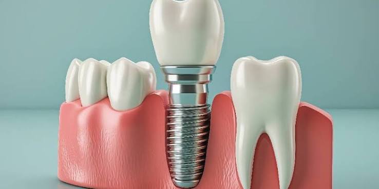
Is a check-up necessary after the completion of implant treatment?
A check-up after completing implant treatment is medically essential to maintain the long-term functionality and tissue health of the new biomechanical system formed by the titanium material placed into the jawbone and the porcelain prosthesis made over it. Even if surgical repair and prosthesis steps are complete, the stabilization of bone tissue, the tension of internal mechanical screws, and the health of surrounding gum tissue can only be objectively monitored through regular dental check-ups.
The procedures applied in dental surgery enter a new biological stage not when the treatment ends, but when the patient actively starts to use that area. The human jaw is a dynamic area, constantly moving, influenced by the mechanical forces created by muscles and rich in bacterial activity. The most significant difference between artificial roots placed in the jawbone and natural teeth is the absence of a stimulating and protective connective tissue (periodontal ligament) around them. This anatomical difference makes artificial roots more sensitive to mechanical pressures and plaque accumulation. Therefore, after the permanent porcelain teeth are fixed in the mouth, the patient does not stop needing clinical follow-ups; on the contrary, this initiates protective dental care that will preserve the success of the treatment for years.
Why are routine check-ups after implant treatment in Izmir a medical necessity?
In routine checks at the clinic in Izmir, it is medically necessary to detect early invisible loosening in the internal mechanical parts that support the prosthesis, to professionally remove bacterial plaque that cannot be cleaned with a toothbrush around the gum borders, and to balance the dynamic changes in jaw closure (occlusion) over time. A small biomechanical deviation in the system, if not addressed, can lead to significant tissue losses.
Medical prostheses do not break or become inflamed on their own; however, the biological foundation supporting them (mucosa and bone) is directly affected by changes in the oral cavity. At the Izmir Avrupadent clinics, it is clearly emphasized during patient consent and information processes that follow-up checks after treatment are not an optional choice but an integral part of the procedure. Natural teeth can wear down over the years and may undergo small positional changes. However, the titanium body, rigidly attached to the jawbone, and the zirconium or porcelain structure on it cannot adapt to this change. This situation causes an uneven chewing load to be placed on the artificial tooth over time. The force imbalance, which can be adjusted by the doctor with just a few minutes of alignment (adjustment) procedure, cannot be felt by the patient and can only be detected during routine checks.
When Should the First Check After the Prosthesis (Upper Structure) Delivery Be Made?
The first clinical check after the delivery of the prosthesis (upper structure) should generally be performed at the end of the first month to monitor the patient’s adjustment process to the chewing function and anatomical adaptation with the new teeth. During this early check session, the cellular response of the gum to the newly placed porcelain crown is observed, and the occlusal contacts during eating are verified with specific articulation tests.
The first several weeks after the prosthesis is delivered is the process of adaptation of the tissues to the new mechanical situation. The patient applies initial chewing force with permanent porcelain teeth for the first time after months of using a temporary prosthesis or experiencing toothlessness. During this adaptation phase, the lip and cheek muscles get accustomed to the shape of the new teeth, while the gum adjusts to the lower edge areas of the covering (emerging profile). During the first month examination, the physician checks for any food trapping under the prosthesis, even if the patient has no complaints. This initial follow-up is a critical medical observation that determines the long-term trajectory of the system.
| Control Phase | Focused Clinical Findings | Physician’s Medical Intervention |
|---|---|---|
| 1st Month Control (Early Phase) | Closure balance, cheek biting complaints, gum edge harmony, condition of interface cleaning. | Adjustment of early contacts with occlusion (chewing) paper, enhancement of hygiene education. |
| 6th Month Control (Mid Phase) | Mechanical screw stability, radiological bone level, plaque and tartar formation. | X-ray bone analysis, dental calculus cleaning if necessary, internal screw torque control. |
How Are Long-Term Implantology Follow-ups Organized in Terms of Periodicity?
Long-term implantology follow-ups are organized at standard intervals of six months or annually, depending on the patient’s systemic health history, oral hygiene habits, and the number of implants in the jaw. These regular intervals ensure the early detection of pathological bone loss or mechanical incompatibilities without causing physical harm to the patient.
Medical literature and the 2026 preventive dentistry protocols require individuals who have undergone implant treatment to have a professional examination at least once or twice a year. In treatments that have completed their first year, the system is considered mechanically and biologically stable. However, this does not mean that the patient should be left to their own devices. Especially in cases where full jaw rehabilitation (All-on-4 or All-on-6 concepts) has been applied, or in systems holding removable dentures, wear may occur over time in plastic and teflon components. During six-month appointments, these small connection parts are renewed, and the retention of the dentures is restored to the initial level.
How Are Jawbone and Gum Evaluated in Check-up Sessions?
During check-up sessions, the jawbone is measured in millimeter levels with periodically taken radiological X-rays (periapical or panoramic films); the gums are evaluated in detail by using medical periodontal probes to monitor tissue pocket depth, possible bleeding points, and color changes.
The doctor does not only examine the visible part during the clinical examination. An X-ray is essential to understand the relationship between the neck part of the titanium root (collar area) and the bone. In the X-ray images, the absence of any dark area (radiolucency) around the titanium grooves proves that the bone is healthy. During the gum examination, a fine millimetric measuring tool called a “probe” is gently placed in the gap between the prosthesis and the gum. In healthy tissue, this depth typically ranges from 1-3 mm and there is no bleeding during the measurement. An increase in depth and the observation of bleeding (BOP – Bleeding on Probing) is the clearest clinical indicator that bacterial plaque has led to inflammation in the area.
Why is the Occlusal Force (Closure Balance) Measured Again in Controls?
The occlusal force (closure balance) is re-measured during controls because natural teeth undergo physiological changes by either slightly shifting over time or by wearing down their enamel, while the porcelain crown over the titanium root remains static and unwearable; this situation may lead to destructive and disproportionate pressure (early contact) on the porcelain prosthesis over the years, which can harm the underlying bone structure.
The dynamic of the jaw is based on the principle of 28 or 32 teeth working in a tremendous balance with one another. The connections that attach the natural teeth to the jaw allow these teeth to flex and adapt at the micro level. However, the medical titanium material that is integrated (welded) into the bone has no such flexibility. As years pass, due to millimetric physiological wear occurring in the patient’s own teeth, when the patient closes their jaw, the first contact starts to intensify on the porcelain prosthesis. This condition, called ‘occlusal trauma,’ may lead to vertical resorption in the bone or breakage of internal mechanical screws. The doctor identifies these pressure points using colored heating papers during control sessions and restores overall balance by polishing (filing) the areas with excessive pressure millimetrically.
How Can We Understand That There Is Loosening in the Internal Screws Holding the Prosthesis?
Loosening in the internal screws holding the prosthesis can be understood if the patient feels a slight clicking, a sense of dislocation, or hears a ‘crack’ sound during the act of biting, and during the clinical examination, it is determined through the doctor’s manual or instrumental application of rotational (circular) stability tests and the detection of micro-gaps between the parts in radiological examinations.
Screw or construction (implant) systems, porcelain teeth attached to the structure inside the bone are subjected to continuous vertical and lateral mechanical pressures. Especially in patients with the habit of teeth grinding or clenching (bruxism) during sleep, the connection screws (abutment screws) may lose their torque (tightening strength) over time due to extreme lateral stresses. The loosening of the screw does not necessarily mean that the titanium root inside the bone has failed. This is entirely a prosthetic, mechanical situation. When the patient notices the loosening, they should avoid playing with the tooth constantly with their tongue and urgently consult a physician. The physician can solve the problem in seconds by opening the prosthesis (or removing it if it is a construction) and bringing the internal screw to standard tightening level with special medical torque wrenches.
- What the Patient Observed: A sound when eating, slight rotation or vertical movement of the porcelain when pushed with the tongue.
- Clinical Findings: The porcelain bends between two dental devices, leakage or redness at the limit of the gum tissue.
- Radiological Findings: The gray gap that appears on the taken periapical film between the titanium root and the connecting part (abutment).
Does Home-Based Individual Oral Care Replace Routine Clinical Checks?
Home-based individual oral care, while extremely vital for tissue health and compulsory for the sustainability of treatment, can never replace routine clinical checks because it cannot clean the hardened dental plaque accumulating under the microscopic borders where the prostheses merge with the gums, nor detect radiological changes inside the bone.
Regular brushing, using an interface brush and floss successfully removes soft bacterial plaque from porcelain surfaces and the accessible limits of the gums. However, due to human nature, anatomical blind spots, or the effect of all-on systems and similar full-arch prosthetics, mineralized hard plaques (tartar) can develop over time. These hard deposits cannot be removed with home toothbrushes. Moreover, due to their porous structure, tartar holds more bacteria, triggering gum inflammation (mucositis). During clinical examinations, the physician completely cleans the tissue using special tools (plastic, Teflon, or carbon-based) that do not harm these prosthetics and ultrasonic devices. The two processes are not competing but are complementary steps in maintaining treatment.
How is Peri-implantitis (Surrounding Tissue Infection) Detected in the Early Stage?
In the early stage of peri-implantitis infection; the appearance of slight or spot bleeding from the gum pocket during the dentist’s examination (BOP), the color of the mucosa changing from pink to red-purple tones, and the detection of millimetric bone resorptions around the grooves (implants) in the obtained radiological sections indicates the presence of the condition.
In cases of periodontitis (gum disease) observed in natural teeth, patients typically notice pain, sensitivity, or gum recession. However, due to the absence of nerve fibers around titanium roots, patients rarely feel any pain until the peri-implantitis infection progresses and causes serious bone loss. This is the most insidious aspect of this silently progressing disease. The initial stage of the infection, known as “peri-implant mucositis”, only affects the gums and can be completely healed with professional cleaning. However, the situation becomes serious once the infection penetrates the bone. The only way to catch the disease while it is still at the mucosal level is through periodic check-ups, regardless of complaints.
| Clinical Picture | Mucositis (Early Stage – Reversible) | Peri-Implantitis (Advanced Stage – Destructive) |
|---|---|---|
| Tissue Involvement | Only the soft gum tissue (mucosa) is affected. | The infection has not penetrated the bone cells. |
| Radiological Findings | No erosion is observed at the jawbone level on X-ray. | Crater-like vertical erosions are observed in the bone surrounding the material. |
| Symptoms | Minor bleeding during brushing or probing. | Deep tissue pockets, sometimes with inflammation (pus) drainage, and rarely exudation. |
| Treatment Approach | Clinical surface cleaning, mouth rinse, and improved home hygiene are sufficient. | Surgical bone cleaning, grafting, or removal of material may be necessary. |
How Do Systemic Diseases (Diabetes, etc.) Affect Control Frequency?
Systemic diseases; especially diabetes (sugar disease) that disrupts microvascular circulation, osteoporosis that alters bone metabolism, or autoimmune disorders, increase the frequency of monitoring as these conditions reduce tissue resistance to bacterial attacks and slow down the repair capacity, requiring the patient to be followed up more frequently in three or four-month intervals instead of the standard bi-monthly periods.
The immune system of a healthy individual can combat the small bacteria plaques surrounding the prosthesis. However, in a diabetic patient experiencing fluctuations in blood sugar (HbA1c), infections spread rapidly because the capillaries beneath the gum are unable to carry sufficient immune cells. Additionally, there is a risk of bone necrosis (death) from any pressure or infection on the jawbone in elderly patients using osteoporosis medications (bisphosphonates) or oncology patients who have undergone radiation therapy. A proactive (preventive) health policy is followed for individuals with these medical risk factors. In Avrupadent processes, these patient groups are placed in “high-risk follow-up” files, and their gum and bone health is professionally monitored under strict short intervals, with visual and radiological examinations.
How Are Control Appointments Planned at Avrupadent Clinics in İzmir?
Control appointments at İzmir Avrupadent clinics are systematically planned by being recorded in automation systems through personal medical files created on the day surgical and prosthetic treatment steps are completed, in accordance with the 2026 health legislation monitoring standards, and by automatically notifying the patient through the hospital’s communication channels when the time comes (such as the 1st, 6th, and 12th months).
It may not always be possible for patients to remember a medical check-up appointment months later within their daily life. Therefore, patient follow-up should not be left to chance in clinical operations or to the hospital’s initiative. The foundation of this healthcare service provided in the İzmir location lies in the responsibility of maintaining post-treatment integration. As the appointment date approaches, the necessity of the check-up is reminded to the patient using institutional communication language. The new digital data and radiological images taken from the patient who comes for examination are evaluated with transparent evidence to determine if there are any anatomical or mechanical deviations by overlaying them with the initial day data archived.
What is the Contribution of Regular Doctor Visits to the Implantology Process?
The contribution of regular doctor visits to the Implantology process is to support the biological and mechanical lifespan of the chewing system formed by the titanium structure placed in the jawbone and the porcelain prosthesis on it, to protect the anatomical structure by addressing any small tissue sensitivities or mechanical issues before they grow, and to continually keep the individual’s personal oral hygiene motivation high.