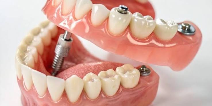
Does plaque form when using implants?
Plaque can form when using implants because even if the porcelain or zirconium coverings on artificial roots placed in the jawbone do not structurally decay, the minerals and bacterial plaque in the mouth combine to form hard dental calculus (tartar) on the surfaces of these prostheses and along the gum line. If regular and specific oral care is not performed, these calcified deposits that accumulate on the prosthetic surface can create a ground for destructive infections that require medical intervention in the surrounding gum tissue and supporting bone structure.
There is a common misconception in the general public that because artificial materials (titanium, porcelain, zirconium) do not decay like natural teeth, they do not require maintenance or will not form tartar on them. From a medical biology perspective, the situation is entirely different. The oral cavity is a dynamic and moist environment that hosts hundreds of different species of bacteria, enzymes, and minerals. The residues of the food we consume combine with these bacteria to create a structure conducive to adhering to all dental surfaces. Regardless of whether it is a natural tooth or a prosthetic, tartar formation on any surface that is not cleaned is a biological and chemical inevitability. Therefore, for a treatment that has been successfully completed with surgical and prosthetic steps to remain healthy for years, the patient must fully comply with daily hygiene disciplines that will prevent tartar formation.
How does plaque accumulate on titanium and porcelain surfaces?
Bacterial plaque on titanium and porcelain surfaces accumulates by forming an invisible and sticky biofilm layer through the combination of microorganisms from the oral flora and food residues. If regular mechanical brushing is not performed, minerals like calcium and phosphate from the plaque chemically deposit into this soft biofilm layer, turning it into hard tartar that cannot be removed with a home toothbrush.
The biofilm layer (plaque) begins with the placement of bacteria on a transparent film called pellicle that starts to reform on the teeth and prosthetics just a few hours after brushing. Regardless of how highly medical restorations like porcelain or zirconia are fired and polished, they are always susceptible to bacterial retention at a microscopic level. Especially, the areas where the prosthetic meets the gums (cervical areas) and the lower parts of bridges are blind spots that the natural cleaning mechanisms of the floss and tongue cannot reach. The soft plaque layer that colonizes these areas starts to mineralize (calcify) within 48 to 72 hours. Once plaque turns into tartar, it becomes porous, serving as an incubator for more bacteria to attach to that area much more quickly.
How Do Individuals in Izmir Notice the Formation of Dental Tartar Around Prostheses?
Individuals in Izmir may notice the formation of dental tartar around prostheses through clinical signs such as small bleeding in the gums during brushing or cleaning, yellow-brown hard exudates felt by the tongue at the lower parts where coverings meet the gums, the mucosa color changing from pink to red-purple tones (inflammation), and the formation of a persistent bad odor in the mouth (halitosis).
In the patient follow-up processes at İzmir Avrupadent clinics, it is frequently emphasized that dental calculus typically does not cause discomfort in the early stages, presenting mainly as pain or sensitivity. Patients can often overlook these attachments with the thought, “There’s no problem with my tooth, it doesn’t hurt.” However, bleeding in the gums around porcelain teeth is actually the body’s first and clearest medical signal of an inflammatory response to bacteria. Healthy gums do not bleed during brushing or flossing. The plaque under invisible prosthetics can only reveal itself through a persistent bad breath caused by gases produced by bacteria. If any of these clinical signs are noticed, it is essential to seek professional evaluation to prevent the condition from progressing to bone loss.
| Clinical Sign (Symptom) | Physiological Cause |
|---|---|
| Bleeding During Brushing (BOP) | Caused by the bacterial plaque irritating the gums and increasing vascularity in the tissue (inflammation). |
| Change in Color of the Gums to Red/Purple | The healthy pink color changes to darker shades due to increased blood flow associated with infection. |
| Persistent Bad Breath (Halitosis) | Production of sulfide gas by anaerobic (oxygen-depriving) bacteria within the dental calculus. |
| Physical Hard Deposits | Crystallization of calcium in the plaque layer at the borders of prosthetics due to deposition (mineralization). |
What Is the Difference Between Tartar Formation Around Natural Teeth and Medical Roots?
The fundamental difference in tartar formation between natural teeth and medical roots lies not in the surface structure of the tooth but in the biological defense mechanism of the surrounding gum tissue. The flexible connective tissue (periodontal ligament) surrounding the natural tooth creates a robust defense against bacteria through the blood circulation, whereas the medical titanium, directly sourced from bone, lacks this protective connective tissue; this leads to a much more rapid tissue destruction as plaque can turn into infection.
When we look at a natural tooth in the mouth, there are periodontal fibers that act almost like a shock absorber and contain dense blood vessels between the bone and the tooth root. These fibers immediately transport defense cells to the area when bacteria enter the gum pocket. Even if tartar forms, this defense line can slow down the resorption of the bone for a certain period. However, a titanium body implanted in the jawbone integrates cell-to-cell (without any fibers in between) with the bone. The surrounding gum tissue of the titanium does not have as many blood vessels and defense cells as in a natural tooth. Therefore, a dental implant formed around a prosthetic tooth faces a much weaker resistance compared to a natural tooth, and bacteria progress towards the jawbone much more swiftly. This anatomical difference proves why hygiene around implant areas should be taken even more seriously than with natural teeth.
What Medical Problems Can Tooth Tartar Buildup in the Implant Area Cause?
The accumulation of foreign material around the implant primarily leads to inflammation of the gum tissue (peri-implant mucositis), and if this inflammation is not treated by the practitioner and progresses, it causes cellular resorption of the jawbone supporting the titanium implant (peri-implantitis). This infection scenario leads to mechanical loosening of the structure due to loss of bone support, ultimately resulting in medically unsuccessful surgical intervention (tissue rejection).
Dental stones (calculus) are not just a layer of physical and unsightly dirt; they are a toxic structure filled with bacteria, porous like a sponge at the microscopic level. These stones cause pressure into the mucosal pocket where the prosthesis combines with the gums. Bacteria continuously secrete acid and enzymes, tearing the weak attachment between the gum and prosthesis apart. When the gum becomes inflamed, ulcers and pockets deepen. Unless cleaned, the process reaches the jawbone. Bone cells (osteoblasts) undergo resorption in an attempt to escape these bacterial toxins (or, as a defensive response, to sacrifice the tissue). The bone around the root that is eroding in a crater shape can no longer support the medical material. This clinical scenario summarizes the consequences of a preventable hygiene problem leading to irreversible tissue loss.
What is Peri-Implantitis (Surrounding Tissue Infection) and How is it Related to Dental Calculus?
Peri-implantitis is a destructive surrounding tissue infection caused by bacteria in dental calculus that accumulates due to insufficient oral hygiene, eroding the jawbone around the titanium root in a crater shape. Dental calculus serves as a reservoir that is porous, safe, and unreachable by the toothbrush for bacteria, forming the primary source of this infection and accelerating the progression of the most serious biological complication encountered in the discipline of Implantology.
The progression of the disease is two-fold. The first stage, peri-implant mucositis, only involves the soft tissue (the gums). At this stage, there is still no damage to the jawbone; the gums are red, swollen, and prone to bleeding. If the dentist cleans the tartar at this stage, the disease can heal completely, and the tissue returns to its original healthy pink form. However, if neglected, the second stage, peri-implantitis, comes into play. The infection has now spread to the jawbone, and X-rays clearly show the bone eroding vertically. At this stage, cleaning the tartar alone is not sufficient; the dentist needs to surgically open the gums to disinfect the bone and often reconstruct the area with additional bone powders. The only way to protect bone tissue is to prevent tartar from settling in this area.
How Should At-Home Care for Dentures (Upper Structure) Be Conducted?
At-home care for dentures involves gently rinsing the gums with soft-bristled brushes at least twice a day, ensuring not only to clean the accessible tooth surfaces but also to apply special thick dental floss and interdental brushes consistently to the joining areas of the dentures. Additionally, using medical mouth rinses that slow plaque accumulation should be included as part of the routine.
The long-term safety of the new biomechanical chewing system established in the human jaw depends entirely on the quality time spent in front of the hospital’s sink. Prosthetics (especially bridges involving multiple teeth or full-arch All-on concepts), which have a more specific geometry compared to natural teeth, cannot be thoroughly cleaned with a standard toothbrush. The purpose of mechanical brushing is to remove food debris from the tooth surface; however, the real danger lies in the bacterial plaque that hides in the fine grooves where the tooth meets the prosthesis (the sulcus area). Therefore, the toothbrush should be used at a 45-degree angle to the gum line rather than horizontally to the tooth, with the bristles lightly massaging into the gum pocket (modified bass technique).
| Care Step | Tools to be Used | Medical Justification |
|---|---|---|
| Surface Cleaning | Soft or Medium Firm Toothbrush | To remove the soft food biofilm from wide surfaces without scratching the porcelain polish. |
| Bridge Base (Body) Cleaning | Special Sponge Thread (Superfloss) | To clean plaque from the lower spaces where connected covers contact the gum. |
| Interface Cleaning | Appropriately Sized Interface Toothbrush | To distribute the plaque that the bristles cannot reach in the triangular spaces where two teeth are adjacent. |
| Chemical Support | Doctor Approved Medical Mouthwash | Slowing down the reproduction rate of free bacteria in the flora where mechanical cleaning is insufficient. |
Does the use of dental floss and interdental brushes prevent plaque accumulation?
The use of dental floss and interdental brushes significantly prevents plaque accumulation by physically removing food debris and soft bacterial plaque from tight (triangular) spaces where prostheses contact neighboring teeth or other coverings. This interim area cleaning, done with proper technique, is the most effective mechanical defense that halts biofilm formation in the blind spots where tartar tends to form, without giving a chance for its accumulation.
There is often a misconception among patients that “the use of dental floss is only for natural teeth.” However, the small gaps between adjacent porcelain restorations are quite conducive to food retention, just like in natural teeth. Traditional dental floss cannot pass over interconnected (bridge-type) prostheses, so specially produced spongy dental floss with an end as firm as fishing line (easily maneuverable) should be used. The stiff end of the floss is threaded beneath the bridge, while the spongy part is gently pulled back and forth against the area where the prosthesis contacts the gum. Interdental brushes, in regions with larger gaps that have opened due to gum recession, can be employed once a day to ensure that the area remains spotless (plaque-free) by inserting and removing the brush head into that space.
Is the use of a water flosser effective against tartar formation?
Using a water flosser helps to effectively remove soft bacterial plaque (biofilm) from the deep pockets under prostheses that toothbrushes or floss cannot physically reach, as it utilizes the pulsating (intermittent pressure) force of water. However, a water flosser cannot remove formed and hardened tartar; it is merely a powerful supportive hygiene tool that prevents mineralized plaque from turning into tartar.
In fixed prosthetic rehabilitations (All-on-4 or All-on-6 systems), the back and lower parts of the prostheses, which are made to fit with the lips, are usually quite thick. Toothbrush bristles or dental floss may not always effectively reach these depths. Water flossers introduce the water jet’s pressure into the gum pocket, washing away food residues and anaerobic bacteria (which thrive in oxygen-free environments) using fluid dynamics. Adding some antibacterial mouthwash along with water in the water flosser increases the chemical effectiveness of the process. The water pressure should be applied at a more horizontal and sweeping angle rather than perpendicularly (90 degrees) to avoid high pressures that could force the gum away from the prosthesis (causing trauma).
How is Dental Tartar Cleaning (Detartrage) Performed in a Clinical Setting?
Dental tartar cleaning (detartrage) in a clinical setting is conducted by a specialist physician using special non-abrasive medical tools (scalers) made of teflon, carbon, titanium, or plastic that will not damage the porcelain structure of the prosthesis and the surface of the underlying titanium root, as well as frequency-adjusted ultrasonic devices. This professional procedure completely removes mineralized (hardened) deposits that the patient could not clean at home with regular brushing, thus stopping the inflammation.
During routine check-ups, the inside of the hospital’s mouth is examined using special instruments (loops) or cameras. When hardened calculus (tartar) is detected at or just below the gum line, the physician initiates a cleaning procedure. The goal of the procedure is not only to break off the stone but also to sterilize that area from bacteria. Ultrasonic instruments, by vibrating at high speed and simultaneously jets of water, cause the stone to break apart at a microscopic level the moment it makes contact and separate it from the surface. If the tissue pocket is deeper than normal (if there is initial bone loss), the inside of the gum pocket is completely cleaned through a local anesthetic (curettage procedure), and the area is rendered free of impurities, paving the way for the gum to reattach to the prosthesis in a healthy manner.
Can Cleaning Methods Applied to Natural Teeth Be Applied to Prostheses?
Classic metal-tipped (steel) curettes and high-power standard ultrasonic cleaning methods applied to natural teeth cannot be directly used on prostheses; because hard stainless steel instruments can scratch the smooth neck area of medical titanium and create microscopic scratches that disrupt the glaze layer of the porcelain surface, making it much easier for bacteria to adhere in the future. Therefore, cleaning should be performed with material-specific, tissue-friendly medical equipment.
The enamel layer of the tooth is the hardest tissue in the human body and is quite resistant to small abrasions that can be caused by steel instruments. However, titanium bodies and connecting parts (abutments) placed in the jawbone are produced in a laboratory setting with a smooth (machined surface) to prevent bacterial adhesion. If the dentist scrapes this area with a hard metal tool, that smooth surface gets scratched. A scratched surface means a protected harbor for bacteria; once plaque settles on those scratches, it can never be cleaned out with a toothbrush and becomes a source of infection. Based on this scientific reality, specially designed carbon or Teflon-tipped hand tools and specialized piezo-electric cleaning devices are used during the cleaning of prosthetic bases.
| Cleaning Feature | Application for Natural Teeth | Application for Titanium/Prosthetic Structures |
|---|---|---|
| Hand Tool Tip Material | Stainless steel or hard metal instruments. | Carbon, Teflon, plastic, or pure titanium-tipped instruments. |
| Surface Reaction | The enamel layer does not get scratched due to its hardness and remains intact. | Metal tools scratch the titanium surface and damage porcelain glaze. |
| Ultrasonic Vibration | Can be applied safely at standard or high frequencies. | Applied at low frequencies, using special silicone-tipped instruments or piezo devices. |
Why Should Routine Check-Ups at Avrupadent Clinics Not Be Neglected?
Routine check-ups at Avrupadent clinics should never be neglected to ensure that tartar buildup under the gum line and potential early signs of infection are detected with medical tools and radiological examinations, and professionally cleaned before any damage to the tissue (bone loss) occurs. Success in the discipline of Implantology relies not only on personal home care but also on professional preventive dentistry follow-ups conducted every six months.
At Avrupadent, located in Izmir, the end of treatment is considered not a graduation, but the beginning of a new healthy lifestyle routine. Due to human nature, regardless of how carefully one brushes, small mineralizations (tartar formations) can occur in the anatomical blind spots of the mouth. The patient does not feel these stones and cannot notice the damage to the tissue (bone loss) because it does not cause pain. During the six-month routine check-ups, the doctor measures the jawbone level with radiological X-rays, checks the bleeding index of the gum, and removes any fresh tartar without harming the tissue. Additionally, by testing the tension of the internal mechanical screws carrying the prosthesis, it prevents loosening. These regular visits are the most effective medical insurance for keeping the investment in treatment healthy for years.
How Do Systemic Diseases (Diabetes, etc.) Affect Tartar and Plaque Formation?
Systemic diseases, especially diabetes, increase the sugar level in the saliva, slowing down the flow rate of saliva, leading to much quicker, stickier, and denser accumulation of bacterial plaque on prosthetic surfaces. In these systemic conditions, where the response of the vascular system is weakened, even the slightest tartar formation can trigger a much more severe inflammatory reaction in the surrounding gum.
Any systemic disease in the mouth directly affects the microbial balance (flora) within it. In diabetic patients, the increase in glucose levels in the blood also raises it everywhere else, creating a feast-like environment for bacteria, as they require sugar to thrive. Additionally, because diabetes disrupts the microvascular structure, it diminishes the gum’s ability to defend itself. A slight tartar buildup, which could be easily managed with mild redness in a healthy individual, can quickly lead to deep tissue pockets and accelerated bone resorption (peri-implantitis) in a diabetic patient. Therefore, individuals with chronic conditions should be twice as meticulous about plaque control compared to healthy individuals and should increase the frequency of clinical control periods (for example, reducing the interval from six months to four months) as a medical necessity.
How Do Tobacco Products and Smoking Affect Oral Tissue Health?
Tobacco products and smoking impair the blood supply (oxygenation) of the gums by narrowing the microvessels in the mouth due to the toxic chemicals and nicotine they contain, weakening the defense mechanism and accelerating bacteria’s attachment to the tooth (leading to tartar formation). The reduction in blood flow masks early infection signs, like gum bleeding (symptoms of mucositis), causing the disease to insidiously progress directly into the jawbone.
The gums of a smoking patient do not appear pink like those of a healthy individual; instead, they might look pale or grayish due to lack of oxygen. An inflamed gum that normally should bleed during brushing does not bleed due to the vasoconstrictor effect of nicotine. The patient misinterprets this lack of bleeding as “my gums are healthy” and delays visiting the doctor. However, there may already be massive tartar accumulation beneath the gum, eroding the bone. Moreover, smoking significantly thickens the plaque structure, reducing the mouth’s mechanical cleaning capacity and drastically accelerating the process of calcium depositing and turning into tartar. Continuing to smoke after jaw surgery is the most dangerous environmental factor accelerating biological damage.
How to Develop Long-Term Hygiene Habits in Implantology Treatments?
Long-term hygiene habits in treatments are achieved through the patient’s full understanding of the medical nature and maintenance requirements of the structure placed in their jawbone, thanks to the clear, evidence-based information provided by the physician, learning proper brushing and interface cleaning techniques through practical application in clinical sessions, and integrating oral care as a mandatory medical discipline into their daily routine with patience.