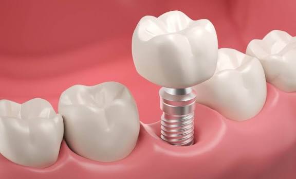
Why is the gum important in anterior dental implants?
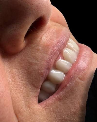
The gum in anterior dental implants is extremely important because it creates an aesthetic framework (pink aesthetics) that ensures the porcelain or zirconium crown appears in natural harmony with the symmetry of the lips and face. Additionally, it serves as a biological barrier that protects the medical titanium material placed in the jawbone from bacterial factors in the mouth. The natural anatomical form of the junction between the tooth and gum directly affects both the visual success of the application and the long-term health of the tissue.
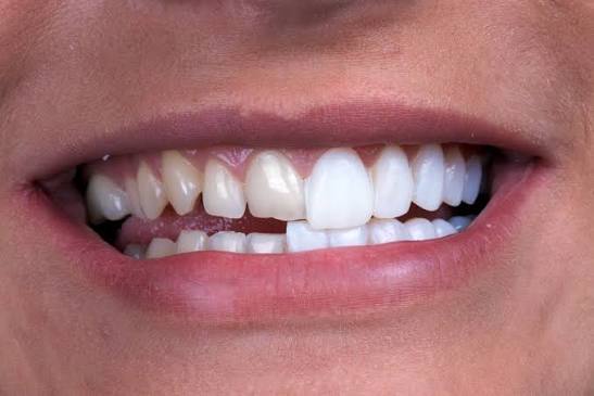
In compensating for dental deficiencies, especially in the front incisor area, which is immediately noticeable from the outside, treatment cannot be deemed complete by merely replacing the missing hard tissue (tooth). The human eye focuses not only on the whiteness of the teeth when evaluating a smile but also on the color, thickness of the gum surrounding those teeth, and the existence of the tissue called papilla that fills the triangular spaces between the teeth. Even if a highly successful porcelain tooth is made over a titanium root, if the surrounding gum appears retracted, grayish, or asymmetrical, that tooth will look artificial from outside.
What does pink aesthetics mean in planning anterior dental implants in İzmir?
In the planning of anterior dental implants in İzmir, pink aesthetics refers to the healthy gum surrounding the new tooth being a light pink color, having a textured appearance (like orange peel), and achieving millimetric symmetry with the gum levels of neighboring natural teeth. Pink aesthetics is considered the most fundamental visual basis that ensures the natural and organic appearance of white aesthetics (the tooth itself).
In the clinics of İzmir Avrupadent, while evaluating patients, the “smile line” is examined with special attention. For individuals with a high smile line, meaning those whose gums are prominently visible when smiling, the perfection of pink aesthetics carries critical importance. If there is a 1-millimeter asymmetry (either above or below) compared to neighboring teeth at the gum level, this creates a mismatch that draws the attention of the opposite party during speaking and smiling. To ensure this harmony, before surgical procedures, doctors analyze the existing level of the gums through three-dimensional digital scans and photographs, creating a tissue shaping (crown contouring) map towards the final goal.
| Aesthetic Component | Medical / Visual Feature | Condition Encountered in Absence |
|---|---|---|
| Gingival Papilla | It is the soft tissue filler in a triangular shape between two teeth. | If the papilla is lost, conspicuous dark areas called “black triangles” appear between the teeth. |
| Cervical Line | It is the curved boundary line where the tooth meets the gum. | If asymmetrical, one tooth appears longer or shorter than the other. |
| Tissue Color and Thickness | The tissue should have a healthy light pink color and be thick enough to conceal the underlying material. | In thin tissues, the underlying titanium material reflects, appearing as a gray/purplish spot on the gum. |
How Does Gum Biotype (Thickness) Affect Anterior Region Treatments?
The gum biotype (thickness); the risk of gray titanium pieces placed in thin gums reflecting from the outside is high, and due to the tendency of the tissue to show recession in response to surgical procedures or trauma from brushing, it directly influences the surgical approach for anterior region treatments, the base materials to be used, and the waiting periods.
In the dental literature, gums are fundamentally categorized into two classes: “thick biotype” and “thin biotype.” Gums with a thick biotype are anatomically denser in fibrous structure, making them resistant to dental trauma; they do not easily recede and are quite successful in masking the color of the underlying medical materials. However, gums with a thin biotype (closer to translucent) are extremely fragile. To avoid the metal color reflecting through a thin gum, dentists usually prefer white zirconium abutments instead of titanium ones. Additionally, a protective medical method frequently employed for patients with a thin biotype is transferring soft tissue grafts taken from the palate area to increase the thickness of the gum during surgical procedures (connective tissue graft).
What Anatomical Changes Occur in the Gum After Tooth Extraction?
After tooth extraction, the gum undergoes structural anatomical changes such as volume loss parallel to the natural resorption of the underlying jawbone, vertical recession (upwards), flattening of the triangular papilla form between the teeth, and overall shrinkage of the tissue. Once the support of the tooth root is removed, the soft tissue loses its former three-dimensional dome shape.
A natural tooth not only serves to chew food but also acts as a mechanical scaffold (support) for both the jawbone and the surrounding tissue from within. When a tooth is extracted from the front area, the body’s first physiological response is to close the wound. As the wound heals, the gum tissue gets crushed. During the months-long healing period, especially due to the resorption of the thin bony wall facing the lip, the gum also migrates inward (toward the palate) and upward. As a result, a significant tissue crater (deficiency) occurs in that area. If this tissue loss is not accounted for and supported during treatment planning, the new porcelain tooth to be placed in the cavity will have to emerge disproportionately long and artificial compared to the adjacent natural teeth.
What Methods are Used to Protect the Gum During Surgical Procedures?
To protect the gum during surgical procedures; techniques that avoid extensive tissue opening (flap raising), flapless surgical methods, immediate surgical protocols where titanium material is placed in the same session as tooth extraction, immediate application of temporary prosthetics, and the use of digital surgical guides are employed to maintain tissue support (atraumatic techniques).
The best way to protect the gum tissue is to cause it as little surgical trauma as possible. In traditional methods, the jawbone is exposed by making a wide incision in the gum tissue, which leads to the gum being peeled away from the bone. This peeling process (disruption of periosteal blood supply) forces the gum tissue to return to its former shape and accelerates bone resorption to some extent. In clinics where modern technologies are applied, thanks to digital tomographies, the bone structure is known in advance in three dimensions, so the procedure can be performed by making a small hole just large enough for the material to enter without cutting the gum. Additionally, placing an artificial root immediately into the socket created when the tooth is extracted ensures that the gum tissue remains taut with a new support before it has a chance to collapse. These protective approaches are crucial for preserving pink aesthetics.
- Flapless Surgery: Large incisions are not made in the gum tissue, minimizing the risk of retraction since the blood supply to the tissue is not disrupted.
- Immediate Placement: By immediately providing support to the extraction site, it prevents the tissue collapse and the flattening of the papillae.
- Soft Tissue Grafting: In necessary situations, the gum tissue is thickened by adding connective tissue simultaneously.
How Do Healing Caps (Gingiva Formers) Shape the Gum Tissue?
Healing caps (gingiva formers) are cylindrical medical pieces that are attached to the gum surface after the titanium root biologically integrates with the jawbone (osseointegration) and they physically guide the tissue to heal in a circular and healthy form, creating a natural collar (band) around this cap, preparing the tissue for the placement of the porcelain tooth.
When the bone marrow healing is completed, the titanium root that remains covered will be opened with a millimetric intervention. Various titanium caps produced in different diameters and lengths will be screwed in to prevent the gum tissue from closing unevenly in the opened area. The body tissue possesses an extraordinary adaptability; the gum will take the shape of this round cap within a few days and tightly wrap around it like a sealing gasket. Generally, these caps, which remain in the mouth for one or two weeks, leave behind a flawless and healthy mucosal cavity reminiscent of the natural tooth neck when they are removed on the day the porcelain crown will be placed. The correct shaping of this cavity is the most important anatomical barrier that prevents bacteria from leaking into the lower parts while brushing teeth.
How Does Temporary Tooth Use Contribute to Gum Shape Formation?
The use of temporary teeth contributes to the formation of gum shape; by applying a sweet and controlled pressure from the inside of the temporary tooth’s underside (extraction profile) prepared in the laboratory, it directs the tissue like a sculptor, forces the flattened papillae to return to a triangular shape, and provides a unique biomechanical contribution by creating the most ideal, natural anatomical cavity for the permanent porcelain tooth.
Especially in anterior area treatments, standard cylindrical healing abutments may not provide sufficient aesthetic results. This is because the root section of a natural anterior tooth is not perfectly round, but rather oval or triangular. To achieve this natural shape for the gum, the dentist places a temporary acrylic or composite crown on the patient’s mouth. Over the weeks, millimetric additions or reductions are made to the part of this temporary tooth that remains within the gum. The gum reacts to these adjustments and gets the desired growth or shape. This process is called “emergence profile”. Once the targeted perfect gum curves are achieved, a three-dimensional measurement of this form is taken and transferred to the laboratory, where the permanent zirconium tooth is produced to fit precisely into this prepared mold.
What Causes Gum Recession in Implantology Treatments and How Can It Be Prevented?
Implantology gum recession occurs due to factors such as erosion of the bony wall facing the lip (buccal bone), hard and incorrect brushing habits, insufficient support of the thin gum biotype, or the titanium root being placed anatomically too far out (towards the lip). To prevent this complication, it is crucial to perform accurate surgical positioning with three-dimensional digital analyses and, when necessary, to strengthen the area with bone powder (graft) and soft tissue transfers during the surgery.
Gum tissue is above the underlying bone. When there is bone resorption in the jawbone, the gum tissue follows that resorption, moving downwards (or upwards). Especially in the anterior teeth, since the bony layer facing the lip is very thin, it is crucial that the titanium structure does not remain outside (in the open) during surgical procedures. If the material is positioned even one millimeter outside the ideal axis, the mechanical pressure on that thin bone will gradually destroy it over time; when the bone is lost, the gum tissue retracts and the gray metal neck underneath the prosthesis appears. To prevent this situation from happening from the beginning, doctors aim for an accurate opening using surgical guide plates. Even though patients vigorously brush horizontally with hard toothbrushes after treatment, this can irritate the tissue mechanically and cause retraction; learning the proper brushing (scrubbing) technique is among the primary protective measures.
| Reason for Retraction | Physiological/Mechanical Explanation | Medical Measures Taken |
|---|---|---|
| Misplacement | Placing the material too close to the lip thins the bone. | Millimetric placement with three-dimensional digital surgical guides. |
| Insufficient Bone | The bone with a very thin wall begins to resorb. | Thickening the wall with biocompatible bone grafts (powder). |
| Mechanical Trauma | Brushing horizontally and vigorously with hard toothbrushes. | Using soft brushes and the scrubbing technique directed from tooth to gum. |
Why Are Zirconium Crowns Advantageous in Terms of Gum Compatibility?
Zirconium crowns have a significant biological and aesthetic advantage in anterior region aesthetics because, unlike traditional metal alloys, they are tissue-friendly (biocompatible), do not cause allergic reactions or irritation in the gum, and do not result in a gray/purple shadow at the gum line due to their light permeability.
In older generation porcelain teeth, gray metal frameworks were used to ensure durability. These metals reflected a dark shadow outside the thin gums and could even cause minor tissue irritations (redness) related to the metal in some patients. Zirconium dioxide (ZrO2) is a medical material that provides the resistance of porcelain but is completely white and does not oxidize. Gum cells establish a highly compatible contact with the zirconium surface and do not trigger any inflammatory reactions in the tissue. In anterior region surgeries, choosing a zirconium (or zirconium-coated titanium) for the abutment, which connects the root in the jawbone to the crown, is one of the most important prosthetic choices to maintain the fullness of pink aesthetics.
How Are Gum Levels Balanced in Aesthetic Smile Design?
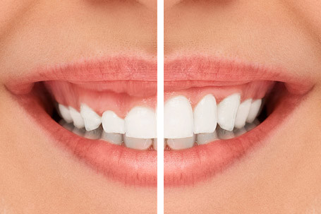
In aesthetic smile design, gum levels are balanced by designing the relationship of the anterior teeth with the lip through photographs digitally (mock-up), shaping asymmetric gum levels millimetrically with laser or cautery devices (gingivectomy/gingivoplasty), and aligning the lengths of the new crowns to this balanced foundation to achieve a symmetrical structure.
Gum design is akin to an architectural restoration process. If the tissue of the patient’s upper front incisor is more than 2 millimeters lower than the tissue of the left front incisor, even if the lengths of these teeth are made the same, one tooth will appear shorter than the other. The dentist identifies these level differences before starting the surgical procedure. During the gum design, the position of the lip line is taken as a reference (zenith points), and the curves of the gums are redrawn. If necessary, minor tissue shaping is done to ensure all tooth silhouettes are parallel and symmetrical. The titanium bodies to be placed in the jawbone are positioned at depths that fully match this aesthetic level, preventing the porcelain structure from exceeding the natural tooth length (appearance of a long incisor).
How is the Health of the Anterior Region Gum Evaluated in Avrupadent Clinics?
In Avrupadent clinics, the health of the anterior region gum is evaluated through the use of medical periodontal probes in the patient’s examination to measure tissue depth and bleeding index (BOP), identifying the biotype of the gum (its thickness) through clinical observation and digital photographs, and by conducting an objective risk analysis by correlating any existing dental calculus and infection foci with radiological findings.
In Avrupadent’s processes in the Izmir region, the understanding of “only looking at the bone to perform the procedure” has been abandoned, and the overall health of the tissue is prioritized. Performing direct surgical intervention on an area without healthy gum, with significant plaque accumulation, bleeding, and lesions (gingivitis), greatly increases the risk of complications. Before surgical planning, doctors completely eliminate the infection in the area and ensure the tissue returns to a tight, pink, non-purulent, and healthy form. In cases with an initially biotype, this situation is shared with the patient in a transparent manner, aligning possible aesthetic expectations with biological realities.
How Should Home Care Be to Protect Gum Health After Treatment?
To protect gum health after treatment, home care should include gentle rinsing at least twice a day with soft or medium-firm toothbrushes that do not harm the gums, the use of interdental brushes and thick (spongy) dental floss daily as recommended by the dentist to remove bacterial plaque from the surfaces of porcelain prostheses. The use of mouthwashes to rinse away residues in the tissue pocket is also a strong protective measure.
The delicate junction where the gum and porcelain tooth meet is the area where bacteria (plaque) most prefer to accumulate. If brushing only occurs on the visible surfaces of the teeth, the gum line cannot be cleaned, and the bacteria there can produce acid, leading to gum inflammation. Infected tissue swells, bleeds, and may lead to an infection that progresses toward the jawbone (peri-implantitis). To prevent this, the toothbrush should be angled at 45 degrees to the gum pocket and small vibrations should be used to dislodge the plaque. Interdental brushes should definitely be added to the routine as a complement to mechanical cleaning for residues left in the gaps that the toothbrush cannot reach.
What is the Role of Regular Clinical Check-ups in the Implantology Process?
Implantology involves the role of regular clinical check-ups to monitor the biological balance of the newly installed system in the jawbone and the surrounding gum tissue, to remove stubborn tartar accumulations (which cannot be cleaned at home) from under the gum line using professional devices to prevent potential infections before they start, and to early diagnose and take precautions against mechanical deviations in chewing forces. Long-term aesthetic and functional integrity is ensured through these periodic preventive medical follow-ups.