Why Should One Go for Post-Implant Controls in Izmir?
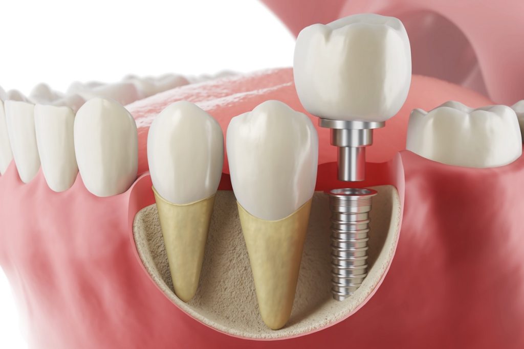
Titanium artificial roots placed in the jawbone to compensate for missing teeth and the prosthetic restorations positioned on them are constantly interacting with the biological dynamics of the organism. The medical structure of this system does not remain static in the state it was placed on the operating table; it is in a continuous cellular adaptation cycle depending on chewing forces, the microbial flora in the oral cavity, and the individual’s systemic health. The question of why one should go for post-implant controls in Izmir is one of the most fundamental medical inquiries for individuals wanting to understand how the anatomical balance is monitored after treatment.
Why Should One Go for Post-Implant Controls in Izmir and How is Biological Adaptation Monitored?
After the surgical procedure is completed, the body’s own repair mechanism becomes active. The titanium element has the potential to form a biological unity as it is surrounded by bone cells (osteoblasts) due to its structural properties. However, this cellular process may be interrupted by external micro-traumas, microbial attacks, or fluctuations in the patient’s metabolic rate. The primary medical rationale for clinical follow-ups is to analyze with concrete data whether this microscopic tissue bonding progresses within the targeted anatomical boundaries.
Appointments made in dental clinics are sessions where the body’s physiological responses are calibrated. Changes in the bone that cannot be seen or felt by the eye, as well as subtle cellular disruptions in the mucous membrane, can only be detected through medical equipment and specific examination techniques used by the doctors. The data obtained in the early stages allows for clinical steps to be taken to ensure the continuation of the process as it typically unfolds.
How is Titanium Material and Jaw Bone Compatibility Evaluated in Clinical Examinations?
Jaw surgeons and prosthetic specialists do not rely solely on visual examination when assessing the stability of the material. How tightly the bone tissue penetrates between the titanium threads is converted into numerical data through mechanical tests. In this way, it is scientifically determined when the prosthetic load can be applied to the material.
| Examination Method | Clinical Procedure | Medical Purpose and Data |
|---|---|---|
| Percussion Test | The frequency of the sound produced is listened to by lightly tapping the material with the metal handle of medical instruments. | A clear and resonant metallic sound indicates tight bonding with the bone; a dull sound suggests the presence of connective tissue residues in between. |
| Resonance Frequency Analysis (RFA) | A small sensor attached to the material sends magnetic waves to measure elasticity at the micro level. | ISQ (Implant Stability Coefficient) provides a numerical value that presents a mathematical table of cellular hardening. |
| Manual Mobility Control | It is a pressure test applied horizontally and vertically to the structure by the physician’s fingers or special press tools (calipers). | Detection of mobility at micro or macro levels informs the clinician of medical complications in the osteointegration cycle. |
What is the Purpose of Periodic Medical Check-ups Conducted within Avrupadent?
The sustainable care philosophy adopted by medical organizations is based on the principle that the work does not end with the completion of surgical operations but rather marks the beginning of a new biological cycle. Interdisciplinary teams working in institutional clinics tie patient care to a periodic schedule from surgical, prosthetic, and periodontal (gum health) perspectives. This integrated approach prepares the ground for the simultaneous evaluation of reactions that the organization may develop against the structure by different medical branches.
Especially the reflections of chewing forces on the jaw joints (temporomandibular joint) and neck muscles may change over time. Millimetric shifts occurring in jaw closure are detected during clinical checks with precise measurement records and balanced through adjustments on the prosthesis. This helps in limiting muscle spasms and mechanical stresses on the structure.
What is Peri-implantitis and How Do Regular Doctor Check-ups Relate to This Condition?
Just as periodontitis (gum recession) occurs when dental calculus accumulates around natural teeth, the same pathological mechanism operates around artificial materials. Since titanium screws do not have periodontal ligaments (connective tissue fibers) like natural roots, the bacterial inflammation initiates a much faster destruction (resorption) process when it reaches the bone. The presentation of peri-implantitis is the primary physiological reason that leads to the long-term loss of stability of the material.
The insidious nature of the disease typically does not create a noticeable discharge or acute symptoms in its early stages. An individual cannot detect this microscopic destruction in the depths of the gums while looking in the mirror at home. Detailed gum pocket measurements and microbial flora analysis conducted by the doctor during six-month clinical periods can detect this inflammatory reaction in its initial phase, thus limiting the progression of destruction.
Why is Radiological Imaging (X-ray) Used in Routine Check-ups?
Oral inspection provides data to the doctor only about the surface of the prosthesis and the color of the gum. Bone tissue, which is the main supporter of the biomechanical system, is hidden beneath the mucosal layers. X-ray technologies are key diagnostic tools that illuminate the invisible parts of medical processes. Routine checks involve taking periapical (small) or panoramic (large) films, which are compared millimetrically with films taken in previous years.
-
\t
- Measurement of Marginal Bone Loss: Detection of cellular bone resorptions that can develop below the neck part of the titanium screw (crest top) in millimeter scale.
- Bone Density Analysis: Evaluation of the appearance of the bone remaining between the screw threads as radiolucent (dark/empty) or radiopaque (light/dense).
- Alignment of Connection Components (Abutment): Identification of microscopic gaps that may occur between the components connecting the prosthesis to the screw, and examination of areas of bacterial leakage.
\t
\t
How are Assessments of the Prosthetic Superstructure (Zirconium/Porcelain) Conducted in Examinations?
Although titanium roots remain fixed in the jawbone, the teeth inside the mouth (especially natural teeth) can exhibit millimetric positional changes (physiological migration) over the years. This situation can disrupt the contact balance with a previously perfectly designed zirconium or porcelain prosthesis in relation to the opposing tooth. The disrupted balance may lead to asymmetrical (uneven) pressure on a specific point of the prosthesis, causing material fatigue.
In clinical appointments, the dentist may ask the patient to simulate chewing using special heating papers that are calibrated to micron thickness. The colored marks left by the paper on the prosthesis map out where the forces are applied. When asymmetrical pressure is detected, small adjustments made with milling on the surface can limit the potential for the prosthesis to break. In the same session, the gloss layer on the porcelain surface (glaze) is checked to evaluate if it has dulled or not, assessing the plaque retention potential.
How Are Deficiencies in Oral Hygiene Detected in Clinical Checks?
Individuals often think they brush their teeth regularly, but toothbrush bristles or dental floss cannot perfectly reach every area of the anatomy. Especially the lower parts of titanium-supported bridges contain micro cavities that facilitate bacterial organization. Dentists uncover these biological accumulations that the patient cannot see during clinical appointments using medical agents.
When plaque coloring tablets or solutions are applied to the teeth, they only color the areas where bacterial colonies are dense. This allows the patient to see in the mirror which areas they missed during brushing, updating their oral hygiene habits. Additionally, blunt-tipped medical instruments called periodontal probes are gently maneuvered between the gums and prosthetics to determine if there is a pocket (pathological space) formed and to numerically record the tissue’s susceptibility to bleeding.
How Should Control Periods be Planned for Individuals with Systemic Diseases?In individuals with systemic diseases such as diabetes, hypertension, or osteoporosis, since tissue repair capacity varies, control periods are generally planned with narrower intervals (every three or four months) compared to standard patients; changes in blood values are monitored alongside medical practitioners to shape the medical chart.
The jawbone and mucosal structure are an integral part of the body’s general circulation and connective tissue system. An increase in blood glucose levels or fluctuations in thyroid hormones directly impact the cellular health within the mouth. Especially in uncontrolled cases of Type 2 diabetes, as protein synthesis is disrupted, the resistance of gum tissue against bacteria significantly decreases.Individuals with a fragile systemic profile are classified by medical organizations as a “risk group” because they possess a physiology more prone to infections. Instead of a standard six-month monitoring schedule, doctors prefer to increase the frequency of examinations to capture subtle changes in the mucosal structure. This enhanced monitoring schedule is an essential medical requirement for limiting potential adverse reactions within the oral cavity related to systemic diseases.
What Medical Criteria Are Followed to Determine Follow-up Appointments for Patients Using Tobacco Products?
The use of tobacco products weakens tissue perfusion, so follow-up appointments for these patients are organized to monitor mucosal health and bone levels more closely, typically scheduled at narrowed intervals of three months to closely observe potential inflammatory processes at a cellular level.Nicotine and thousands of toxic substances found in cigarette smoke have a vasoconstrictor effect, narrowing capillaries. When sufficient blood does not reach the gum tissue surrounding titanium implants, the tissue’s immune cells also cannot access the area. This condition prepares the ground for bacterial plaque to progress directly into the bone structure without encountering any resistance.
An even more critical medical scenario is the suppression of gum bleeding in tobacco users. In a healthy individual, when plaque accumulates, the gums bleed, providing a “warning”. However, in smokers, the narrowed blood vessels prevent bleeding; patients may believe their gums are healthy, leading them to neglect clinical appointments. Dentists are aware of this misleading physiological condition, and thus they more frequently include patients who use tobacco in their medical follow-up radar, aiming to detect insidious bone destruction with radiological and clinical tools.
What Months Are Included in the Control Schedule Within the First Year?
In the first year following the surgical operation, the routine clinical schedule is typically planned to include radiologically supported examinations in the first week (acute tissue control), the first month (soft tissue adaptation), the third month (bone healing progression), the sixth month, and the twelfth month.The first fifty-five days from the beginning of the treatment is the most dynamic time frame where the jawbone establishes a biological connection with the newly encountered material and the loads of prosthetic implants are organized by bone trabeculae (strands). The medical schedule applied during this transitional phase exhibits a flexible structure according to the reactions of the tissues.
Initial Year Clinical Follow-Up Schedule Examination Period Clinical Focus Point Medical Justification 1st Week (Post-Op) Suture and Edema Control The epithelialization (closure) of the wound lips is monitored, and if present, surgical threads are removed to relieve the tissue. Between 1st and 3rd Month Early Stage Osteointegration The stability of cellular healing before or after prosthetic loading is measured through frequency analyses. 6th Month Control Mechanical and Radiological Monitoring The effects of prosthetic loading on the jawbone are monitored radiographically to obtain physiological approval for chewing function. 12th Month (Annual Review) Plague Index and Occlusion Detection The individual’s annual oral hygiene performance is noted, and potential deviations in the relationship between the prosthesis and the opposing tooth are mapped.
How are Clinical Controls Shaped According to Climate and Dietary Habits in İzmir Location?
The chemical-mechanical effects created by the acidic citrus fruits, shelled dry food, and fibrous food consumption prevalent in the Izmir region on prosthetic surfaces and dental tissues require physicians to more closely examine marginal fit (edge sealing) and porcelain surface wear during clinical checks.The nutritional culture offered by the local geography directly affects the biochemical and mechanical cycle within individuals’ mouths. In the Aegean region, the frequent consumption of high acid fruits (such as mandarins and figs) lowers the intraoral pH value, modifying the structure of salivary enzymes. These acidic attacks may prepare the ground for microscopic dulling in the polish of prosthetic materials.
Additionally, the long-term chewing of local hard foods (such as boyoz, crispy bread, or fig seeds) creates mechanical stress accumulation on the masticatory (occlusal) surfaces of porcelain restorations. Physicians assess regional dietary habits in medical history forms; during clinical checks, they guide medical maintenance by scanning the occlusal thicknesses of prosthetics, contact points with neighboring teeth, and the formation of micro-cracks under special light sources.
What Medical Procedures are Implemented in Long-Term (Annual) Maintenance Treatments?
In annual maintenance treatments, professional plaque cleaning is performed using special teflon or carbon curettes that do not damage the titanium surface, the connection (abutment) parts of screws are clinically checked, and the individual’s personal oral hygiene practices are updated based on medical data to support the sustainability of long-term stability.Even years after a surgical procedure, medical materials continue to be exposed to biological mobility in the oral ecosystem. During this phase, referred to by medical organizations as the “maintenance phase,” the aim is to preserve the current state of health (status quo). In the blind spots (in the depths of the sulcus) inaccessible through routine brushing and flossing at home, plaques hardened by calcium in the tartar revert to dental stone.
Expert physicians use special periodontal devices made of plastic, Teflon, or carbon that do not scratch the titanium surface instead of metal tools during annual visits, breaking these deposits using the cavitation (sound wave) method. A non-scratched surface is inhospitable for bacteria to adhere. Simultaneously, the inner screws (abutment screws) connecting the titanium prosthesis are tested with torque wrenches to check for micro-loosening over the years. Loose connections are re-tightened, preventing fluids and bacteria that could seep in from being medically avoided.
The jawbone and mucosal structure are an integral part of the body’s general circulation and connective tissue system. An increase in blood glucose levels or fluctuations in thyroid hormones directly impact the cellular health within the mouth. Especially in uncontrolled cases of Type 2 diabetes, as protein synthesis is disrupted, the resistance of gum tissue against bacteria significantly decreases.
Individuals with a fragile systemic profile are classified by medical organizations as a “risk group” because they possess a physiology more prone to infections. Instead of a standard six-month monitoring schedule, doctors prefer to increase the frequency of examinations to capture subtle changes in the mucosal structure. This enhanced monitoring schedule is an essential medical requirement for limiting potential adverse reactions within the oral cavity related to systemic diseases.
What Medical Criteria Are Followed to Determine Follow-up Appointments for Patients Using Tobacco Products?
Nicotine and thousands of toxic substances found in cigarette smoke have a vasoconstrictor effect, narrowing capillaries. When sufficient blood does not reach the gum tissue surrounding titanium implants, the tissue’s immune cells also cannot access the area. This condition prepares the ground for bacterial plaque to progress directly into the bone structure without encountering any resistance.
An even more critical medical scenario is the suppression of gum bleeding in tobacco users. In a healthy individual, when plaque accumulates, the gums bleed, providing a “warning”. However, in smokers, the narrowed blood vessels prevent bleeding; patients may believe their gums are healthy, leading them to neglect clinical appointments. Dentists are aware of this misleading physiological condition, and thus they more frequently include patients who use tobacco in their medical follow-up radar, aiming to detect insidious bone destruction with radiological and clinical tools.
What Months Are Included in the Control Schedule Within the First Year?
The first fifty-five days from the beginning of the treatment is the most dynamic time frame where the jawbone establishes a biological connection with the newly encountered material and the loads of prosthetic implants are organized by bone trabeculae (strands). The medical schedule applied during this transitional phase exhibits a flexible structure according to the reactions of the tissues.
| Examination Period | Clinical Focus Point | Medical Justification |
|---|---|---|
| 1st Week (Post-Op) | Suture and Edema Control | The epithelialization (closure) of the wound lips is monitored, and if present, surgical threads are removed to relieve the tissue. |
| Between 1st and 3rd Month | Early Stage Osteointegration | The stability of cellular healing before or after prosthetic loading is measured through frequency analyses. |
| 6th Month Control | Mechanical and Radiological Monitoring | The effects of prosthetic loading on the jawbone are monitored radiographically to obtain physiological approval for chewing function. |
| 12th Month (Annual Review) | Plague Index and Occlusion Detection | The individual’s annual oral hygiene performance is noted, and potential deviations in the relationship between the prosthesis and the opposing tooth are mapped. |
How are Clinical Controls Shaped According to Climate and Dietary Habits in İzmir Location?
The nutritional culture offered by the local geography directly affects the biochemical and mechanical cycle within individuals’ mouths. In the Aegean region, the frequent consumption of high acid fruits (such as mandarins and figs) lowers the intraoral pH value, modifying the structure of salivary enzymes. These acidic attacks may prepare the ground for microscopic dulling in the polish of prosthetic materials.
Additionally, the long-term chewing of local hard foods (such as boyoz, crispy bread, or fig seeds) creates mechanical stress accumulation on the masticatory (occlusal) surfaces of porcelain restorations. Physicians assess regional dietary habits in medical history forms; during clinical checks, they guide medical maintenance by scanning the occlusal thicknesses of prosthetics, contact points with neighboring teeth, and the formation of micro-cracks under special light sources.
What Medical Procedures are Implemented in Long-Term (Annual) Maintenance Treatments?
Even years after a surgical procedure, medical materials continue to be exposed to biological mobility in the oral ecosystem. During this phase, referred to by medical organizations as the “maintenance phase,” the aim is to preserve the current state of health (status quo). In the blind spots (in the depths of the sulcus) inaccessible through routine brushing and flossing at home, plaques hardened by calcium in the tartar revert to dental stone.
Expert physicians use special periodontal devices made of plastic, Teflon, or carbon that do not scratch the titanium surface instead of metal tools during annual visits, breaking these deposits using the cavitation (sound wave) method. A non-scratched surface is inhospitable for bacteria to adhere. Simultaneously, the inner screws (abutment screws) connecting the titanium prosthesis are tested with torque wrenches to check for micro-loosening over the years. Loose connections are re-tightened, preventing fluids and bacteria that could seep in from being medically avoided.