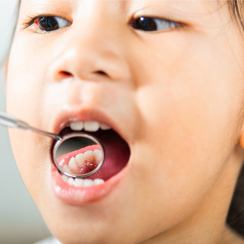
Should Cavities in Milk Teeth Be Treated in İzmir?
Cavities in milk teeth should definitely be treated without delay to ensure that the child can continue chewing properly, complete their speech development, and support the healthy growth of the jawbone. These teeth are not just temporary structures; they are vital organs that guide the correct positioning of the permanent teeth that will come up from below and directly affect the overall body health in case of infections.
In dental practice and public health perception, the role of milk teeth is often mistakenly seen as merely being “a temporary stage that will give way to new ones.” However, biological and medical realities show that milk teeth bear significant physiological responsibilities during the most critical developmental stage of human life. When a child’s milk tooth decays, it not only compromises the enamel of that tooth; the muscles, bones, and systemic circulatory system linked to that tooth also suffer from this infection. In clinical practices serving in the İzmir location, treating cavities in milk teeth is not presented as an option but as a scientific necessity to protect the child’s current comfort and future anatomical integrity. Cavities are not a self-healing condition; if left untreated, it becomes a chronic microbial disease that continually deteriorates surrounding tissues.
What Medical Issues Can Arise from Not Treating Cavities in Milk Teeth?
Failure to treat cavities in milk teeth can lead to the infection spreading from the tooth root to the jawbone, causing an abscess, severe pain, and completely disrupting the child’s feeding and sleeping patterns. Progressing infections can cause erosion in the jawbone, leading to the early loss of the milk tooth and irreversible structural damage to the underlying permanent tooth germ.
Although a cavity may look like a black spot from the outside, it is actually a biochemical battle where acid-producing bacteria have hollowed out the tooth from within. The enamel layer of milk teeth is extremely thin, and the nerve (pulp) chambers are very close to the surface. An untreated cavity can penetrate this thin layer and reach the nerves in just a few months. When the nerve tissue becomes inflamed, the child will experience severe throbbing pain, especially waking from nightly sleep. When bacteria travel from the nerve chamber to the tip of the root, it creates a pus (abscess) pocket within the jawbone. This condition is referred to as a dental abscess. An abscess can cause serious swellings (cellulitis) that can be noticeable from the outside on the child’s face. The pressure from the abscessed tooth during chewing turns eating into a torture, hindering the child’s physical development.
Why the “Milk Tooth Will Fall Out Anyway” Notion is a Medical Misconception?
The belief that the milk tooth will fall out regardless ignores the fact that these teeth can remain in the mouth until the age of 12 in the back molar area, forming a physical foundation for the skeletal development during the child’s most intense growth phase, thus representing a significant medical misconception. Each milk tooth is a biological structure programmed to complete its function by a specific age.
The change of teeth in a person’s mouth does not happen all at once. The primary incisors, which emerge during infancy, are lost around the ages of 6-7, while the primary molars (first and second primary molars), which perform eighty percent of the chewing function, must remain in the mouth until the child reaches the ages of 10, 11, and sometimes 12. If a cavity occurs in the primary molar of a 5-year-old child, and the family avoids treatment saying, “it will fall out anyway”, the fact that the infected tooth must stay in the mouth for at least another 6-7 years is often overlooked. Living with an infected tooth for years undermines every mechanism from the child’s digestive system to their immune system. Therefore, the temporariness of the teeth does not mean that their health is insignificant; it means that they must function perfectly for a certain period.
| Type of Primary Tooth | Average Loss (Change) Age | Duration of Issues if Untreated |
|---|---|---|
| Incisors | Between 6 – 8 Years | Short-term chewing and speech disorders. |
| Cuspid Teeth (Canines) | Between 9 – 11 Years | Jaw asymmetry and loss of guidance. |
| Primary Molars | Between 10 – 12 Years | Prolonged abscess, digestive difficulty, and severe pain. |
How Do Untreated Primary Teeth Affect Permanent Teeth?
Untreated primary teeth negatively affect permanent teeth by allowing bacterial abscesses to seep from their roots, coming into contact with the permanent tooth’s developing follicle, leading to yellow or brown spots (Turner tooth) and deformities in the enamel layer. In the case of early extraction, the permanent teeth may remain unerupted due to lack of space.
The roots of primary teeth and the buds of developing permanent teeth are only a few millimeters apart, nestled within the jawbone. When the nerve in the center of the primary tooth reaches an infection, it flows towards the roots and creates an acidic inflammation pocket there. The enamel layer of the permanent tooth works hard to harden (to mineralize) precisely at this stage, that is, within that acidic environment. If the permanent tooth is subjected to this acid bath, it cannot fully synthesize its enamel layer. Years later, when that tooth erupts into the dental arch, instead of having a healthy white appearance, it bears developmental hypoplastic defects (brown pits or absent enamel structures). So, not intervening in a disease of the primary tooth permanently damages the adult tooth the child will use for life.
How Do Primary Tooth Cavities Disrupt a Child’s Jaw and Facial Development?
Cavities in primary teeth cause a child to develop a tendency to chew on one side (asymmetrical chewing), avoiding the side where they feel pain; this results in uneven loads on the jaw joint, causing disproportionate growth of the facial muscles, narrowing of the jaw curve, and in later years, skeletal asymmetries, which seriously disrupts the development of the jaw and face.
In order for the jaw bones of a child to grow normally and broadly in a U shape, the muscles on both sides (masseter and temporal muscles) must work equally and transmit the chewing pressure evenly to the bone. However, a child with a deep cavity in the right side will reflexively transfer all bites to the left side to avoid the sensation of discomfort when food touches that area. This unilateral chewing habit, lasting for months or years, leads to the muscles on the left side developing and hypertrophying more, while the right side remains weaker. The temporomandibular joint (jaw joint) will be subjected to asymmetric pressure. A visible deviation will occur in facial symmetry. An untreated simple tooth decay can create a biomechanical domino effect, exerting a powerful and destructive influence capable of completely altering the vector of bone growth in the face.
How Do Cavitary Infections of Milk Teeth Threaten the Child’s Overall Health?
Cavitary infections of milk teeth, by allowing bacteria proliferating in the oral flora to enter the bloodstream through swallowing and circulation (bacteremia), constantly occupy the immune system, reducing the child’s overall bodily resistance, leading to chronic fatigue, appetite loss, growth retardation, and opening the risk of focal infections in distant organs such as heart valves, thereby directly threatening overall health.
Oral and dental health is not an isolated island independent of the body’s overall systemic health. The body of a child with an abscessed tooth root constantly directs immune cells (leukocytes) to that area to contain and deal with the infection. The persistent battle of the immune system against this active chronic infection, 24/7, drains the child’s energy. These children are often picky eaters (due to chewing pain) and do not get enough sleep. Nutritional deficiencies and poor sleep quality can hinder the secretion of growth hormone, causing delays in weight and height development (in percentile charts). Furthermore, when the bacteria from the tooth root enters the bloodstream through chewing pressure, it creates a condition known as “focal infection” in the body. In children at systemic risk, such as those with rheumatic heart disease, kidney failure, or joint disorders, these infections in the mouth can lead to much more serious complications in distant organs, which is why the medical field requires these infections to be cleared urgently.
What Treatment Methods Are Applied in Milk Tooth Cavities?
In milk tooth cavities, depending on the depth of the cavity, cleaning the cavity tissue and filling it with biocompatible materials (glass ionomer or composite), partial root canal treatment (amputation) in cases where the infection has reached the nerves, or using pediatric stainless steel/zirconium crown restoration methods in cases of severe tissue loss are applied. The main goal is to keep the tooth in the mouth until it reaches the age of shedding by eliminating the infection.
The philosophy behind treatments applied in pediatric dentistry seeks to maintain teeth in the least invasive manner possible (minimal invasive approach). If decay is still on the surface, that is, at the enamel and dentin boundary, the dentist removes the decayed, softened, and bacteria-laden tissue with special instruments. In the cleaned cavity, biological fillings containing glass ionomer or aesthetic composite materials that especially release fluoride, nourishing the tooth and being more tolerant to moisture, are placed. In cases of moderate decay where the progression has reached the nerve (pulp) chamber but has not fully surrounded the roots, partial canal interventions referred to as amputation come into play. The common goal of all treatment options is to successfully preserve the position of the tooth in the jawbone until the underlying permanent tooth emerges.
| Decay Level (Depth) | Clinical Symptoms | Applied Treatment Method |
|---|---|---|
| Initial Level (Enamel) | White matte spots, no sensitivity, no cavity. | Remineralization using only Fluoride Varnish without using a drill (burr). |
| Moderate Level (Dentin) | Sensitivity to sweet/cold stimuli, brown cavities. | Cleaning of decay and restoration with Glass Ionomer / Composite Filling. |
| Advanced Level (Pulp/Nerve) | Pounding pain that wakes you from sleep at night, formation of abscess. | Amputation (Partial Canal Treatment) and Pediatric Crown Coverage. |
Why is Root Canal Treatment for Primary Teeth Different from Adults?
Root canal treatment (amputation) for primary teeth is different from that for permanent teeth because, instead of completely clearing out the root up to the apex and filling it with non-resorbable materials, the process involves just removing the diseased nerve in the upper part, allowing the roots to remain alive, and using special medical pastes that can naturally dissolve (resorbable) as the underlying permanent tooth emerges alongside the roots of the primary tooth.
When performing root canal treatment (endodontics) on an adult patient, the doctor’s goal is to effectively seal that tooth within the bone. Therefore, channels are filled with solid rubber materials (gutta-percha) that never dissolve in the body. However, this procedure is completely inadvisable for primary teeth. When the time comes for a primary tooth, the underlying permanent tooth slowly dissolves its root, causing the primary tooth to become loose and fall out. If a non-resorbable adult filling is placed in the canals of a primary tooth, the permanent tooth coming up from below may not be able to overcome that barrier and will remain buried within the jawbone. Due to this biological difference, when doctors remove inflamed nerves in primary teeth (pulpotomy/amputation), they typically use special absorbable pastes based on zinc oxide-eugenol or iodoform that can dissolve spontaneously at the same time as the canal development stage.
What is the Approach to Primary Tooth Extraction at Izmir Avrupadent Clinics?
The approach to primary tooth extractions at Izmir Avrupadent clinics is a comprehensive process that focuses on the child’s biological and psychological integrity, aiming to clean and treat the extraction site with minimal intervention (minimally invasive) using current preventive dental protocols, preventing dental phobia by gamifying the treatment process, and ensuring that the tooth remains functionally in the mouth until the age of shedding through transparent communication established with families.
Our expert team in Izmir does not view cavities in milk teeth merely as a mechanical defect to be filled; they conduct medical investigations that analyze the underlying nutrition, genetics, and hygiene habits. From the child’s entry into the clinic to their getting up from the chair, the scary concepts of traditional dentistry (needle, extraction, pain) are set aside, and communication is based entirely on abstraction (playful interaction). Even in cases where local anesthesia is required, the procedure is presented to the child as “the tooth is going to sleep.” Meanwhile, zero-error diagnostic procedures are carried out using digital imaging devices that comply with 2026 health standards, and transparent, objective treatment plans are provided to the family through radiological images.
What Restorations (Crowns) Are Preferred for Milk Teeth with Severe Material Loss?
In milk teeth with severe material loss, where there is not enough solid wall to hold a filling, prefabricated stainless steel crowns that encompass the tooth entirely and physically prevent the tooth from breaking, or biocompatible pediatric zirconium crowns are preferred, especially when aesthetic expectations are high. These restorations restore the anatomical dimensions of the tooth exactly.
Sometimes, the cavities in primary teeth spread to such wide areas that by the end of the cleaning process, nearly only the root or very thin walls of the tooth remains. Even if a standard filling material (such as composite) is placed into such a tooth, when a child bites into hard food (applying pressure), that filling can detach from the tooth and break from the weakened tooth root. To save these types of “damaged” teeth and keep them in the mouth until the age of shedding, “pediatric crowns” are used. Stainless steel crowns (SSC) are placed like a cap on the tooth and fixed with dental adhesives, providing incredible resistance against chewing forces. For the anterior incisors or back teeth where aesthetics are important, “zirconium pediatric crowns” with a tissue-friendly, completely white color and a glass-like smooth surface that prevents the retention of caries bacteria are applied to achieve an excellent anatomical restoration.
What Can Be Done to Preserve the Space of Primary Teeth That Need to Be Extracted Early?
To preserve the space of primary teeth that need to be extracted early, dental devices known as “space maintainers” are applied. These can be fixed or removable dental appliances that mechanically prevent the neighboring teeth from tilting into the gap created by the extraction. These devices maintain the necessary anatomical space for the permanent tooth to erupt correctly.
The primary goal of preventive dentistry is to maintain the tooth; however, if a root infection has severely damaged the root tip, eroded the underlying permanent tooth bud, or if the tooth is so decayed that even a covering cannot be applied, that tooth is extracted as a medical necessity. However, treatment does not end at the time of extraction. If a primary molar that should have fallen out at age 11 is extracted at age 6, the adjacent teeth will shift forward and completely close that gap. The dentist will measure the dimensions of the gap immediately after the extraction procedure and prepare a custom “space maintainer” in the laboratory. These fixed appliances, consisting of a metal band shaped like a crown and a wire extension that protects the gap, do not affect the child’s eating or talking. On the day the underlying permanent tooth is visible in the jaw, the dentist quickly removes this appliance to ensure the process is completed safely and without skeletal loss.
How Does Pediatric Dentistry (Pedodontics) Manage Behavior in Treatment of Deciduous Teeth?
Pediatric Dentistry (Pedodontics) specialists manage behavior in treatment of deciduous teeth by introducing medical instruments with names appropriate for children’s imaginary world (by making it playful), overcoming anxiety about the unknown through the demonstrate-explain-do technique, and giving the child a sense of control during the procedure. In advanced infection cases where cooperation cannot be established, general anesthesia or conscious sedation techniques are used to completely prevent psychological trauma.
The most challenging part of treating children with dental issues is not the drilling or filling of teeth, but ensuring that the child remains calm and still (cooperative) in the chair. While it is sufficient to tell an adult to simply open their mouth, a child requires a step-by-step approach to this process. During the procedure, dentists maintain continuous communication. Directives such as “I am cleaning your tooth with a wind machine, I am painting your tooth with magic dough (filling)” redirect the child’s attention to a different focus (distraction). However, if the child is a special needs individual, such as one with autism or cerebral palsy, or if they are too young (for example, 2-3 years old) to understand verbal directions, treating them by forcefully keeping them in the chair is incompatible with modern medical ethics. In such cases, dentists consult with anesthesiologists to safely treat all dental issues in a single session under general anesthesia, in fully equipped operating room conditions where the child is in deep sleep and feels no pain or fear.
How Long Are Primary Tooth Fillings Designed to Last in the Mouth?
Primary tooth fillings are designed to last in the mouth until the underlying permanent tooth completes its development in the jawbone and erupts properly (around 11-12 years of age for back molars) and can withstand chewing forces, using biocompatible materials that are fully aligned with the biological lifespan of that tooth (root resorption process).
The chemistry of dental materials must be suitable for the dynamics of the tissue they are applied to. In fillings applied to adults, the goal is to achieve long-lasting durability, while the primary goal of a filling applied to a primary tooth is to ensure functionality for a period of 3-5 years until the tooth naturally sheds and to resist acidic attacks in that environment. Due to the high saliva production and mobility in children, dentists prefer special materials called “glass ionomer cements” or “compomers” (polyacid modified composite resin) that continue to nourish the tooth from the inside against decay by creating a fluoride release in the applied area. These intelligent materials ensure tight adhesion by forming a chemical bond with the tooth enamel. A properly applied filling in a primary tooth has high technological durability, remaining in place until the natural root resorption is completed and the tooth is expected to shed naturally by the child.
How Does the Treatment of Primary Teeth Directly Affect Speech (Phonetic) Development?
The treatment of primary teeth directly affects speech (phonetic) development because these teeth serve as a mechanical barrier, ensuring the correct articulation of sounds such as “s, f, v, t, z” when the tongue rests against them. Early loss of these teeth due to decay can lead to lisping and persistent articulation (pronunciation) disorders in children, hindering social communication.
Speech is not only the result of the vocal cords but also the flawless coordination of the tongue with the palate, lips, and teeth (articulation). The preschool period (ages 3-6), when the child begins to understand speech and letters, is also the period when the upper front teeth are most frequently lost due to “bottle rot.” A child who has completely lost their front teeth and whose roots remain will not be able to find the physical area where their tongue can rest while producing the sounds “s” or “t.” The tongue will push through this gap (resulting in slurring). This pronunciation mistake, which initially appears to be temporary, can settle into the child’s speech pattern over time and evolve into a permanent speech disorder. Doctors treat not only the child’s smile but also their diction and self-confidence by reconstructing this mechanical barrier with aesthetic zirconia or composite overlays applied to the lost front teeth.
How Can Families Understand That Primary Teeth Need Treatment?
Families can recognize that primary teeth need treatment by the presence of chalky white spots or brown-black cavities on the tooth enamel, by signs of sensitivity when the child consumes sweet or cold foods, by the child avoiding the use of a specific side while chewing, and by small pimple-like swellings (fistula/abscess) appearing around the gum line. However, the most accurate diagnosis is made through routine dental check-ups.
Children may hide toothaches from their parents due to the high sensitivity of their teeth and sometimes fear of going to the doctor. However, body language and eating habits always reveal a dental issue. If a child suddenly flinches while drinking their favorite cold water, consistently chews on the same side, or refuses to let a specific tooth be touched during brushing, it indicates that a cavity has likely formed there. Families should examine their children’s teeth monthly under good lighting (at home) by slightly lifting their lips. Food particles stuck between two teeth that do not come out, reddish-purple lesions on the gum edges, or dark shadows on the chewing surface of the tooth are alarm signals that require urgent intervention. For cavities that cannot be seen with the naked eye, X-rays taken in a clinical setting by the dentist are the gold standard.
What Preventive Measures Should Be Taken at Home for Treated Baby Teeth?
For treated baby teeth at home, regular brushing twice a day with child-safe fluoridated toothpaste under parental supervision, the use of interdental brushes every evening to prevent bacterial buildup at filling margins, and completely eliminating habits of consuming carbohydrate-rich sticky foods (especially nighttime feedings) are preventive and hygienic steps that must be taken diligently.