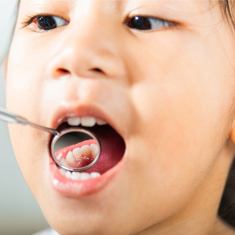
When Does Oral and Dental Health in Children Begin?
Oral and dental health in children begins with the formation of milk tooth buds while the baby is still in the womb and turns into an active care process immediately after birth with the cleaning of the palate in the toothless period. With the emergence of the first milk tooth in the mouth, this process becomes a lifelong health routine through dental brushing and preventive care measures. The steps taken in the early period form the basis of healthy jaw development.
The concept of oral and dental health is generally perceived in society as a process that starts with “the first tooth falling out” or “the child reaching an age to take their toothbrush.” However, scientific and biological reality proves that the foundations of dental health are established in the earliest stages of life, during the cellular coding phase in the mother’s womb. A healthy oral structure is not just made up of the presence of teeth. The proper closure of jaws, correct positioning of the tongue, nasal breathing, and the functioning of the swallowing reflex are all components of a vast biomechanical system that works together. For this reason, the answer to the question of “when does it start” encompasses a complicated biological chronology that extends into the first weeks of a baby’s cellular development.
Does Oral and Dental Health in Children Start in the Womb?
Oral and dental health in children begins directly in the womb when the milk tooth buds start to form within the jawbone around the average sixth week of pregnancy. During this period, the calcium, phosphorus, and vitamins that the mother takes define the quality of the baby’s dental enamel at the cellular level. The nutritional pattern during pregnancy and the mother’s overall health is the first step in building the future risk profile for the baby.
The pregnancy period is the time frame during which the most intense organogenesis (organ formation) occurs. Although a baby’s milk teeth are not visible at the moment of birth, they have already taken their place within the jawbone long ago, beginning to calcify (harden). Maternal illnesses during pregnancy, some specific medications used (for example, tetracycline antibiotics), or malnutrition conditions can lead to lasting structural defects (hypoplasia) on the developing enamel of the baby’s teeth. Additionally, since the bacteria load in the mother’s own mouth and existing cavities will be transferred to the baby through the oral route after birth, it is a preventive medical approach for the expectant mother to complete her dental treatments before pregnancy to protect the baby’s oral health.
How Should Oral Care Be Done During the Toothless Period After Birth?
Oral care during the toothless period after birth should be done after each feeding or at least twice a day, using a clean, boiled, and cooled drinking water-moistened cloth to gently wipe the baby’s gums, palate, and the insides of the cheeks to remove residual breast milk or formula. This process helps balance the oral flora and prevents the formation of thrush.
The mouth of a newborn is completely sterile at birth, but with the first feeding, the oral flora gets introduced to environmental factors. While breast milk contains unique antibodies that strengthen the baby’s immune system, it also contains natural lactose (milk sugar). The remnants of this milk left on the palate and tongue after feeding create a suitable environment for the growth of fungi (especially Candida albicans) and bacteria in the warm and moist oral environment. In clinical trainings conducted at the İzmir location, parents are advised to continue this physical cleaning process until the baby’s teeth erupt. Regularly cleansing and massaging the gums protects against infections and facilitates the baby’s adaptation to oral interventions, making the transition to tooth brushing extremely easier in the following months.
| Care Step | Application Method | Biological/ Physiological Benefit |
|---|---|---|
| Tissue Cleaning | A gas-filled cloth or a clean cloth is dipped in warm water and wrapped around the finger. | By removing milk residues, it stops the formation of fungi (candidiasis). |
| Gum Massage | Light circular pressure is applied to the crests (the bumps where the teeth will emerge). | It increases blood circulation in the tissue and relieves the discomfort during teething. |
| Desensitization | Routine interventions are made in the baby’s mouth at regular intervals. | It helps the baby get accustomed to having something in their mouth and adapts them to brushing. |
What Symptoms Are Observed During The Eruption Process Of The First Tooth?
During the eruption process of the first tooth, completely physiological symptoms such as swelling and redness of the gums in babies, increased drooling linked to heightened salivary secretion, the desire to bite hands or hard objects, loss of appetite, mild restlessness, and disruption of sleep patterns are observed. These symptoms are a natural result of the cellular inflammation that occurs when the tooth’s mucosa begins to break through into the mouth.
Tooth eruption (eruption) usually begins with the emergence of the lower front incisors around the sixth month, but this timeline can vary depending on genetic factors, occurring as early as the fourth month or as late as the twelfth month. The sharp enamel of the tooth forces the gum tissue outwards, causing tension in this tissue, which results in localized sensitivity in the baby. Salivary glands begin to produce excessive saliva to protect the gums and cool the inflammation in that area. Although families often associate this period with fever and diarrhea, clinical literature emphasizes that a slight increase in body temperature during tooth eruption does not indicate a fever above 38 degrees or severe diarrhea arising from teething, but rather from another systemic infection. In such cases, the process must be monitored under medical supervision.
What is the Ideal Time for a First Dental Examination in Izmir?
According to current health approaches in the Izmir region, the ideal time for the first dental examination is between the sixth month, when the first milk tooth appears in the baby’s mouth, and the first birthday. This early visit serves as a protective guidance step aimed at teaching families proper nutrition and hygiene habits within the framework of Pediatric Dentistry.
The vision often expressed in the processes of the Avrupadent clinic is to step into the clinic before any development occurs. By the time a baby reaches one year of age, the lower and upper front teeth typically emerge. During the first examination, the baby is not subjected to any surgical or instrument-based intervention. While sitting in the parent’s lap, the doctor examines the enamel structure of the erupted teeth (to see if there are any congenital disorders) and the length of the lip/tongue tie (frenulum) with the help of a mirror. A short tongue tie detected in early stages can negatively affect the baby’s sucking function and future speech, making this examination highly important. Additionally, parents are shown how to clean the baby’s teeth with age-appropriate brushes.
When and How Should Teeth Brushing Habits Be Developed in Babies?
Teeth brushing habits in babies should be developed under parental supervision using a soft-bristled, small-headed toothbrush suitable for the baby’s age, as soon as the first milk tooth appears in the mouth. Until the age of two, a grain-size amount of toothpaste is sufficient, while after the age of three, a pea-sized amount should be used to establish a brushing routine twice a day, which is a fundamental rule of mechanical cleaning.
Young children’s hand and motor skills are not developed enough to effectively reach every point of their mouth with the toothbrush. These motor skills usually mature around the age of 7-8, when the child can tie their own shoelaces. Therefore, giving the toothbrush to the child and expecting them to clean their teeth is a significant mistake that puts dental health at risk. Until school age, the tooth brushing process should be carried out under the supervision and active assistance of a parent. In the early months, silicone brushes that attach to the finger can be used, and standard baby toothbrushes should be transitioned to once the back molars erupt. Rather than presenting brushing as a punishment or obligation, it can be introduced as a game where the teeth are counted with music in the background or in front of a mirror, helping the child internalize this routine.
How Does Bottle Use Affect Oral and Dental Health?
Bottle use, especially when flavored milk or formula is given during bedtime, can lead to the pooling of liquids on the tooth surfaces and a slowdown in the swallowing process, which negatively affects oral and dental health by causing rapid acid production by bacteria, resulting in the common and destructive “bottle decay.” Not cleaning the teeth after feeding significantly increases this effect.
During sleep, the activity of salivary glands slows down in human physiology, and the amount of saliva that maintains (buffers) the acidity balance in the mouth decreases. When a baby falls asleep with a bottle containing milk, formula, or a mixture of honey or molasses in their mouth at night, this liquid remains on the teeth for long hours. The bacteria in the oral flora immediately convert this sugary liquid into lactic acid. The enamel of milk teeth is anatomically much thinner compared to adult teeth; therefore, in the presence of this acid film, the enamel layer breaks down quickly and begins to crumble in a porous manner. Early childhood caries, which initially appear as brown/yellow spots on the upper front teeth, can lead to severe abscesses and early tooth loss if not prevented, destroying the entire dentition. This biological scenario indicates that bottle usage should be switched to sippy cups at the latest between the ages of 1-1.5.
| Application Error | Medical Outcome and Risk | Correct (Preventive) Behavior Model |
|---|---|---|
| Putting to Sleep with a Bottle | Rapidly progressing bottle caries due to acid retention. | To stop the bottle before sleep, give water afterward, and wipe the teeth. |
| Dipping the Pacifier in Sweet Food | Quick dissolution of the enamel layer by exposing bacteria to pure sugar. | Giving the pacifier clean and absolutely not dipping it in honey or molasses. |
| Use Until Age Transition (3+ Years) | Orthodontic deformities, palatal narrowing, and open bite. | Gradually wean off the pacifier and bottle completely by the age of 2.5. |
When Do Thumb Sucking and Pacifier Habits Threaten Jaw Development?
If thumb sucking and pacifier habits continue after the age of two or two and a half, they seriously threaten jaw development by applying continuous vertical and orthodontic pressure to the upper palate. This mechanical pressure can lead to narrowing of the upper jaw, protrusion of the front teeth (causing flaring), and open bite disorders between the upper and lower jaws.
In infants, the sucking reflex is the most natural physiological way to cope with life and to soothe oneself psychologically. Sucking on fingers or pacifiers is part of a developmental process during the first months. However, during the ages when skeletal structure is forming, jaw bones are quite flexible and soft. Fingers or pacifiers apply a lever force toward the outside of the upper teeth and the inside of the lower teeth for hours while in the mouth. If these habits are not ended by age 3, a significant gap remains between the front teeth even when the child closes their jaw completely (open bite). This gap is not only an aesthetic problem; it leads to functional loss that can cause the tongue to protrude through that gap when speaking (slurring) and prevents effective biting. In cases where habits cannot be broken, doctors use simple habit breaker appliances placed in the mouth to eliminate this mechanical pressure.
How Does Jaw Development Get Affected in Children Who Breathe Through Their Mouth?
The development of the jaw in children with mouth breathing is adversely affected due to the loss of the physiological position the tongue must adopt on the palate, causing it to lower into the mandible. This situation leads to maxillary constriction, skeletal asymmetries, a long facial structure, and prepares the ground for orthodontic discrepancies, thereby disrupting the normal anatomical functions of oral and dental health.
The initiation of oral and dental health is not only related to brushing but also directly linked to respiratory function. In a healthy individual, breathing occurs through the nose, and at rest, the tip of the tongue rests against the front part of the hard palate. This position of the tongue creates an inward pressure, allowing the maxilla to expand in a ‘U’ shape. However, a child who cannot breathe through the nose due to adenoids or tonsil enlargement must inevitably breathe through the mouth. To breathe through the mouth, the jaw must open, and the tongue must lower. The maxilla, lacking support from the tongue, cannot resist the pressure applied by the cheek muscles from the outside and narrows into a ‘V’ shape, resulting in collapse. When the jaw constricts, the permanent teeth cannot find sufficient space and erupt in a stacked manner (malocclusion). To prevent this skeletal deformation (adenoid facial shape), early intervention through collaboration between otolaryngology and dentistry is essential.
How Should the Dietary Pattern Be for the Healthy Development of Milk Teeth?
For the healthy development of milk teeth, the dietary pattern should include foods rich in calcium, phosphorus, and vitamins; sugary and sticky carbohydrates (from packaged snacks) should be avoided and not consumed frequently between meals. Foods like cheese, yogurt, and milk, which buffer acid attacks and balance the structure of the enamel, should be preferred as snacks, as this is a critical biochemical defense for enamel surface protection.
Cavities in primary teeth are essentially a battle for pH (acid-base) balance in the mouth. When children frequently consume snacks, biscuits, sweets, or fruit juices, the cavity-causing bacteria in the mouth (Streptococcus mutans) immediately break down these carbohydrates and produce acid. When the pH level in the mouth drops below 5.5, the enamel begins to dissolve (demineralization). If a child continuously consumes items, it becomes impossible for the saliva to neutralize the acid (which takes about 20-30 minutes on average), leading to cavities under the acidic bath of the oral environment. Therefore, it is safer to consume sweet foods immediately after the main meals since the saliva flow is at its maximum during primary meals. Eating cheese or dried fruits as snacks helps rapidly change the acidic environment to a basic one, facilitating the repair of the enamel.
How Do Cavities in Primary Teeth Affect Permanent Teeth?
Cavities in primary teeth directly affect the permanent tooth germs developing right beneath them by impairing the enamel calcification due to infection seeping from the tooth root into the jawbone, leading to structural stains (Turner’s tooth) and shape abnormalities in the permanent teeth. Early extraction of primary teeth can cause narrowing of the gap, resulting in the misalignment of the permanent teeth.
Although baby teeth are perceived as temporary and insignificant guests in society due to their name; they actually play a much more critical guiding role than permanent teeth in terms of their functions in the mouth. When a baby tooth is lost, it does not merely signify the loss of that tooth. Inside the jawbone, there is a permanent tooth bud (follicle) waiting in attendance nestled right between the roots of that baby tooth. Bacteria spread from the nerve canal of the baby tooth downwards, causing an abscess to form at the root tip. This abscess sac is highly acidic. The enamel of the permanent tooth makes an effort to form right inside this acid. As a result, years later, the new tooth that emerges bears developmental brown lesions or missing enamel layers. For this reason, the primary aim of root canal treatments performed on baby teeth is not only to save that tooth, but actually to protect the real permanent tooth inside the bone.