Is an implant performed for those who have been edentulous for a long time?
Tooth loss is not just a simple mechanical issue; rather, it is a physiological condition that initiates progressive degradation at the cellular level within the jawbone. The human body shows resorption of tissues that are not used and deprived of mechanical function over time. In cases where tooth extraction has occurred for decades, the vertical height and horizontal thickness of the jawbone significantly decrease, limiting the viability of conventional prosthetic procedures.
Is an implant performed for individuals who have been edentulous for a long time and how is it biologically possible?
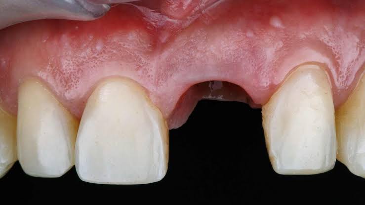
The stability of any artificial material to be placed within the jawbone depends on having a healthy bone wall that encircles it with a minimum thickness (at least 1.5 – 2 mm). Over the years following tooth extraction, the bone tissue thins due to the inability to bear chewing pressure and takes on a sharp knife-edge shape. This does not mean that the procedure cannot be performed; it merely requires the use of “advanced surgical” techniques that go beyond standard surgical procedures.
The discipline of jaw surgery in dentistry is based on the principle of reconstructing the lost tissue by utilizing the body’s repair mechanisms (regeneration). Cellular scaffolds (bone powders) applied to long-empty areas trigger the organism to send calcium and repair cells to that area. As a result of this biological maturation that lasts for months, a solid and dense base is medically created where titanium implants can anchor.
What Anatomical Changes Occur in Jawbone That Has Been Empty for Many Years?
The human skeletal system is a reactive structure that shapes itself according to the load imposed on it (Wolff’s Law). Natural tooth roots stimulate osteoblast (bone-forming) cells by transmitting the pressure generated while chewing food to the jawbone. When a tooth is extracted, this stimulatory signal is interrupted. The body codes this bone area, which it considers no longer functional, as structurally unnecessary and withdraws calcium and minerals from that area into the bloodstream.
| Elapsed Time | Physiological Change in Bone | Clinical and Aesthetic Reflections |
|---|---|---|
| 0 – 12 Months | A rapid resorption is observed, particularly in the cortical bone on the lip-cheek side (buccal). | Significant narrowing in bone width (thickness); deterioration in the gums. |
| 1 – 5 Years | In addition to horizontal narrowing, vertical (from top to bottom) height losses accelerate. | Sunken areas in the lips, deepening of nasolabial lines (wrinkles at the corners of the nose). |
| 5 Years and Over | The jawbone melts down to the basal (main) structure, taking on a slender strip form. | The inability of removable dentures to hold, the proximity of the jaw nerve to the surface, leading to the necessity for advanced surgery. |
What Advanced Surgical Methods Are Planned for Patients with Resorbed Jawbone in İzmir?
- Block Graft (Autogenous Transfer): This procedure involves fixing small bone blocks taken from the tip of the patient’s jaw or the wisdom tooth area to the resorbed area using micro-screws. It is the protocol that gives the highest cellular response.
- Guided Bone Regeneration (GBR): This involves placing particulate (powder form) bone materials in the resorbed area, covered with resorbable or non-resorbable titanium-supported membranes (barriers) to support the new bone formation from the outside.
- Split Crest (Bone Separation): This method involves the special tools used to stretch the very thin crest of bone apart and place titanium roots in between, filling the gaps with biomaterials.
How Does Bone Graft (Bone Powder) Application Function as a Medical Procedure in Long-Term Edentulousness?
The use of biomaterials is based on the principle of manipulating the body’s own repair capacity. Bone powders used in jaw surgery can originate from various sources: xenografts (animal-derived), allografts (human-derived), or synthetic (alloplastic) particles produced in laboratories. These materials are sterilized to remove proteins, leaving only calcium phosphate structures which consist of the mineral skeleton.
When the material is placed in the toothless area, the organism perceives it as a wound and accelerates blood flow to the area. The porous structure of the biomaterial traps blood clots, bringing along osteoblast (bone-forming) cells. Over an approximately six-month physiological cycle, as the added bone powders are gradually absorbed by the body, they leave behind a hard, fresh, and resilient bone mass capable of carrying titanium roots made from the patient’s own cells.
What Medical Criteria Are Used to Evaluate Cases with Bone Insufficiency within the Avrupadent Community?
In modern medical organizations, two-dimensional panoramic X-rays are not considered sufficient diagnostic tools for assessing bone losses. Identifying the thinness of bone tissue in relation to the lips and palate can only be accomplished with three-dimensional imaging systems that provide volumetric data. Physicians determine objectively with software which density class (from D1 to D4) the remaining bone belongs to, using the sections obtained in a virtual environment.
This detailed assessment is combined with the individual’s medical history. If a person has multiple tooth loss and advanced bone loss while also being a diabetes or osteoporosis patient, surgical planning is revised to implement a more gradual treatment protocol (not in a single session, but divided into sessions) to prevent disruption of tissue repair. The aim is to keep local trauma within the physiological limits that the body can tolerate.
How Does Long-Term Use of Removable Palate Prostheses Affect Jaw Bone?
Individuals often fall into the misconception that palate dentures completely replace missing teeth; however, these dentures are supported by soft tissue, not bone. The kilograms of pressure created during chewing directly impact the sensitive gum tissue and the underlying thin bone crest through the denture’s plastic. In human physiology, bone tissue exhibits a withdrawal (resorption) reflex from an area when subjected to direct and uncontrolled pressure in order to protect itself.
Atrophy related to this pressure progresses much more aggressively in the lower jaw. In patients who have used palate dentures for ten years or more, the lower jawbone resorbs to the extent that there are no anatomical peaks (crests) left to hold the denture in place; the denture moves freely in the mouth while speaking or eating. In such cases of severe tissue loss, the primary medical necessity becomes performing extensive bone repair (grafting) in order to place fixed titanium structures.
What Changes Will Long-Term Tooth Loss in the Upper Jaw Cause in Sinus Cavities?Long-term toothlessness in the back of the upper jaw allows for the downward expansion and sagging of the maxillary sinus air cavities (due to the effect of pneumatization). As the resistance created by tooth roots diminishes, the sinus membrane bone thins, forcing a decrease in vertical bone distance to millimeter levels, which necessitates sinus lifting (elevation) surgery.
Anatomically, maxillary sinuses are cavities that lighten the skull’s weight and warm the inhaled air. The base of these cavities is very thinly covered by a bone layer that is in contact with the roots of the upper jaw’s molars. When a tooth is extracted and the area heals, the air pressure within the sinus cavity exerts an expansion pressure on the underlying bone. Since it does not receive chewing stimulation, the weakened jawbone cannot resist this air pressure, causing the sinus membrane to sag downward (pneumatization).As a result of this physiological change, although it may appear that there is a thick jawbone when viewed from the mouth of the hospital, it is determined through tomography that the inside of the bone is hollow and as thin as paper. Attempting to directly place a titanium root in these cases causes the material to fall into the sinus cavity. With the “Sinus Lifting” operation, the membrane is pushed upwards and filled with bone powder (graft) to overcome this anatomical limitation within medical frameworks.
How Does the Prolongation of the Edentulous Process in the Lower Jaw Narrow the Nerve Canal Distance?As a result of the lower jawbone (mandible) being resorbed for many years, the lower jaw nerve (Nervus Alveolaris Inferior) that passes below the bone approaches dangerously close to the oral mucosa (surface). Since contact of the titanium screw with this nerve can lead to permanent numbness, in cases where vertical distance loss is significant, special short materials or vertical grafting are planned.
The nerve canal that runs through the lower jaw (mandible) like a pipeline is a vital anatomical structure that transmits the sensation from the lips and teeth to the brain. In a normally healthy anatomy, there is a safe bone distance between the tooth roots and this nerve canal. However, as the edentulous process prolongs, the bone melts and descends, and the safe distance between it and the nerve canal erodes millimeter by millimeter.In severely resorbed lower jaws, doctors may prefer to use special “short titanium roots” (short implants) whose lengths vary between 4-6 mm to avoid touching the nerve. In extreme resorption situations where short materials cannot be used, complicated medical protocols such as vertical bone grafting or, in very rare cases, surgical alteration of the position of the nerve (nerve transposition) may have to be implemented.
Can Surgical Intervention Be Planned for Individuals Who Have Been Edentulous for a Long Time in the Older Age Group?In elderly (geriatric) patients, chronological age is not the only criterion; rather, the individual’s cellular renewal capacity and blood values are taken into account. Surgical and titanium root procedures can be planned for individuals in their eighties with acceptable systemic health and completed medical checks, despite having a long history of bone quality providing mechanical resistance.
Although the aging process slows down cellular metabolism in the body to some extent, it does not eliminate the biological potential for bone tissue integration (osteointegration). Many individuals in advanced age enter medical treatment due to the inability of the dentures they have used for decades to retain, leading to nutritional deficiencies. This deficiency in the mouth, which is the starting point of the digestive system, directly threatens the general health and quality of life of elderly individuals.
In clinical assessments, doctors meticulously examine the medical medications used by elderly individuals (especially blood thinners and osteoporosis medications). Surgical stress is limited by using minimally invasive (least traumatic) surgical methods. The biological repair process is allowed a few months longer than that for younger patients, providing the body the necessary physiological time for cells to organize the bone grafts and titanium surfaces.
Why Does the Healing Timeline Differ in Treatments Performed After Long-Term Toothlessness?
Advanced surgeries aimed at increasing tissue volume, such as bone grafts or sinus lifting, require time for the body to transform the inorganic structures in that area into living human bone. This biological repair need adds an additional physiological healing timeline of an average of four to eight months to the standard osseointegration period.Titanium roots applied to an individual with newly extracted or preserved bone volume attach directly to the existing living bone walls. In this case, the osteointegration cycle (two-three in the lower jaw, three-four in the upper jaw) progresses at a standard level. However, in a patient who has been edentulous for a long time and has experienced bone resorption, the biomaterials (bone powders) applied are not alive themselves; the body must grow blood vessels into those materials to vivify them.
Biological Mechanisms That Extend the Healing Timeline:
\t- Angiogenesis (Vascularization): The process by which the body’s new capillary networks weave into the graft materials added is a physiological process. Cellular repair cannot occur without vascularization.
\t
- Bone Remodeling (Consolidation): The hardening process through the bonding of calcium particles, resulting in a bone block that firmly grips the titanium grooves.
\t
- Two-Stage Surgery: If bone resorption is very advanced, the physician does not place the screw on the same day. Initially, only bone powder is added to the area, and a waiting period is observed, followed by the placement of the screw in the newly formed bone with a second surgery; this can spread the overall timeline over a year.
How Do All-On-4 or All-On-6 Systems Provide a Prosthetic Alternative in Cases of Insufficient Bone Volume?
All-On-4 or All-On-6 systems allow for the placement of opening materials into the denser bone areas in the front by bridging severely resorbed bone areas and anatomical voids (sinus/nerves) in the posterior regions. This way, fixed prosthesis options can be created in a single session without the need for extensive bone powder surgeries in patients who have been edentulous for a long time.In cases of complete edentulism for many years, especially in the posterior regions, there may be almost no bone left. Elevating these areas with graft operations that last for months creates a tiring clinical schedule for the patient and also carries the risk of disrupting tissue repair. “All-on” concepts are designed to overcome these biomechanical and anatomical limitations.
The anterior regions (parts where incisors and canines are located) are areas where bone resorption occurs the least and slowest, preserving the cortical (hard) structure. The physicians create a wide load-bearing area (polygon) by positioning two titanium screws at angles ranging from 30 to 45 degrees, without touching the anatomical voids in the posterior region. The opening technique supports the mechanical tightening of the screws (primary stability), medically ending the period when the patient had to use a removable prosthesis.
How is the medical process managed in individuals with systemic diseases and those who have been edentulous for a long time?
In cases where systemic diseases such as diabetes, hypertension, or osteoporosis are associated with long-term edentulism; medical consultation is obtained from relevant branch doctors before advanced surgical procedures are performed. Keeping blood values and wound healing capacity within reference ranges is an essential approach for the normal course of cellular regeneration.Advanced surgical operations used to correct bone resorption create a widespread demand for tissue repair in the body (inflammatory response). The mechanisms of blood supply and perfusion in individuals with systemic disease may struggle to respond to this demand. For instance, in patients with uncontrolled diabetes, the vascularization of bone grafts added to the area may slow down, increasing the risk of graft infection. Limiting blood sugar levels (HbA1c) to medical thresholds prior to surgery reduces this risk.
Similarly, in individuals with a history of osteoporosis and those using heavy medications that slow bone destruction (such as bisphosphonates), extensive bone segments can lead to jawbone necrosis (cell death). Physicians prefer to avoid extensive graft surgeries in these medical conditions, opting instead to limit trauma with fewer but exposed titanium roots (like All-on-4) or temporarily modify medication regimens under physician supervision.
What Medical Findings Does Pre-Operative Radiological Diagnosis (3D Tomography) Reveal?
Three-dimensional dental tomography (CBCT) establishes the foundation for virtual surgical planning by revealing the vertical-horizontal millimeter dimensions of the jawbone that has been toothless for a long time, the precise distance to nerve canals, the anatomical sagging rates of sinus cavities, and the regional hardness (density) index of the bone in Hounsfield units.Looking at a toothless jaw through a mirror or panoramic (two-dimensional) X-ray for many years only reflects the visible part of the iceberg. While the gum may appear as a thick tissue, the underlying bone can be as thin as a razor blade. Two-dimensional films do not show the horizontal thickness facing the tongue and adjacent teeth. To minimize error margins in advanced surgical plans, obtaining volumetric data is a medical necessity.
Radiological sections are transferred to digital software to create surgical mapping. The physician determines on the computer screen before the surgery where and how much bone graft should be placed, and which millimeter screw will be used. The bone density measurement shows how much of the bone is spongy and how much is hard cortical tissue; it provides the physician with a clinical prediction about the mechanical resistance they will encounter during surgery.
How is the Long-Term Maintenance of the Functionality of the Bite Achieved After Treatment?
After titanium-supported prosthetic restorations are completed, it is essential to thoroughly perform daily hygiene maintenance (oral rinsing, superfloss) to maintain the volume of bone and tissue health obtained. Radiological checks will be carried out by physicians every six months, and medical plaque cleaning will prevent the development of peri-implantitis (inflammation of the surrounding tissue) and maintain the structural integrity of the system.The newly reconstructed bone tissue and prostheses, resulting from months of effort with advanced surgical techniques, cannot be excluded from medical follow-up once the operation process is completed. The new jawbone obtained with the support of biomaterials is quite sensitive to bacterial infections (plaque accumulation). If food residues and bacterial plaque accumulate around the prosthesis, the body will begin to rapidly dissolve that vital bone (peri-implantitis) while fighting infection.
The only physiological way to prevent this is regular mechanical cleaning. The undersides of zirconium or porcelain prostheses create areas that standard brushes cannot reach. The microbial cavities must be cleaned with pressurized water and special hygiene threads. During annual doctor check-ups, the balance of closure (occlusion) between the prosthesis and the opposing tooth is evaluated to prevent abnormal biting pressures that could trauma the jawbone.
Anatomically, maxillary sinuses are cavities that lighten the skull’s weight and warm the inhaled air. The base of these cavities is very thinly covered by a bone layer that is in contact with the roots of the upper jaw’s molars. When a tooth is extracted and the area heals, the air pressure within the sinus cavity exerts an expansion pressure on the underlying bone. Since it does not receive chewing stimulation, the weakened jawbone cannot resist this air pressure, causing the sinus membrane to sag downward (pneumatization).
As a result of this physiological change, although it may appear that there is a thick jawbone when viewed from the mouth of the hospital, it is determined through tomography that the inside of the bone is hollow and as thin as paper. Attempting to directly place a titanium root in these cases causes the material to fall into the sinus cavity. With the “Sinus Lifting” operation, the membrane is pushed upwards and filled with bone powder (graft) to overcome this anatomical limitation within medical frameworks.
How Does the Prolongation of the Edentulous Process in the Lower Jaw Narrow the Nerve Canal Distance?As a result of the lower jawbone (mandible) being resorbed for many years, the lower jaw nerve (Nervus Alveolaris Inferior) that passes below the bone approaches dangerously close to the oral mucosa (surface). Since contact of the titanium screw with this nerve can lead to permanent numbness, in cases where vertical distance loss is significant, special short materials or vertical grafting are planned.
The nerve canal that runs through the lower jaw (mandible) like a pipeline is a vital anatomical structure that transmits the sensation from the lips and teeth to the brain. In a normally healthy anatomy, there is a safe bone distance between the tooth roots and this nerve canal. However, as the edentulous process prolongs, the bone melts and descends, and the safe distance between it and the nerve canal erodes millimeter by millimeter.In severely resorbed lower jaws, doctors may prefer to use special “short titanium roots” (short implants) whose lengths vary between 4-6 mm to avoid touching the nerve. In extreme resorption situations where short materials cannot be used, complicated medical protocols such as vertical bone grafting or, in very rare cases, surgical alteration of the position of the nerve (nerve transposition) may have to be implemented.
Can Surgical Intervention Be Planned for Individuals Who Have Been Edentulous for a Long Time in the Older Age Group?In elderly (geriatric) patients, chronological age is not the only criterion; rather, the individual’s cellular renewal capacity and blood values are taken into account. Surgical and titanium root procedures can be planned for individuals in their eighties with acceptable systemic health and completed medical checks, despite having a long history of bone quality providing mechanical resistance.
Although the aging process slows down cellular metabolism in the body to some extent, it does not eliminate the biological potential for bone tissue integration (osteointegration). Many individuals in advanced age enter medical treatment due to the inability of the dentures they have used for decades to retain, leading to nutritional deficiencies. This deficiency in the mouth, which is the starting point of the digestive system, directly threatens the general health and quality of life of elderly individuals.
In clinical assessments, doctors meticulously examine the medical medications used by elderly individuals (especially blood thinners and osteoporosis medications). Surgical stress is limited by using minimally invasive (least traumatic) surgical methods. The biological repair process is allowed a few months longer than that for younger patients, providing the body the necessary physiological time for cells to organize the bone grafts and titanium surfaces.
Why Does the Healing Timeline Differ in Treatments Performed After Long-Term Toothlessness?
Advanced surgeries aimed at increasing tissue volume, such as bone grafts or sinus lifting, require time for the body to transform the inorganic structures in that area into living human bone. This biological repair need adds an additional physiological healing timeline of an average of four to eight months to the standard osseointegration period.Titanium roots applied to an individual with newly extracted or preserved bone volume attach directly to the existing living bone walls. In this case, the osteointegration cycle (two-three in the lower jaw, three-four in the upper jaw) progresses at a standard level. However, in a patient who has been edentulous for a long time and has experienced bone resorption, the biomaterials (bone powders) applied are not alive themselves; the body must grow blood vessels into those materials to vivify them.
Biological Mechanisms That Extend the Healing Timeline:
\t- Angiogenesis (Vascularization): The process by which the body’s new capillary networks weave into the graft materials added is a physiological process. Cellular repair cannot occur without vascularization.
\t
- Bone Remodeling (Consolidation): The hardening process through the bonding of calcium particles, resulting in a bone block that firmly grips the titanium grooves.
\t
- Two-Stage Surgery: If bone resorption is very advanced, the physician does not place the screw on the same day. Initially, only bone powder is added to the area, and a waiting period is observed, followed by the placement of the screw in the newly formed bone with a second surgery; this can spread the overall timeline over a year.
How Do All-On-4 or All-On-6 Systems Provide a Prosthetic Alternative in Cases of Insufficient Bone Volume?
All-On-4 or All-On-6 systems allow for the placement of opening materials into the denser bone areas in the front by bridging severely resorbed bone areas and anatomical voids (sinus/nerves) in the posterior regions. This way, fixed prosthesis options can be created in a single session without the need for extensive bone powder surgeries in patients who have been edentulous for a long time.In cases of complete edentulism for many years, especially in the posterior regions, there may be almost no bone left. Elevating these areas with graft operations that last for months creates a tiring clinical schedule for the patient and also carries the risk of disrupting tissue repair. “All-on” concepts are designed to overcome these biomechanical and anatomical limitations.
The anterior regions (parts where incisors and canines are located) are areas where bone resorption occurs the least and slowest, preserving the cortical (hard) structure. The physicians create a wide load-bearing area (polygon) by positioning two titanium screws at angles ranging from 30 to 45 degrees, without touching the anatomical voids in the posterior region. The opening technique supports the mechanical tightening of the screws (primary stability), medically ending the period when the patient had to use a removable prosthesis.
How is the medical process managed in individuals with systemic diseases and those who have been edentulous for a long time?
In cases where systemic diseases such as diabetes, hypertension, or osteoporosis are associated with long-term edentulism; medical consultation is obtained from relevant branch doctors before advanced surgical procedures are performed. Keeping blood values and wound healing capacity within reference ranges is an essential approach for the normal course of cellular regeneration.Advanced surgical operations used to correct bone resorption create a widespread demand for tissue repair in the body (inflammatory response). The mechanisms of blood supply and perfusion in individuals with systemic disease may struggle to respond to this demand. For instance, in patients with uncontrolled diabetes, the vascularization of bone grafts added to the area may slow down, increasing the risk of graft infection. Limiting blood sugar levels (HbA1c) to medical thresholds prior to surgery reduces this risk.
Similarly, in individuals with a history of osteoporosis and those using heavy medications that slow bone destruction (such as bisphosphonates), extensive bone segments can lead to jawbone necrosis (cell death). Physicians prefer to avoid extensive graft surgeries in these medical conditions, opting instead to limit trauma with fewer but exposed titanium roots (like All-on-4) or temporarily modify medication regimens under physician supervision.
What Medical Findings Does Pre-Operative Radiological Diagnosis (3D Tomography) Reveal?
Three-dimensional dental tomography (CBCT) establishes the foundation for virtual surgical planning by revealing the vertical-horizontal millimeter dimensions of the jawbone that has been toothless for a long time, the precise distance to nerve canals, the anatomical sagging rates of sinus cavities, and the regional hardness (density) index of the bone in Hounsfield units.Looking at a toothless jaw through a mirror or panoramic (two-dimensional) X-ray for many years only reflects the visible part of the iceberg. While the gum may appear as a thick tissue, the underlying bone can be as thin as a razor blade. Two-dimensional films do not show the horizontal thickness facing the tongue and adjacent teeth. To minimize error margins in advanced surgical plans, obtaining volumetric data is a medical necessity.
Radiological sections are transferred to digital software to create surgical mapping. The physician determines on the computer screen before the surgery where and how much bone graft should be placed, and which millimeter screw will be used. The bone density measurement shows how much of the bone is spongy and how much is hard cortical tissue; it provides the physician with a clinical prediction about the mechanical resistance they will encounter during surgery.
How is the Long-Term Maintenance of the Functionality of the Bite Achieved After Treatment?
After titanium-supported prosthetic restorations are completed, it is essential to thoroughly perform daily hygiene maintenance (oral rinsing, superfloss) to maintain the volume of bone and tissue health obtained. Radiological checks will be carried out by physicians every six months, and medical plaque cleaning will prevent the development of peri-implantitis (inflammation of the surrounding tissue) and maintain the structural integrity of the system.The newly reconstructed bone tissue and prostheses, resulting from months of effort with advanced surgical techniques, cannot be excluded from medical follow-up once the operation process is completed. The new jawbone obtained with the support of biomaterials is quite sensitive to bacterial infections (plaque accumulation). If food residues and bacterial plaque accumulate around the prosthesis, the body will begin to rapidly dissolve that vital bone (peri-implantitis) while fighting infection.
The only physiological way to prevent this is regular mechanical cleaning. The undersides of zirconium or porcelain prostheses create areas that standard brushes cannot reach. The microbial cavities must be cleaned with pressurized water and special hygiene threads. During annual doctor check-ups, the balance of closure (occlusion) between the prosthesis and the opposing tooth is evaluated to prevent abnormal biting pressures that could trauma the jawbone.
The nerve canal that runs through the lower jaw (mandible) like a pipeline is a vital anatomical structure that transmits the sensation from the lips and teeth to the brain. In a normally healthy anatomy, there is a safe bone distance between the tooth roots and this nerve canal. However, as the edentulous process prolongs, the bone melts and descends, and the safe distance between it and the nerve canal erodes millimeter by millimeter.
In severely resorbed lower jaws, doctors may prefer to use special “short titanium roots” (short implants) whose lengths vary between 4-6 mm to avoid touching the nerve. In extreme resorption situations where short materials cannot be used, complicated medical protocols such as vertical bone grafting or, in very rare cases, surgical alteration of the position of the nerve (nerve transposition) may have to be implemented.
Can Surgical Intervention Be Planned for Individuals Who Have Been Edentulous for a Long Time in the Older Age Group?In elderly (geriatric) patients, chronological age is not the only criterion; rather, the individual’s cellular renewal capacity and blood values are taken into account. Surgical and titanium root procedures can be planned for individuals in their eighties with acceptable systemic health and completed medical checks, despite having a long history of bone quality providing mechanical resistance.
Although the aging process slows down cellular metabolism in the body to some extent, it does not eliminate the biological potential for bone tissue integration (osteointegration). Many individuals in advanced age enter medical treatment due to the inability of the dentures they have used for decades to retain, leading to nutritional deficiencies. This deficiency in the mouth, which is the starting point of the digestive system, directly threatens the general health and quality of life of elderly individuals.
In clinical assessments, doctors meticulously examine the medical medications used by elderly individuals (especially blood thinners and osteoporosis medications). Surgical stress is limited by using minimally invasive (least traumatic) surgical methods. The biological repair process is allowed a few months longer than that for younger patients, providing the body the necessary physiological time for cells to organize the bone grafts and titanium surfaces.
Why Does the Healing Timeline Differ in Treatments Performed After Long-Term Toothlessness?
Titanium roots applied to an individual with newly extracted or preserved bone volume attach directly to the existing living bone walls. In this case, the osteointegration cycle (two-three in the lower jaw, three-four in the upper jaw) progresses at a standard level. However, in a patient who has been edentulous for a long time and has experienced bone resorption, the biomaterials (bone powders) applied are not alive themselves; the body must grow blood vessels into those materials to vivify them.
-
\t
- Angiogenesis (Vascularization): The process by which the body’s new capillary networks weave into the graft materials added is a physiological process. Cellular repair cannot occur without vascularization.
- Bone Remodeling (Consolidation): The hardening process through the bonding of calcium particles, resulting in a bone block that firmly grips the titanium grooves.
- Two-Stage Surgery: If bone resorption is very advanced, the physician does not place the screw on the same day. Initially, only bone powder is added to the area, and a waiting period is observed, followed by the placement of the screw in the newly formed bone with a second surgery; this can spread the overall timeline over a year.
\t
\t
How Do All-On-4 or All-On-6 Systems Provide a Prosthetic Alternative in Cases of Insufficient Bone Volume?
In cases of complete edentulism for many years, especially in the posterior regions, there may be almost no bone left. Elevating these areas with graft operations that last for months creates a tiring clinical schedule for the patient and also carries the risk of disrupting tissue repair. “All-on” concepts are designed to overcome these biomechanical and anatomical limitations.
The anterior regions (parts where incisors and canines are located) are areas where bone resorption occurs the least and slowest, preserving the cortical (hard) structure. The physicians create a wide load-bearing area (polygon) by positioning two titanium screws at angles ranging from 30 to 45 degrees, without touching the anatomical voids in the posterior region. The opening technique supports the mechanical tightening of the screws (primary stability), medically ending the period when the patient had to use a removable prosthesis.
How is the medical process managed in individuals with systemic diseases and those who have been edentulous for a long time?
Advanced surgical operations used to correct bone resorption create a widespread demand for tissue repair in the body (inflammatory response). The mechanisms of blood supply and perfusion in individuals with systemic disease may struggle to respond to this demand. For instance, in patients with uncontrolled diabetes, the vascularization of bone grafts added to the area may slow down, increasing the risk of graft infection. Limiting blood sugar levels (HbA1c) to medical thresholds prior to surgery reduces this risk.
Similarly, in individuals with a history of osteoporosis and those using heavy medications that slow bone destruction (such as bisphosphonates), extensive bone segments can lead to jawbone necrosis (cell death). Physicians prefer to avoid extensive graft surgeries in these medical conditions, opting instead to limit trauma with fewer but exposed titanium roots (like All-on-4) or temporarily modify medication regimens under physician supervision.
What Medical Findings Does Pre-Operative Radiological Diagnosis (3D Tomography) Reveal?
Looking at a toothless jaw through a mirror or panoramic (two-dimensional) X-ray for many years only reflects the visible part of the iceberg. While the gum may appear as a thick tissue, the underlying bone can be as thin as a razor blade. Two-dimensional films do not show the horizontal thickness facing the tongue and adjacent teeth. To minimize error margins in advanced surgical plans, obtaining volumetric data is a medical necessity.
Radiological sections are transferred to digital software to create surgical mapping. The physician determines on the computer screen before the surgery where and how much bone graft should be placed, and which millimeter screw will be used. The bone density measurement shows how much of the bone is spongy and how much is hard cortical tissue; it provides the physician with a clinical prediction about the mechanical resistance they will encounter during surgery.
How is the Long-Term Maintenance of the Functionality of the Bite Achieved After Treatment?
The newly reconstructed bone tissue and prostheses, resulting from months of effort with advanced surgical techniques, cannot be excluded from medical follow-up once the operation process is completed. The new jawbone obtained with the support of biomaterials is quite sensitive to bacterial infections (plaque accumulation). If food residues and bacterial plaque accumulate around the prosthesis, the body will begin to rapidly dissolve that vital bone (peri-implantitis) while fighting infection.
The only physiological way to prevent this is regular mechanical cleaning. The undersides of zirconium or porcelain prostheses create areas that standard brushes cannot reach. The microbial cavities must be cleaned with pressurized water and special hygiene threads. During annual doctor check-ups, the balance of closure (occlusion) between the prosthesis and the opposing tooth is evaluated to prevent abnormal biting pressures that could trauma the jawbone.