How Are Dental Implants Made? Treatment Process Step by Step
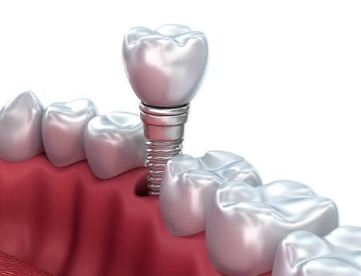
Planned implantology procedures in clinical settings aim to medically support the functions of lost chewing. The method is based on the principle that biocompatible titanium materials placed in the jawbone form a stable infrastructure by integrating with surrounding tissues over time. This method is one of the dental disciplines applied to individuals who have reached adulthood and have a health profile suitable for surgical intervention.
Dental deficiencies in the mouth not only interrupt the digestive process, which is the beginning of the digestive system but can also affect the pronunciation of phonetic sounds and the structural balance of jaw anatomy. These structures, prepared using titanium screws, receive support directly from the bone tissue without placing physical stress on neighboring teeth. In this content we prepared, we will examine all clinical phases of the application in a medical perspective, from the preparatory phase to the placement of the prosthesis, step by step.
How is the Preliminary Examination and Planning Stage of Dental Implants Conducted?
The preliminary examination stage is realized by the clinician evaluating the mouth clinically and examining the anatomical structure of the jawbone through three-dimensional radiological scans. The diameter and length of the titanium material to be used in this process are calculated with millimetric values, creating an individualized surgical map and medical schedule for the person.
The clinical evaluation process begins with a detailed inquiry into the individual’s history of systemic diseases. Blood pressure values, the current diabetes status, use of anticoagulant medications, and other medical data affecting bone metabolism are recorded. During the oral examination, the current condition of the gums is assessed. If active infection signs such as gingivitis or periodontitis are detected, the periodontal treatment for this area is prioritized according to medical protocols. It is a fundamental rule of medical standards not to proceed with surgical intervention without establishing a healthy foundation.
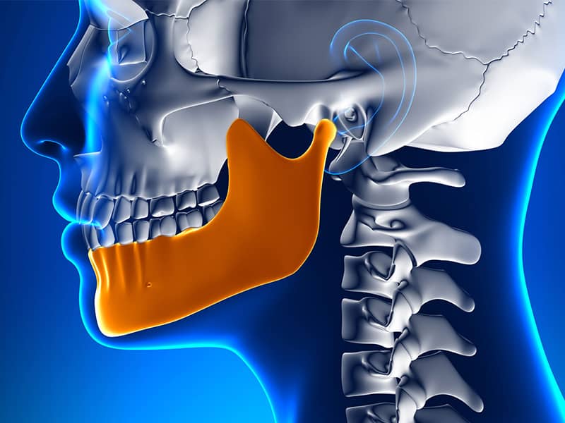
In the imaging phase, digital sections obtained from Dental Volumetric Tomography (CBCT) devices are of great importance. These scans clearly reveal the degree of sagging of the maxillary sinus cavities located in the upper jaw and the position of the mandibular nerve pathway running through the lower jawbone. The physician plans an appropriate positioning scheme in digital form by obtaining measurements of the thickness and density of the jawbone to avoid damaging the anatomical neighbors.
What Steps Are Followed During Surgical Application?
The surgical procedure consists of the steps involving the local anesthesia of the relevant jaw area, the millimetric separation of the gum, and the placement of a titanium screw into the jawbone. After the procedure, the gum is returned to its original position and closed with sutures, allowing the wound tissue to rest biologically under clinical guidelines.
The clinical operation is performed in an environment where asepsis and antisepsis (sterilization) rules are strictly followed. After local anesthesia is injected into the surgical area, nerve transmission is blocked. Once the area is numbed, the physician elevates the gum tissue with a medical scalpel to expose the cortical (outer) surface of the jawbone. Once the bone surface is reached, specialized drills (perforating medical instruments) are used in succession to open a cavity at the designated width and depth.
During the preparation of the cavity, sterilized cooling fluids (saline solution) are dripped onto the area throughout the procedure to prevent thermal damage (necrosis) to the bone tissue. Once the planned depth is reached, the dental implant is taken from its sterile package and fixed to the cavity prepared with special guides. After the upper part of the screw is covered with a small cap, the gum tissue is repositioned over or around the screw and stitched with fine sutures. The surgical phase takes an average of half an hour for a single area, depending on the number of teeth involved.
How Long Does the Healing and Bone Integration (Osseointegration) Phase Take?
The healing and osseointegration phase takes about two to three months on the lower jaw and approximately three to four months on the upper jaw. During this time, the osteoblast cells in the jawbone adhere to the titanium surface, forming a cellular network at a micro level and creating a stable foundation for the prosthetic crown.
Osseointegration is a biological adaptation phase that forms the basis of medical implantology. The body’s repair mechanisms are activated from the moment the screw is placed into the jawbone. Bone cells migrate to the specially treated surface of titanium, creating new bone islands here. Due to the anatomical structure, the mandible, being harder and more cortical, may undergo the cellular integration process more quickly compared to the maxilla. The spongy (cancellous) structure of the maxilla requires extra time for biological maturation.
In this period, the course of the process is monitored with X-rays taken according to the physiological structure of the clinic and the patient. For example, in Avrupadent process management and general clinical protocols, it is requested from the patient that no physiological load (bite pressure) is placed on the screw during this waiting period. Otherwise, pressures applied before the cell structures have solidified may cause micro-movements, leading to titanium being surrounded by soft tissue (fibrous binding) instead of bone. To prevent this situation, it is quite important for the region to rest.
How is the Prosthetic Tooth Placement Process Progressed?
The placement of prosthetic teeth progresses after confirming the biological integration of the screw with the jawbone, by sending measurements taken from the mouth to the laboratory. The prepared porcelain or zirconium superstructures are fixed to the titanium root via intermediate pieces (abutment), thereby medically supporting the patient’s biting function.
When the standard level of soft tissue regeneration is completed, the stage of shaping the gum form is reached. If the implant is covered, a small local intervention is made to open the gum and metallic pieces called “healing abutment” (gingiva former) are placed over the screw. These abutments stay in the mouth for about 7-10 days, allowing the gum to contour naturally according to a normal tooth form and creating an aesthetic emergence profile.
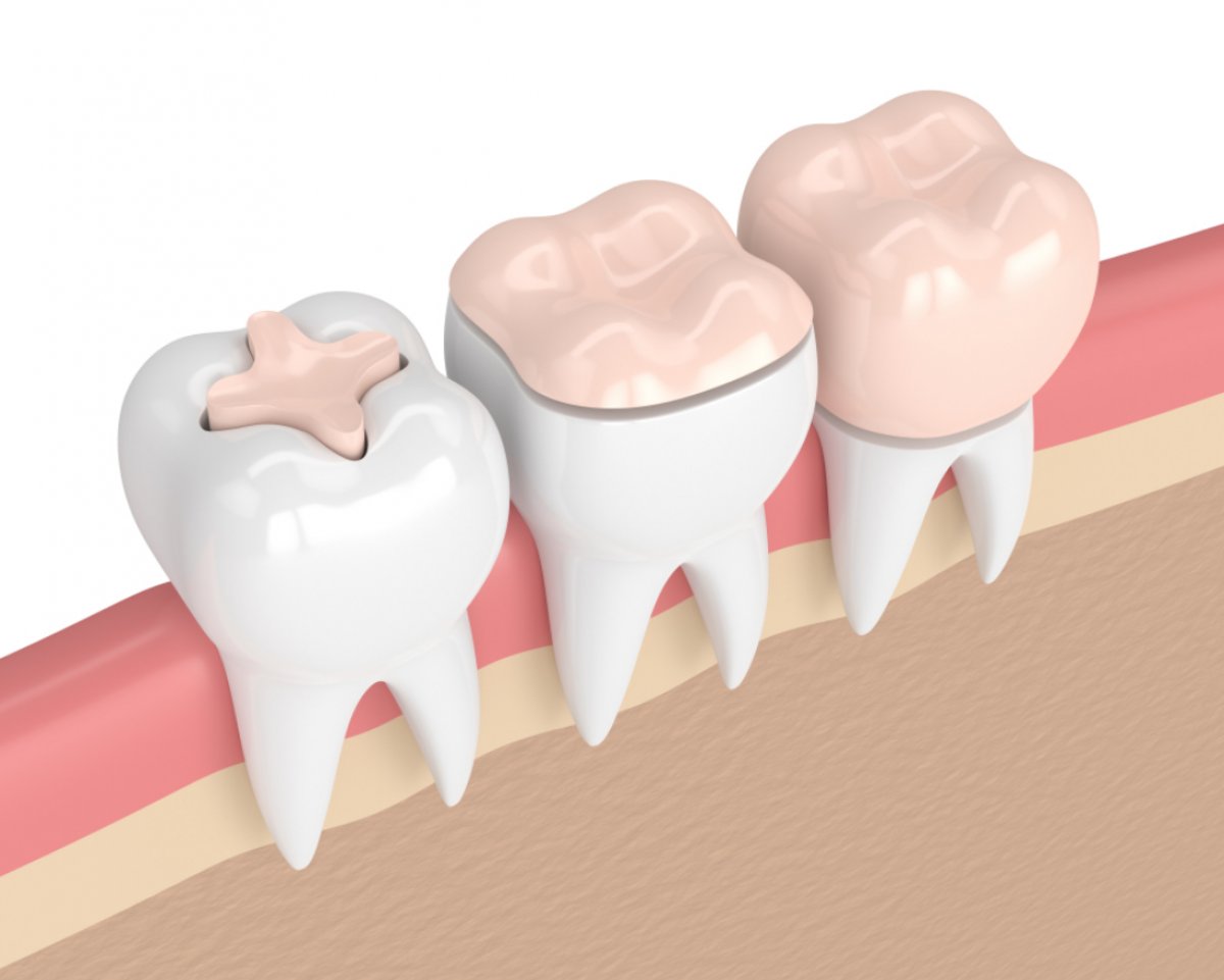
After the gum has been shaped, the measurement process begins. Three-dimensional records of the edentulous area and opposing arch are created using classic silicone materials or intraoral (in-mouth) digital scanners. Dental technicians produce porcelain or zirconium crowns based on the dentist’s prescription from these measurements. When the produced crowns are placed in the mouth, height trials are conducted. The occlusal contacts of the arches are checked with sensitive papers, and if appropriate, the prostheses are fixed with screw systems or dental cements (medical adhesives).
How Does the Process Work When the Jawbone Volume is Insufficient?
In cases where the jawbone volume is insufficient, additional surgical procedures such as adding medical bone powder (graft) or elevating the sinus floor in the upper jaw (sinus lifting) come into play. This allows for the biological depth and thickness necessary for placing the titanium root to be achieved in the area.
After many years have passed since the loss of teeth, the anatomical resorption known as “resorption” begins because the chewing pressure is not transmitted to the jawbone in that area. When the bone volume decreases in height and width, it mechanically becomes difficult to place a standard implant securely in that region. To overcome this obstacle, materials obtained from synthetic, rigid sources (bovine) or the patient’s autogenous (own) bone are surgically applied to the deficient area. To prevent the spread of this added powder structure, it is covered with barrier membranes.
Directly above the area where the upper jaw has the molar teeth, there are anatomical cavities known as “maxillary sinuses.” Due to resorption, when the sinus floor approaches toward the oral cavity, bone height can decrease to millimeters. In a sinus lifting operation, the thin membrane that reflects this cavity is pushed upward with special instruments to prevent damage, and the resulting cavity is filled with a bone graft. The integration of these added grafts into the patient’s own cellular structure, leading to the formation of new bone tissue, may require a waiting period of around 4 to 6 months, depending on the biological speed of the body.
How Do Digital Dentistry Tools Contribute to the Implantology Process?
Digital dentistry tools allow for virtual planning before the operation through three-dimensional scanners and software, contributing to the process. With the use of a surgical guide, incision sizes can be minimized, anatomical risk areas can be analyzed in advance, and the procedure can be managed within the planned limits.
In addition to traditional methods, digital workflows have become an integral part of today’s clinical processes. Intraoral optical scanners transfer precise digital copies of teeth and gums to the computer environment within seconds. This data is superimposed with the hospital’s tomography images in CAD/CAM (Computer-Aided Design/Manufacturing) software. Doctors virtually determine the location where the implant screw will be placed down to the millimeter on the computer screen.
Through this virtual planning, a transparent plate (surgical guide) that fits perfectly on the patient’s oral structure is produced using 3D printers. The openings and directions of the holes where the doctor will apply the drill are prepared on the plate. When this plate is placed in the mouth during the operation, the doctor can perform the procedure without straying from the planned opening and, in most cases, without the need to widely open the gums (flap lifting). This method helps to control swelling and tissue sensitivity situations after the procedure since it narrows the intervention area.
How Should Post-Treatment Oral Care and Controls Be Organized?
Post-treatment oral care should be organized based on the principle of brushing with fluoridated toothpaste at least twice a day, using interdental cleaners (dental floss, interdental brush), and minimizing plaque accumulation. Additionally, radiological and clinical tissue checks should continue with regular doctor visits every six months or annually.
Titanium and porcelain materials do not undergo tooth decay (cavitation) due to their structure; however, this does not mean that the area does not need to be cleaned. The gum tissue surrounding the screw and the underlying jawbone reacts to bacterial plaque just like natural teeth. Insufficient oral hygiene can lead to food residues and bacteria accumulating around the gum tissue, which can cause inflammation. If this situation progresses, a medical condition called ‘peri-implantitis’ may develop, leading to bone resorption in the jaw.
To move past tables like this, a mechanical cleaning routine is essential. The edges of crowns (covers) that the toothbrush cannot reach, specially designed interface brushes, and water flossers should be used for cleaning. During control appointments, the doctor examines whether there is bleeding in the gums or the formation of pockets with probes (measuring tools). In necessary cases, small dental stone cleaning sessions can be conducted to remove attachments from the area using professional methods, thereby medically supporting the health of surrounding tissues.
What are the Distinguishing Features of Implantology Applications Compared to Traditional Prosthetics?
Implantology applications differ from traditional prosthetics by deriving support directly from the jawbone without touching the adjacent healthy teeth. This approach transmits chewing forces directly to the bone, maintaining physiological stimulation in the area and eliminating the need for support tooth reduction seen in classic bridge systems.
In treatment planning, the method to be chosen is clarified based on the size of the existing edentulousness and the condition of the anatomy as a result of a clinical examination. The table below summarizes the biomechanical relationship of both systems with the tissues and the differences in their functioning:
| Comparison Criteria | Titanium Screw Supported Applications | Traditional Bridges and Dentures |
|---|---|---|
| Effect on Healthy Teeth | No cutting/alteration procedures are performed on the enamel or anatomical structure of adjacent teeth. | To construct a bridge, the healthy teeth on the right and left of the gap must be reduced. |
| Force Applied to Bone Tissue | Pressure is transmitted directly to the jawbone, aiding in bone metabolism through physiological stimulation. | Vertical and horizontal melting may occur over time in the edentulous area due to the unsupported condition. |
| Structural Independence | The edentulous area is rehabilitated with an independent crown and root system on its own. | The existing prosthesis works in conjunction with neighboring teeth (as a block), and there is no independent structure. |
| Oral Cleaning Process | It can be cleaned in all directions with standard brushing and dental floss, just like an anatomical tooth. | Special bridge cleaning threads should be used for food that gets stuck in the gap beneath the bridge body. |
| Clinical Process Expectation | The phase of biological osseointegration is expected to be spread over a few months in a timeline. | After tissue sampling, the laboratory phase is completed within a few weeks. |
Frequently Asked Questions (FAQ)
1. Is there a possibility of allergic reactions to titanium material?
Pure titanium and medical alloys are metals that show high biocompatibility with human tissue. Cases of allergic reactions developing against titanium have been reported to be quite rare in the literature. For patients who have concerns about metal sensitivity, special medical tests may be conducted before the procedure, or alternatively, ceramic-based zirconia materials may be considered.
2. What type of anesthesia is applied during the surgical procedure?
Surgical procedures are carried out under local anesthesia of the relevant area in accordance with standard protocols. As the medication penetrates the tissues, transmission along the related nerve pathway is blocked. If the doctor deems it necessary or if the patient has a severe dental phobia (fear of dentists), sedation or general anesthesia methods may be considered under the supervision of medical practitioners.
3. Can this treatment be applied when there are gum diseases?
Surgical procedures are not undertaken if there is active inflammation, bleeding, or infection in the gums, such as periodontitis. Placing medical material in an inflamed area risks the integration of the tissue. First, periodontal treatments such as tartar cleaning or curettage are applied to bring the oral flora to a healthy level.
4. What should I pay attention to for wound healing after the operation?
The area where surgical stitches are present should be protected from mechanical impacts and hard objects. Minor bleeding in the form of light ooze within the first 24 hours after the procedure is a normal response of the body. It is necessary to apply a cold compress externally to the area, follow the medical prescription given by the doctor, and avoid activities that increase blood circulation, such as hot baths, for a few days.
5. How does the procedure work for patients with heart or blood pressure issues?
For patients with cardiovascular diseases or those using blood thinners (anticoagulants), the process is carried out in consultation with the cardiology specialist overseeing the patient. Temporary adjustments in medication usage may be required to manage the risk of bleeding during the surgical procedure, depending on the approval of the medical doctor.
6. Can multiple titanium screws be placed within the same day?
Yes, if the anatomical structure of the jawbone is found to be suitable based on radiological assessments, multiple dental implants can be placed in the bone during the same clinical session. This allows for the management of both the surgical process and the healing period within a single time frame.
7. What are All-on-4 or All-on-6 systems?
In cases of complete edentulism (absence of any teeth), strategic locations of the jawbone are used to place 4 or 6 titanium screws at specific angles, onto which a fixed prosthesis covering all teeth is anchored. Special placement techniques are used in order to overcome anatomical difficulties or bone deficiencies in the posterior areas.
8. What is the purpose of the healing abutment (gingiva former)?
Once osseointegration is complete, these are cylindrical metal pieces that are attached to the upper part of the screw. Their purpose is to create an anatomical roundness on the gum tissue that has undergone a surgical procedure. Thus, when transitioning to the prosthetic phase, the crown tooth that will be attached looks aesthetically like it emerges from the gum tissue like a natural tooth.
9. Is the use of bone powder (graft) a necessary procedure?
No, it is not mandatory. It is only included in the plan by the physician in cases where the length or width of the jawbone does not meet medical standards. In individuals with sufficient bone volume, the main surgical phase can proceed directly. The use of grafts is a supportive procedure that entirely depends on the extent of existing anatomical deformity.
10. Why are trials necessary during prosthesis preparation?
It is necessary to check whether the teeth prepared in the laboratory are compatible with the patient’s jaw closure relation, lip support, and phonetic (speech) habits. During the trials, color harmony, height balance, and functional contacts are evaluated, and necessary revisions are made. Only when all criteria are medically suitable is the simulation (construction/screwing) process initiated.
11. How is this process managed in individuals with temporomandibular joint discomfort?
In individuals with temporomandibular joint (TMJ) discomfort or night teeth grinding (bruxism) issues, the goal is to prevent uncontrolled load on newly made crowns. In these patients, after the procedure, the production of joint-protective night plates or medical relaxing treatments aimed at chewing muscles (botulinum toxin applications, etc.) can also be considered.
12. Do zirconium and porcelain materials cause odor in the mouth?
Materials like zirconium or porcelain used in crowns are odorless and have special surfaces that do not harbor bacteria. However, if sufficient brushing is not done on the edges of the prosthetics, food residues that accumulate may decompose over time and cause odor. The source of the odor is not the material but the level of oral hygiene maintained in the surrounding area.
13. Are screws or prosthetic connectors used to connect titanium parts?
Both methods can be preferred according to the clinic’s requirements. In cement-retained prostheses, the upper structure is fixed to the intermediate piece with a medical agent. In screw-retained systems, the prosthesis is attached to the body by passing through a tiny screw hole and its upper part is covered with composite filling. Screw-retained systems allow the prosthesis to be easily removed and cleaned by the physician when needed.
14. When should the stitches in the operation area be removed?
Depending on the type of suture material and the patient’s wound healing rate, stitches are generally removed in a clinical setting 7 to 10 days after surgery. If self-dissolving (resorbable) sutures were used, it is expected that the thread will disappear through hydrolysis within the tissue. During the time the stitches are present, it is important to protect that area from hard brush impacts for healing.
15. Is there a problem entering X-ray imaging or MRI devices?
Titanium is not a magnetic (ferromagnetic) material. Thanks to this property, it does not get affected or heat up when entered into the magnetic field of MRI (Magnetic Resonance) devices. Similarly, it does not emit a signal in the security scanners at airports. Therefore, it does not pose any obstacle for individuals’ future medical imaging needs.