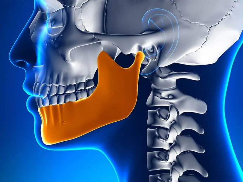
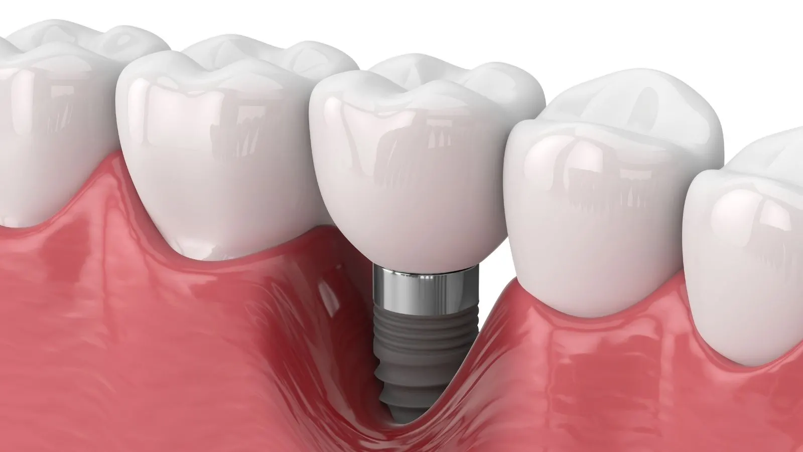
The short answer to “what is the maxilla” is this: the maxilla is the upper jaw bone, located in the middle of the face and holding the upper teeth. Unlike the mandible, the lower jaw bone, it is fixed to the skull and does not move.
The maxilla is the upper jaw bone
The maxilla is the bone that houses the roots of the upper teeth, forms most of the palate and surrounds the nasal cavity and the maxillary sinus. The upper teeth sit in sockets within this bone. Because it joins the cheekbone and other facial bones, the maxilla gives shape to the middle part of the face. The bone structure of the upper jaw is especially important in implant planning; you can read more in our article on sinus lifting before implants.
How it differs from the mandible
The mandible is the lower jaw bone, and its most obvious difference from the maxilla is that it moves. It attaches to the skull through the jaw joints, and it is the part that opens and closes when you speak, swallow and chew. The maxilla, by contrast, stays still. Together the two bones allow the teeth to close against each other.
- Maxilla: Upper jaw, fixed, holds the upper teeth, sits next to the sinuses.
- Mandible: Lower jaw, mobile, holds the lower teeth, connects to the skull through the jaw joint.
Its role in chewing and facial structure
During chewing the maxilla gives the upper teeth a steady base and determines the structure of the middle of the face. While the lower jaw moves, the upper teeth rest against this stationary bone, so food can be ground. The lines around the nose, cheeks and upper lip also depend largely on this bone structure.
How it looks on an X-ray
On an X-ray the maxilla appears as the bone structure above the mandible, seen together with the sinus cavities. Bone density and air-filled spaces show up in different shades on the film; by looking at these differences the dentist assesses the tooth roots, bone loss or findings such as a cyst. For more detail on imaging, see our page on what a dental X-ray is and how it is taken.
Panoramic and periapical X-ray terms
A panoramic X-ray (orthopantomogram) shows the upper and lower jaws, all the teeth and the jaw joints in a single frame, while a periapical X-ray shows only one or two teeth in detail, including the root tip. Neither replaces the other; the dentist requests one or both depending on the purpose.
- Panoramic X-ray: Gives a general overview; impacted teeth, the overall condition of the jaw bone and the sinuses can be seen.
- Periapical X-ray: Used for details of a single tooth, such as the root and surrounding bone, decay or inflammation at the root tip.
When a CT scan is requested
When two-dimensional films do not give enough detail, a CT scan is requested for a three-dimensional image. For example, it is used when bone volume must be measured for implant planning, when the position of impacted teeth must be determined, or when closeness to the sinuses and nerves must be assessed. For details, see our page on dental tomography (dental CT scan).
When the dentist refers you
In cases of swelling, long-lasting pain, sinus complaints, a suspicious appearance in the jaw bone or a need for surgical planning, the dentist may request additional imaging or refer you to a specialist field. For surgical topics related to the jaw bone, you can read our article on what oral, facial and jaw surgery looks at. Rather than interpreting X-ray findings on your own, it is better to review them with the dentist during an examination; to make an appointment, you can reach us through our contact page.
Frequently asked questions
What is the maxilla?
The maxilla is the upper jaw bone that holds the upper teeth, forms most of the palate and is fixed to the skull.
What is the mandible?
The mandible is the lower jaw bone. It holds the lower teeth and moves by connecting to the skull through the jaw joints.
What is the difference between the maxilla and the mandible?
The maxilla is on top and fixed; the mandible is below and mobile. Together they let the teeth close during chewing and speaking.
What is a panoramic X-ray?
A panoramic X-ray is a wide image that shows both jaws, all the teeth and the jaw joints in a single frame.
What is a periapical X-ray?
A periapical X-ray is a small film that shows one or two teeth in detail, together with the root tip and the surrounding bone.