What is Sinus Lift Surgery and When Might It Be Necessary?
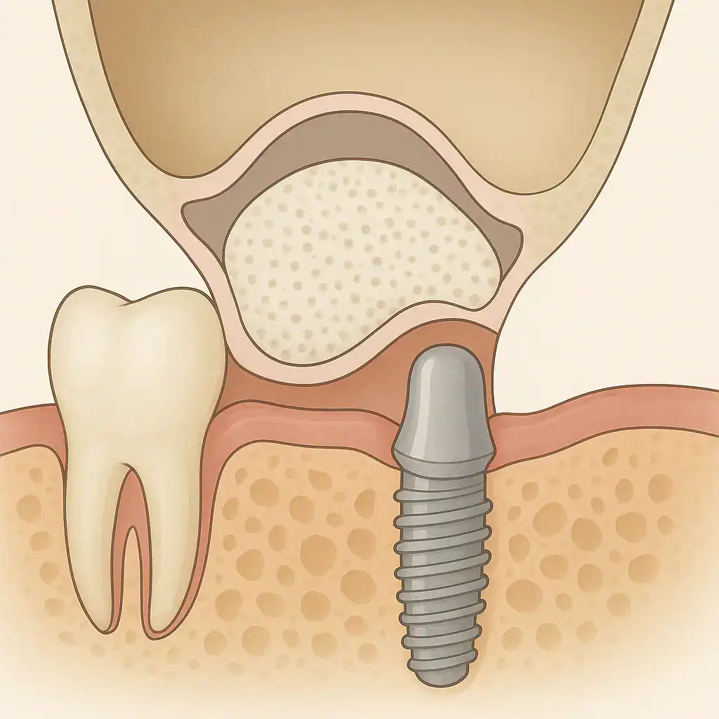
One of the fundamental anatomical constraints encountered while planning restorations of the upper jaw in dental clinics is the downward extension of the air spaces known as maxillary sinuses into the jawbone. These air chambers, which have physiological functions such as relieving the weight of the skull and humidifying the inhaled air, exhibit a tendency to expand downward toward the bone base due to gravity and atmospheric pressure, especially after the loss of molar teeth. This expansion complicates the anatomical positioning of the artificial root forms intended to be placed in the area by narrowing the vertical volume of the jawbone.
The advanced surgical approach applied to manage this anatomical narrowing medically and restore a functional bone height to the area is called sinus lifting (sinus floor elevation). This procedure, which occupies an important place in oral surgery practice, is based on the principle of gently mobilizing the delicate membrane that covers the interior of the air space upward and supporting this newly formed biological cavity with a bone graft (powder). In this context, the biological background of the sinus lift procedure, the medical techniques applied (both open and closed), surgical processes, management of complications, and its clinical integration with Implantology procedures will be discussed in detail.
Where Are Maxillary Sinus Cavities Anatomically Located?
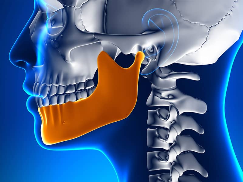
There are four pairs of air cavities known as paranasal sinuses in the human skull. Among these, the maxillary sinuses are the largest structures directly related to dentistry. Anatomically, there is a very thin bony wall between the roots of the upper small (premolar) and large (molar) teeth and the floor of the maxillary sinus. In some individuals, these tooth roots may extend into the sinus cavity. As long as the relevant teeth remain healthy in the mouth, mechanical stimuli arising from chewing forces nourish the bone and support the floor of the sinus to stay stable in its natural position.
These cavities, lined with respiratory epithelium and covered by a thin membrane (Schneiderian membrane), undertake vital medical tasks such as adjusting sound resonance and warming and filtering the inhaled air. In medical evaluations in dentistry, this anatomical proximity requires great precision. Before any surgical intervention, the sizes of the sinuses in the posterior region of the upper jaw, the thickness of the membrane, and whether there are polyps (soft tissue growths) or sinusitis in the area are examined through radiological investigations.
What Biological Necessity Does the Sinus Lifting Procedure Address?
When tooth loss occurs, the alveolar bone (the part of the jaw that supports the teeth) enters a physiological shrinkage process because it is not stimulated by biting pressure. In the upper jaw, this shrinkage occurs not only upwards from inside the mouth but also downwards from within. The air pressure inside the maxillary sinus pushes the unsupported bony floor downwards, causing what is known as “sinus pneumatization,” an anatomical sagging. Over the months and years, the bone height in the posterior area of the upper jaw can decrease to a thin shell layer of 1-2 millimeters.
Such a thin layer of bone cannot provide biomechanical support for an artificial root to be added on top. If intervention is made without adding volume to this area, the placed material may fall into the sinus cavity, or a rupture of the membrane can create a ground for chronic infections (sinusitis). Doctors elevate the sinus membrane to its original place in order to overcome this physiological limitation and place a bone graft underneath, thereby regaining vertical bone volume in the area. The table below summarizes the changes experienced in the upper jaw following tooth loss:
| Anatomical Structure | Condition with Tooth Present | Developing Condition After Tooth Loss (Pneumatization) |
|---|---|---|
| Crest Height | The height is maintained through biting stimulation. A minimum of 10-12 mm of bone is present. | With the elimination of stimulation, bone resorption accelerates vertically. |
| Sinus Floor | It remains suspended at a fixed level due to support from dental roots. | Air pressure reduces the bone volume by sagging downwards. |
| Bone Density | Osteoblastic activities are active, and vascularization within the bone is high. | The cortical bone thins and weakens, while spongy bone gets filled. |
In Which Situations is the Open Sinus Lifting Technique Preferred?
In the medical literature, open sinus lifting, also known as the “lateral window technique,” is a standard medical procedure performed when bone atrophy reaches advanced stages. The physician surgically elevates the gum tissue (flap) in the relevant area to access the buccal side of the maxilla. On this surface, a small bone window is created in an oval or rectangular shape using ultrasonic surgical devices (piezosurgery) or very thin diamond tools.
From this created window, access is directly made to the Schneiderian membrane that lines the sinus. Using specially designed curatorial hand instruments, the membrane is pushed upwards in a dome shape by gently stripping it from the bony base. Sterile bone powders are carefully packed into the void created under the membrane. The window area is covered with a barrier membrane, and the gum is returned to its original position and sutured. Performing the procedure openly offers a clinical advantage that allows the physician to immediately observe and manage any potential tears (perforations) in the membrane.
In Which Anatomical Conditions is the Closed Sinus Lifting Technique Applied?
In the closed sinus lifting technique, defined as osteotomy or crestal approach, a wide surgical area on the lateral bone surface is not opened. The physician prepares a vertical socket from the back of the jaw (the crest) like a standard placement. The drilling process is advanced to the last thin bone layer at the base of the sinus. This remaining thin layer is broken using special perforating tools (osteotomes) or devices employing water pressure, allowing the membrane to stretch upwards correctly.
With the upward stretching of the membrane, a millimetric void is created, into which bone graft is injected from the socket (hole) below upwards. After the procedure, a titanium unit is placed in the same socket, completing the operation. Since no incision is made from the cheek side, tissue trauma is much less, and the swelling (edema) that occurs post-operatively on the face remains at a minimal level compared to the open technique. The table below compares the medical differences between open and closed techniques:
| Comparison Parameter | Open Sinus Lifting (Lateral Approach) | Closed Sinus Lifting (Crestal Approach) |
|---|---|---|
| Application Area (Bone Height) | In advanced resorption cases where the remaining bone amount is between 1 – 4 mm. | In mild sagging cases where the remaining bone amount is between 5 – 8 mm. |
| Surgical Incision and View | The window is opened from the cheek side. The doctor has a wide field of direct view. | A hole is only made from the crest apex. The procedure is performed blindly, with the sense of touch. |
| Post-Operative Recovery | Since a wide tissue flap has been raised, there is a possibility of mild edema and bruising. | Because tissue trauma is minimal, the healing routine progresses more quickly and comfortably. |
What Materials Are Used After Sinus Floor Elevation?
When the air void is pushed up, the lower void does not fill with bone on its own; this area needs to be supported by a medical scaffold. In dental clinics, the most frequently used materials for this purpose are xenografts (bone powders from animal sources). These minerals from bovine sources are prepared under sterile laboratory conditions after being stripped of all proteins and cellular waste. Since xenografts maintain their volume for a very long time, they create a strong physical barrier against the collapse of the sinus void.
In some cases, a doctor may accelerate bone cell formation (osteogenesis) by mixing small amounts of bone particles (autogenous graft) harvested from the patient’s own jaw area with ready-made powders, and applying this mixture to the site. These particles, which come into contact with blood cells, will revert to the patient’s original bone tissue during the healing period that lasts for months. The membranes used to cover the opening made on the cheek side in the open technique are usually derived from pig or cow collagen and are absorbed by the body via enzymatic pathways within 3 to 6 months.
Why Are Radiological Analyses Important Before the Operation?
This procedure, which is among advanced jaw surgery procedures, cannot be planned with only two-dimensional panoramic films. Since panoramic X-rays overlap anatomical structures, they do not reflect the width of the sinus cavity in the cheek-palate dimension and the actual shape of the bone. Therefore, doctors scan the area cross-sectionally using Cone Beam Computed Tomography (CBCT – Three-Dimensional Dental Tomography).
The first parameter examined in the tomography data is the health condition of the membrane (Schneiderian membrane) that indicates sinusitis. A healthy membrane appears as a thin line on an X-ray; however, a thickened membrane due to chronic sinusitis, allergies, or root apex cysts appears as a thick gray mass on tomography. If active inflammation is detected in the membrane, the lifting procedure is postponed until after a medical consultation with an Ear, Nose, and Throat (ENT) specialist or until the inflammation subsides. Additionally, whether there are bony partitions called ‘septum’ inside the sinus cavity is diagnosed via tomography, and the surgical approach is organized accordingly.
How Does the Process Work in Case of Sinus Membrane Rupture (Perforation)?
The Schneiderian membrane has a very thin and delicate structure, anatomically varying between approximately 0.3 and 0.8 millimeters, reminiscent of the inner membrane of an egg. During the process of scraping the bone base to remove this membrane, small tears (perforations) may occur due to the narrowness of the area or physiological thinning in the membrane. In medical literature, this situation is considered a manageable surgical complication rather than a side effect of the operation.
The doctor evaluates the size of the tear in the operating area in millimeters. In micro-tears of 1-2 millimeters, the membrane is closed by folding over itself or by using collagen barrier membranes applied externally. Following this repair, the routine protocol continues with the placement of a bone graft (powder). However, if the tear has formed with a larger diameter, there is a risk of the added powders escaping into the sinus cavity and subsequently into the respiratory tract. In such large perforations, the doctor safely concludes the procedure by cleaning and closing the area. An average biological rest period of 3 to 4 months is allowed for the membrane to heal itself; after healing, the operation can be re-planned.
When Can We Transition to Implantology After Adding Bone Powder?
The timing strategy entirely depends on the original bone volume of the patient in the area where the procedure is performed. Even if closed techniques are used, a certain amount (4 mm and above) of cortical bone must be present in the tables where the material will be compacted at the crest tip; therefore, a “simultaneous approach” is adopted. In this method, the sinus membrane is lifted upwards, the grafts are filled, and titanium implants are screwed in on the same day, completing the operation in one surgical session. During the healing period that will last for months, both the integration of the graft with the bone and the osseointegration of the titanium with this new tissue occurs simultaneously.
In response to this, a “gradual approach” is a medical necessity in cases where the vertical thickness of the jawbone ranges from 1 to 3 millimeters due to advanced resorption. In this case, if the bone remains only as a thin shell, the artificial root to be placed cannot remain mechanically stable (primary stability cannot be achieved). In this scenario, the doctor simply performs sinus lifting and augmentation (material addition) and closes the area in an upright manner. It takes 6 to 8 months for the added biomaterials to become vascularized and turn into a solid mass. After this period, when bone maturation is confirmed through radiological measurements, the placement of the artificial roots is carried out as a second step.
How are Sinus Lifting Steps Conducted in Avrupadent Clinical Processes?
In the organization of a medical treatment process, the analysis of patient-specific anatomical data is the key to success. In Avrupadent processes, during the first examination, not only dental profiles but also any allergic rhinitis, chronic sinusitis, or past ENT (Ear, Nose, Throat) surgery histories of the patient are included in the system. In digital sections obtained with Dental Volumetric Tomography devices, the sinus membrane, anatomical septa (bony partitions), and vascular pathways (for example, the antral artery) are individually identified to create a virtual operation plan.
In surgical procedures, a completely sterile surgical environment is prepared to prevent cross-contamination. Modern ultrasonic surgical systems are used during the stages of soft tissue incision and membrane elevation to minimize tissue trauma. The medical lot numbers of the bone grafts (powders) and membranes used are recorded in the patient’s file, ensuring traceability of materials used in the treatment process. Inviting the patient to the clinic at regular intervals during the cellular repair phase after surgery, performing radiological confirmations, and advancing the treatment schedule based on this data is an inseparable part of clinical practice.
What Should Be Considered in Daily Life After Surgery?
After sinus lifting surgery, the added bone graft in the area is still in a spongy form and may shift. The sinus membrane on top works to preserve its integrity with fine sutures and healing mechanisms. Because this area is directly connected to the airways (nasal cavity), sudden changes in air pressure inside the nose can exert mechanical stress on the surgical area. Patients are clinically advised to leave their mouths open when they feel the urge to sneeze, allowing the pressure to escape outward.
In the first weeks of the recovery period, actions such as blowing balloons or drinking through a straw (which creates negative pressure) should be avoided. To prevent strain on the stitches in the gums, the lip and cheek tissues in the operation area should not be pulled too much. In terms of dietary habits, pureed, soft, and non-grain liquid foods should be preferred at room temperature to prevent chewing pressure. Keeping the head elevated with an extra pillow during sleep helps to minimize fluid accumulation and tissue swelling in the area. Generally, activities that alter atmospheric pressure, such as air travel or scuba diving, are medically restricted due to the risks they pose to the physiological state of the graft in the first two weeks.
How Does Smoking Affect Sinus Grafting Process?
Any tissue repair or bone calcification process requires a rich blood circulation network (vascularization) in the relevant area. The oxygen carried in the blood, growth factors, and osteoblast (bone-forming) cells filter through the added powder material (the graft) and initiate biological integration. Chemicals found in cigarette smoke, such as nicotine and carbon monoxide, cause an effect called “vasoconstriction”. When blood vessels constrict, it becomes impossible for a sufficient blood supply to reach the newly formed bone bed.
This physiological deficiency prolongs the process of organizing the added graft material into a living bone by the body or exerts pressures at the cellular level. Additionally, the heat generated by cigarette smoke can cause tensions in the sutures over the surgical area and lead to the opening of the wound (dehiscence). Data in the medical literature indicates that the rate of local complications in smokers is higher compared to non-smokers in advanced surgical and augmentation (bone graft) cases. For these reasons, it is clinically advised that patients refrain from smoking products a few weeks prior to surgery and during the subsequent healing months to facilitate the course of cellular reactions.
Is Sinus Lifting Applicable in the Elderly Age Group?
In the past, advanced age was considered a risk factor for dental surgery; however, in current medical approaches, age alone is not regarded as a contraindication (an obstructive condition). Particularly in individuals of advanced age who have used palatal prostheses (removable dentures) for many years, bone resorption and sinus sagging become much more pronounced. For these individuals, the sinus floor elevation procedure may become an anatomical necessity to support their masticatory function.
The medical distinction here involves not just aging itself but also accompanying systemic diseases (comorbidities). The cardiovascular diseases the patient has previously experienced, the anticoagulants (blood thinners) they are using, an uncontrolled diabetes condition, or the bisphosphonate medications taken due to osteoporosis can influence the surgical decision. The physician evaluates the patient’s clinical condition by requesting a consultation (medical opinion) from cardiology, endocrinology, or orthopedics before planning the operation. For elderly individuals whose blood values are within safe ranges and whose cell renewal capacity is deemed sufficient, advanced surgical support can be provided within medical limits.
Frequently Asked Questions (FAQ)
1. Is the sinus lifting surgery performed under local anesthesia? The majority of the procedures are carried out in accordance with standard jaw surgery protocols, with only that area being anesthetized locally. Since nerve transmission is interrupted in the relevant area, the patient does not experience pain during the procedure. 2. How long does the operation take? The duration varies depending on whether the technique to be applied is open or closed, as well as whether a titanium implant will be placed the same day. However, a unilateral procedure generally requires a clinical time of between 45 minutes and 1.5 hours. 3. Will the body reject the bone powder added to the sinus defect? The proteins and organic components in the used filler, whether synthetic or derived from allografts, are completely eliminated at high temperatures. The pure mineral structure that emerges is not recognized as foreign by the body, so tissue rejection does not occur medically. 4. Does bone powder (graft) completely dissolve and disappear over time? The added xenograft (animal-derived) particles are designed to maintain their volume for years. The body grows its own cells among these particles, and the grafts are replaced very slowly over months or years by the patient’s original bone (remodeling). 5. Can patients with sinusitis undergo this surgery? This surgery is not performed in cases of acute (active) sinusitis or when there is pus in the sinus. Initially, sinusitis is treated with medication or surgically, under the supervision of an ENT specialist; surgery can be planned once the sinus mucosa is healthy. 6. Is it normal to see swelling (edema) on the face after surgery? Especially with the open sinus lifting technique, since the gum flap is lifted, it is natural for the body’s physiological inflammatory response to result in mild to moderate edema (swelling) and bruising on the skin in the first 2-3 days after the procedure. 7. Is the sinus lifting procedure performed on the lower jaw? No, the anatomical cavities known as maxillary sinuses are located only in the posterior regions of the upper jawbone (maxilla). Therefore, this type of surgery is specifically planned for the posterior missing teeth of the upper jaw. 8. Why do we wait for months for bone maturation? The incorporation of the added powdered minerals into the blood requires an average of 6 months of cellular calendar for osteoblast (bone) cells to migrate to the area, encapsulate the particles, and create a hard, mechanically durable bone by accumulating calcium. 9. Can a temporary prosthesis be used after the procedure? The use of hard movable prostheses that put direct pressure on the sutured area is medically restricted in the early stages of healing. However, after the tissue has completely closed (usually after 2-3 weeks), temporary prostheses with soft inner linings can support aesthetic function. 10. Is there a medical alternative to sinus lifting? If the patient has advanced resorption and does not want to undergo sinus lifting, the use of much shorter titanium implants can be considered, or concepts such as open (All-on-4) can be planned, directing towards the healthy bones surrounding the gap. 11. Does a torn sinus membrane heal on its own? If millimeter-sized tears occur during the procedure, they can be repaired in the same session with a collagen patch. However, if the tear expands significantly and the operation is postponed, the membrane tissue biologically heals itself back to its original form within 3-4 months. 12. Does it make it difficult for me to breathe through my nose? The membrane inside the maxillary sinus cavity is only stretched slightly upwards from its base. This localized elevation minimally affects the overall air volume of the sinus and does not lead to an anatomical narrowing or difficulty in breathing through the nasal passage. 13. When should the stitches be removed after the procedure? Stitches are usually removed within 7 to 14 days depending on the condition of the healing (epithelization), with a short clinical appointment by the physician to prevent tissue irritation. In some cases, dissolvable sutures are used. Is a CT scan mandatory for this procedure? Two-dimensional standard X-rays (panoramic) do not show the thickness of the sinus membrane, any existing infections, or the volume of the bone in the cheek-palate direction. Therefore, CBCT (three-dimensional tomography) analysis is a medical necessity to map the boundaries of the procedure. Is there pain when the sinus membrane is lifted up? No, since the area is completely numbed with local anesthesia, there is no physical pain perceived by the patient when the sinus membrane (Schneiderian membrane) is detached from the bone base; only slight pressure and noise may be felt. |