

What Should Be Considered When Getting Single Tooth Implant Treatment? İzmir Process Analysis
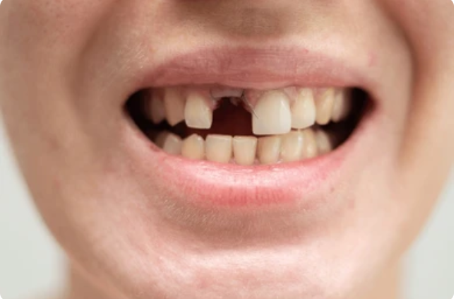
Tissue losses occurring in oral anatomy are clinical pictures that directly affect many physiological functions, from chewing mechanics to speech articulation. In the medical literature, one of the inorganic restoration methods applied to compensate for missing tooth gaps is the integration of titanium alloy artificial roots into the alveolar bone tissue. The stability and biological lifespan of this surgical intervention depend on numerous anatomic, cellular, and systemic variables that must be considered before and after the operation.
What Medical Criteria Are Taken into Account When Making a Decision for Single Tooth Implant Treatment?
During the decision-making phase, the skeletal maturity of the patient, the trabecular density of the jawbone, and the individual’s systemic health profile are examined. The absence of an active infection in the relevant area and the supporting cellular volume of the bone structure for the titanium screw constitute the fundamental medical criteria for surgical planning.
The human skeletal system has a dynamic structure. The anatomy of the jaw-face (maxillofacial) region enters a continuous remodeling cycle due to tooth extractions, chronic periodontal diseases, or trauma. When making an intervention decision for a single tooth deficiency, the skeletal development process of the patient is evaluated first. In individuals where growth plates have not yet closed and skeletal growth is ongoing, titanium artificial roots are not preferred as the jawbone continues to expand and elongate. The conclusion of growth is usually confirmed by radiographic analyses conducted after the age of 18.
The second important criterion is the microbial flora of the operation area. If there is a cystic formation in the extraction site, a root tip lesion, or active purulent (inflammatory) drainage originating from neighboring teeth, this situation will hinder cellular integration (osseointegration). In order for the titanium screw to be integrated into the bone matrix, it is essential that the involved area’s soft and hard tissues are free from inflammation. Therefore, the expectation is to clean existing local infections and allow the tissue to progress into the physiological healing phase. In the procedural steps at the Avrupadent clinics located in İzmir, not only the area where the defect is present but also the overall oral hygiene of the patient and their overall dentition structure is subjected to analysis.
How Does Jawbone Volume Affect Single Tooth Implant Application?
In order for the titanium screw to adhere to the bone matrix, it is expected that the alveolar ridge in the area of the missing tooth has sufficient vertical height and horizontal width. If physiological resorption (loss) of bone volume is detected, reconstructive medical procedures such as bone augmentation (graft application) are applied before the procedure.
The jawbone receives cellular stimulation from the masticatory forces transmitted by the teeth on it. If a single tooth is lost, the bone cells (osteoblasts) in that area are deprived of mechanical stimulation, and the resorption (loss) process begins according to Wolff’s Law of the skeletal system. A significant loss is particularly observed in the horizontal (buccal-lingual) width of the alveolar bone within the first six months after extraction. In implant planning, it is clinically necessary for the bone wall surrounding the screw to be at least 1.5 – 2.0 millimeters thicker than the diameter of the screw to be used.
In cases where bone narrowing is detected, the procedure of directly creating a channel may lead to the formation of fenestration (opening) in the bone wall, so a step called “augmentation” is added to the treatment process. Depending on the anatomical defect of the patient, bone particles from allogenic (xenograft), human (allograft), or from the patient’s own chin (autogenous) sources are transplanted to the deficient area. These biomaterials serve as a scaffold that the body’s cells can proliferate on, facilitating the formation of new living bone tissue in the area. These reconstructive steps enhance the cellular foundations of the process by adding months of anatomical recovery to the overall timeline of the treatment.
Why Are the Anatomical Features of the Area to Be Implanted Important?
The distance of the surgical area to neighboring anatomical structures determines the route of the surgery. The mandibular nerve canal, which carries lip sensation in the lower jaw, and the maxillary sinuses with air cavities in the upper jaw are critical anatomical boundaries that must be maintained during the positioning of the screw.
If a single tooth deficiency is located in the back region of the lower jaw (molar area), the primary focus of the physician is the thick nerve cord called “inferior alveolar nerve” that runs through the bone tissue. This nerve conveys the sensation of touch from the tip of the jaw and the lower lip to the central nervous system. The drills (perforating medical instruments) used when opening the surgical cavity must remain at least 2 millimeters away from this canal. The existing vertical (height) bone length between the two points provides objective data that determines the maximum length of the artificial root to be placed.
In the upper jaw background, there are air cavities known as “maxillary sinuses” that contribute to respiratory physiology and provide resonance to sound. With tooth loss, the mucous membranes of these sinuses show a tendency for downward sagging (pneumatization). When there is only a few millimeters of bone layer left between the ground and the oral mucosa, placing the implant directly in this area anatomically contradicts medical protocols. In such cases, the ‘Sinus Lifting’ operations are performed, where the sinus membrane is carefully pushed upwards and the space is supported with biological bone grafts. These anatomically sensitive medical approaches form the backbone of the clinical evaluation processes at İzmir Avrupadent.
What Should the Structural Properties of the Titanium Material to be Used be?
The material to be used as an artificial tooth root must be made of titanium or titanium alloys that are biocompatible and resistant to corrosion. The surface porosity of the material is selected to support the bone cells’ ability to wrap around the screw and aid in biological integration (osseointegration).
The organism develops an immunological defense mechanism (foreign body reaction) against any substance that is foreign to it. The reason titanium (Ti) is commonly used in dentistry and orthopedics is that the thin titanium oxide layer that forms on its surface does not chemically interact with bodily fluids and is not rejected by the binding system. This inorganic material has the ability to bond with alveolar bone cells at a microscopic level due to its biocompatibility.
The microscopic surface structure of the implant is a key engineering factor that determines the quality and speed of cellular integration. A standard titanium screw is porous during the manufacturing phase. However, medical literature shows that osteoblasts (the bone-forming cells) tend to attach more easily to porous surfaces. Therefore, materials are subjected to special surface treatments such as sandblasting and acid etching (SLA) to convert them into a micro-porous (spongy) format. The materials to be used in areas where chewing force is extremely high, such as the lower jaw, should ideally be of Titanium Grade 4 or Grade 5 alloy quality to increase resistance to mechanical fatigue and fractures, which is of great importance from a biomechanical maintenance perspective.
| Material Property | Clinical Function | Impact on Operation |
|---|---|---|
| Biocompatibility | The system does not recognize it as a foreign body. | Prevents tissue rejection and minimizes the risk of inflammation. |
| Surface Porosity (SLA vs.) | Creates micro indentations that cells (osteoblasts) can adhere to. | Supports cell migration by increasing osseointegration (fusion) speed. |
| Alloy Mechanical Resistance | The adaptability of the elastic modulus against high occlusal (chewing) forces. | Limits exposure of the abutment screw or implant body to micro-fractures. |
How Are Systemic Diseases Evaluated Before Single Tooth Implant Surgery?
Chronic diseases that affect the hospital’s blood pressure, blood sugar levels, and blood coagulation mechanisms directly guide cellular wound healing. Medical intervention can be performed when systemic disorders such as diabetes, hypertension, or osteoporosis reach reference values through pharmacological adjustments by the relevant specialty physicians.
Intraoral surgical interventions not only create a localized tissue injury, but also integrate with the overall physiology of the organism. The patient’s existing systemic diseases and regularly used medications are thoroughly examined during the medical history (anamnesis) taking phase. For example, in cases of diabetes (diabetes mellitus), the chronic elevation of blood glucose levels can impair microcirculation (microangiopathy), limiting the transport of sufficient oxygen and defense cells to the surgical area. This situation, which slows down the closure of the wound, leads to an HbA1c test being requested from diabetic patients before the operation, with values expected to be within specific medical norms (around %7).
However, in patients using bisphosphonate group medications diagnosed with osteoporosis (bone loss), the remodeling cycle of the jawbone has been suppressed. Particularly with intravenous intensive use of these drugs, cellular healing in the area of surgical intervention may be hindered, leading to a risk of bone necrosis. In individuals using anticoagulants (blood thinners) due to cardiovascular blockages, it is essential to manage the bleeding profile. In the İzmir Avrupadent clinic protocols, medication doses are adjusted for a short period through consultation with cardiologists or internal medicine specialists to ensure bleeding is controlled during the operation.
Why is Radiological Examination Necessary in İzmir Avrupadent Procedures?In the Izmir location, analyses are performed with three-dimensional dental volumetric tomography (CBCT), going beyond two-dimensional films. By creating a digital model of the jawbone, the angle and depth of the screw’s entry into the bone are calculated in a computer environment before surgery.
In the Izmir location, analyses are performed with three-dimensional dental volumetric tomography (CBCT), going beyond two-dimensional films. By creating a digital model of the jawbone, the angle and depth of the screw’s entry into the bone are calculated in a computer environment before surgery.
When viewed from the mucosa surface of the gum, the thickness of the underlying bone matrix and its anatomical formations cannot be evaluated by the naked eye. Although two-dimensional panoramic X-rays are functional in detecting cysts and general root lengths, they cannot reflect the front-back (bucco-lingual) thickness of the jaw and contain distortion on the image. To eliminate this uncertainty, three-dimensional Cone Beam Computed Tomography (CBCT) systems integrated into dentistry are put into operation.
Computerized tomography scans the jawbone in hundreds of millimetric sections, measuring the density of the bone structure in Hounsfield Units (HU). The bone hardness scale ranging from D1 (hardest) to D4 (softest) guides the surgical techniques to be used by the physician during the operation. During this ‘backward planning,’ carried out in a digital environment, the form of the porcelain tooth to be placed in the gap is virtually designed, and the titanium screw is positioned with an approach that will transfer mechanical forces to the bone in the most suitable vector. If necessary, surgical guides are produced with 3D printers to ensure that the drills only move in the planned axis during the operation.
How is Local Anesthesia and Surgical Comfort Managed During the Operation?Local anesthetic agents temporarily suspend impulse transmission by blocking sodium channels in nerve fibers. With this medical protocol, physiological stimuli occurring at the surgical site do not reach the central nervous system. While the patient remains conscious, they only perceive the pressure in the tissue and the vibrations of the device.
Local anesthetic agents temporarily suspend impulse transmission by blocking sodium channels in nerve fibers. With this medical protocol, physiological stimuli occurring at the surgical site do not reach the central nervous system. While the patient remains conscious, they only perceive the pressure in the tissue and the vibrations of the device.
Local anesthesia management in oral surgery works on the principle of injecting pharmacological agents around specific nerve branches. The physician isolates the nerve pathways that provide sensation to the relevant single tooth gap using infiltration in the upper jaw and mandibular block techniques in the lower jaw. These agents interrupt the transmission of electrochemical stimuli generated when a cut is made in the tissue or when there is intervention in the bone through pharmacological means. While anesthesia is in effect, the procedures performed on the tissue are anatomically sustained.
In the stage of preparing the implant socket, micromotor systems are used to cool with sterile physiological serum to prevent the tissue temperature from exceeding 47 degrees Celsius. While these devices rotate, they create a certain amount of mechanical vibration (oscillation) in the jawbone. Although the local anesthesia blocks the fine nerve fibers that carry pain sensation, it may not be equally effective on the thick fibers that carry the sensation of pressure; therefore, the patient’s experience of pressure or vibration in the jaw is a biological response expected from the tissue. For patients with severe dental phobia or those who cannot tolerate stress systemically, alternatives for conscious sedation administered via intravenous access can also be considered by consulting with anesthesia prior to the operation.
What to Expect During the Osseointegration (Bone Healing) Phase?
Osseointegration, which is the fusion of titanium screws with the alveolar bone at the cellular level, is a biological tissue maintenance phase that lasts about two to three months in the lower jaw and three to six months in the upper jaw. In this phase, osteoblast cells fill the spaces between the titanium grooves, creating the mechanical resistance that will support the prosthesis.
When the surgical procedure is completed, the initial tightness of the screw inside the bone (torque value) is referred to as “primary stability.” This hold is purely a mechanical compression. Over time, as this mechanical compression decreases, it gives way to a cellular healing process known as “secondary stability.” The initial blood clot that forms in the operation area transforms over time into fibroblasts and subsequently into osteoblast cells that build bone, allowing for the mineral deposition into the titanium surface’s porous microstructure.
This chain of biological reactions is the body’s own physiological repair timeline, which cannot be accelerated by an external pharmacological agent. The anatomical structure of the lower jawbone (mandible) being very hard and dense (cortical) allows for a faster transition of cellular organization to mechanical stability. In contrast, the upper jaw (maxilla), being adjacent to air-filled sinus cavities and possessing a spongy (trabecular) bone structure, takes anatomically longer to reach the same mineral density. In clinical practices in İzmir, Avrupadent advises patients to avoid hard foods during this waiting period and to protect the treatment area from trauma as a medical directive.
How is the Selection of Zirconium or Porcelain Crown in Prosthetic Stage Made?
After the bone healing is completed, digital or conventional measurements are taken for the upper structure prosthesis. In the posterior regions where chewing forces are intense, metal-supported ceramics with mechanical strength are preferred, while in anterior regions where aesthetics are prioritized, zirconium crowns with high light permeability are chosen.
Once the osseointegration phase has successfully completed (after radiological confirmation), the cover screw inside the implant is removed and healing abutments (gingiva formers) that shape the gum are placed in its place. After the gum takes on a natural form, using impression transfer pieces, an exact replica of the jaw is transferred to the laboratory. In current digital dentistry, this impression process can also be performed in three-dimensional data formats using intraoral optical scanners (CAD/CAM systems).
The selection of crown material depends on the position of the missing tooth within the mouth, the vertical distance to the opposing tooth, and the patient’s biting habits. The back molars are subjected to significant horizontal and vertical pressures (hundreds of Newtons of force) during chewing and crushing actions. In this area, material toughness (elasticity resistance) is prioritized over aesthetic concerns, hence porcelain crowns with a high mechanical resistance metal base are frequently applied. In the anterior tooth area, it is aesthetically necessary for the material to not leave a gray shadow on the gum and to reflect light similarly to natural enamel. In these areas, complete ceramic crowns with a zirconium base, which have high biocompatibility and translucency (light permeability), are positioned to complete the functional restoration.
| Material Type | Medical/Mechanical Advantage | Recommended Anatomical Area |
|---|---|---|
| Metal Supported Porcelain | The resistance to fracture against compressive and crushing forces is very high. | Back Molars (Molar and Premolar regions) |
| Zirconium Based Crown | With its white/whitish structure, it does not create a metallic reflection on the outer surface, providing high biocompatibility. | Anterior Teeth (Aesthetic Zone) |
| Monolithic Zirconium | It minimizes the risk of porcelain chipping (breaking) due to being milled as a single block. | In patients with teeth grinding habit (Bruxism), all areas |
What rules should be followed during the recovery period after surgery?
To control the physiological swelling that occurs after the surgical procedure, cold compresses are applied for the first two days. It is essential to use the prescribed antibiotics and antimicrobial mouthwashes on time, to consume soft and warm foods in the initial days, and to avoid tobacco products for wound healing.
Local incisions made on the gum and alveolar bone initiate an inflammatory (non-inflammatory, biological repair purpose) reaction within the tissue. During the normal course of this reaction, the observation of fluid accumulation (swelling) in the adjacent cheek or lip tissues to the surgical area is a physiological expectation. To keep this fluid accumulation and any possible slight capillary leak after surgery to a minimum, it is recommended that the patient apply cold compresses (ice) intermittently for the first 48 hours. Cold helps to constrict the vessels and maintain control over the blood volume in the area.
Modifications in dietary habits are essential to prevent the tautness of Yara’s lips and to keep the stitches stable. Excessively hot and acidic foods can disrupt the coagulation structure and irritate the tissue; therefore, it is recommended that pureed, lump-free foods be consumed at room temperature, and that the chewing action is performed on the side of the jaw where the procedure was carried out. It is a medical necessity to use the antibiotics prescribed by the doctor for infection prophylaxis (protection) in the determined dose and duration without fail. Additionally, since the nicotine in tobacco smoke constricts capillaries and significantly reduces tissue oxygenation, smoking is considered the most prominent external factor that delays healing and increases the risk of infection. Therefore, limiting tobacco consumption during the recovery period is communicated to the patient as a medical directive by doctors.
How is the Long-Term Care and Clinical Maintenance of a Single Tooth Implant Provided?
The maintenance of the functionality of artificial teeth in the mouth depends on regular brushing, the use of inter-dental brushes, and dental floss to prevent tartar buildup. Regular clinical check-ups every six months to measure the depth of gum pockets and to maintain tissue health is a medical rule.
The inorganic structure of titanium material protects against corrosion or tooth decay (caries) by not being affected by acid attacks in the oral flora. This anatomical difference can create a misconception among patients regarding the need for prosthetic dental care. However, the biological tissues surrounding the implant body, such as the gum (peri-implant mucosa) and jawbone, are extremely sensitive to microbial dental plaque. Periodontal attachments, which surround the roots of natural teeth and slow the progression of inflammation to the bone, do not form around the titanium screw. Therefore, when the bacterial plaque that accumulates in the gum pocket is not cleaned, the infection can rapidly penetrate deeper tissues, leading to the resorption of the jawbone (peri-implantitis).







