What Should Be Known About Bone Grafting and Implant Treatment?
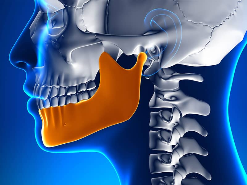
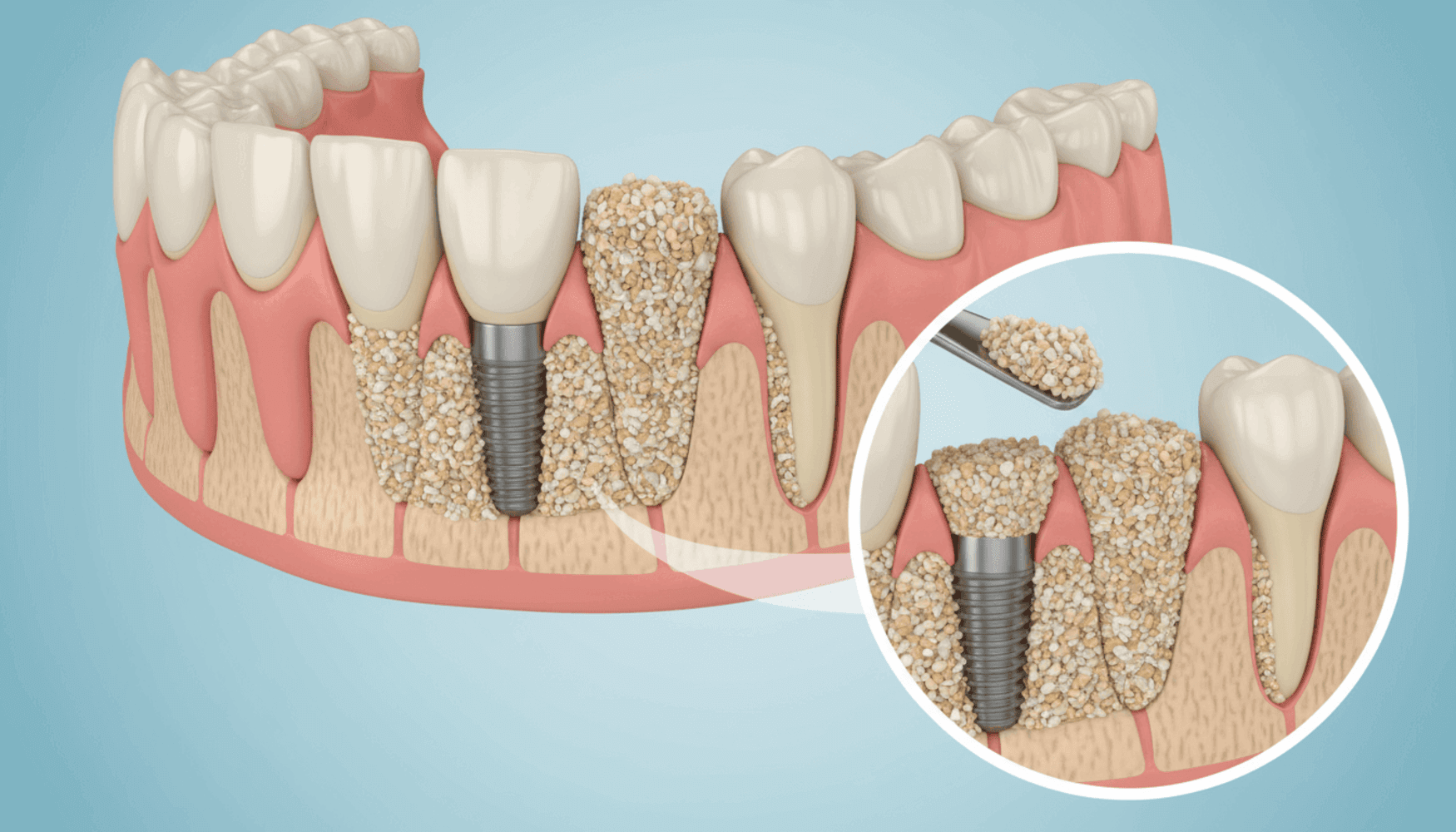
The procedures of implantology, which involve placing artificial roots into the jawbone to compensate for tooth loss, progress directly depending on the horizontal and vertical volume of the bone tissue in the area where the procedure will be performed. Long-term tooth loss, as a result of periodontal inflammations or cystic formations, can lead to reductions in the cellular dimensions of the jawbone, which may anatomically restrict the stable positioning of medical titanium materials. In medical literature, the procedures referred to as “bone grafting” (commonly known as bone powder) are advanced surgical approaches aimed at repairing these anatomical deficiencies at the cellular level, with the goal of increasing the form and biological volume of the jaw ridge, known as the alveolar crest.
Bone grafting does not merely mean filling the existing void with a physical substance; it is a complex regeneration process that guides the formation of live and vascularized new jawbone tissue in that area by mobilizing the patient’s own physiological renewal mechanisms. Advances in medical technologies have made it possible to develop graft materials obtained from different biological sources, thus rendering many medical scenarios that were previously considered physiologically unfeasible for fixed prosthesis construction manageable. This guide will detail how bone graft applications work with various cellular principles, the properties of the medical materials used, surgical planning strategies, and the medical processes related to the healing period.
What Medical Needs Arise for Bone Grafting (Bone Powder) Application?
The application of a bone graft is necessitated by medical needs such as reversing the cellular resorption process that occurs over time in the area where the tooth root has been lost, obtaining sufficient bone wall thickness to completely encase the artificial titanium material, and creating a solid biological foundation that will absorb the mechanical stresses during chewing function. When teeth are present in the mouth, the chewing forces transmitted to the jawbone through the roots help to maintain volume by stimulating the bone tissue at a cellular level. The cessation of this mechanical stimulus with the extraction of the tooth leads the body to perceive the relevant area as non-functional, allowing osteoclast (bone-resorbing) cells to dominate, resulting in a reduction in both vertical and horizontal dimensions of the bone.
This anatomical narrowing occurs significantly in the first months following tooth extraction and can assume a form as thin as the crest (the back of the jaw). The diameter of the titanium screw to be placed in implantology procedures generally requires at least 1 to 1.5 millimeters of healthy bone tissue thickness to remain around it. Intervening forcefully on a jaw with insufficient bone thickness can lead to the screw remaining exposed, the gum tissue receding, and ultimately losing the stability of the system due to the inability to withstand chewing forces in the long term. Bone grafting procedure is performed to overcome these limitations medically.
Among the other medical conditions that cause the need, periodontal diseases (periodontitis) are included. The bacterial flora, by penetrating deeper from the periodontal pocket, erodes the bone around the root in a crater-like manner, leaving extensive bone defects after tooth extraction. Similarly, large spaces formed in the jaw after cyst or tumor operations, or tissue losses caused by mechanical traumas such as traffic accidents, necessitate the clinical involvement of volumetric augmentation (lifting and expansion) procedures. Bone powder functions as a scaffold, added to these areas to initiate the body’s own bone construction.
How Are Grafts Added to the Jawbone Accepted by the Body?
Grafts added to the jawbone are accepted by the body through three fundamental biological mechanisms known as osteoconduction (structural conduction), osteoinduction (cellular stimulation), and osteogenesis (direct bone formation). The body’s immune system does not perceive these sterilized particles, completely freed from its proteins, as foreign substances; rather, it views them as a substrate for migrating blood vessels and cells to advance into the area.
Osteoconduction refers to the situation where the graft particles behave like a scaffolding structure. Bone-forming cells (osteoblasts) that reach the area through circulation attach to the porous structure of the graft, proliferate, and embed themselves into these pores. The graft does not produce new bone on its own, but it preserves the physical space necessary for the body to weave its own bone network and prevents soft tissues from collapsing into that area.
The osteoinduction process involves the proteins present in certain specialized graft materials (such as Bone Morphogenetic Proteins – BMP) stimulating stem cells to encourage their conversion into bone-forming osteoblast cells. The final mechanism, osteogenesis, occurs only in live bone blocks taken from the patient’s own body (autogenous); the living cells within the transferred bone piece begin to synthesize new bone directly. After months of this biological cycle, a significant portion of the added powder particles is absorbed at the cellular level by the body, replacing it with living, vascularized, and mechanically resistant original jawbone that carries the patient’s own DNA.
What Biological Materials are Used in Bone Graft Procedures?
The biological materials used in bone graft procedures are obtained from the patient’s own tissue, sterilized animal sources such as cadavers, human tissue banks prepared in laboratory settings, or calcium-based synthetic components. Each group of materials differs in terms of medical absorption duration, porosity, and medical application area, and is chosen based on the specific needs of the case.
In medical literature, these materials are categorized into four main types according to their origins. The type of graft selected is determined by the size of the bone defect to be repaired, the vascularization capacity of the region, and the targeted biological maturation period, as decided by the physician. The table below summarizes the origins and structural properties of the grafts used in medical processes:
| Medical Classification of the Graft | Source and Method of Acquisition | Medical Properties and Areas of Application |
|---|---|---|
| Autogenous Grafts (Patient’s Own Bone) | Harvested from the patient’s chin tip (symphysis), wisdom tooth area (ramus), or iliac bone. | Biological integration for living cells is the highest material. It is the clinical preference in large-volume reconstructions (as block grafts). |
| Xenografts (Animal-Derived) | Produced by removing all organic structures from bovine or equine (horse) sources at high temperatures. | Only mineral residue remains, maintaining its volume for a very long time. It is the most commonly used powder; it is absorbed very slowly by the body over the years, turning into your own bone. |
| Allografts (Human-Derived) | Obtained by sterilizing bones taken from healthy donors through medical testing in tissue banks. | Does not require opening a second surgical site. It can contain proteins that encourage bone formation (osteogenic) in its structure. |
| Alloplastic Grafts (Synthetic) | Forms such as tricalcium phosphate, hydroxyapatite, and bioglass that are chemically produced in the laboratory. | Mineral powders produced under laboratory standards that pose no infection risk for patients who do not wish to use animal or human-derived materials. |
How are Surgical Steps Planned in Augmentation (Bone Addition) Treatments?
Surgical steps in augmentation (bone addition) treatments are planned with local anesthesia to numb the area, accessing the underlying thin bone layer by separating the gingival flap, creating small micro-holes (decortication) on the cortical bone surface to increase bleeding, placing the prepared sterile graft in the area, and isolating the area from surrounding tissues using barrier membranes.
During the preparation phase of the procedure, nerve conduction in the area is temporarily halted and small incisions are made in the mucosa of the gum, allowing for the flap (gum tissue) to be set aside. The outer surface (cortex) of the jawbone is generally hard and its vascularization is reduced. To nourish the added bone powder, rich blood vessels in the inner part of the bone are needed. Therefore, the physician creates small bleeding points on the bone surface using fine-tipped instruments. This procedure is called decortication; thus, the growth factors in the blood reaching the area infiltrate the graft.
The prepared graft material is moistened with physiological saline or PRF (Platelet-Rich Fibrin) obtained from the patient until it reaches a paste-like consistency, and it is carefully placed into the deficient jaw area. However, the procedure does not stop here; since gum cells have a very rapidly proliferating structure, if the graft is left exposed, the gum cells will seep into the bone powder and disrupt the osseointegration. To restrict this cellular seepage, the surface of the graft is tightly covered with a barrier membrane. Finally, the retracted gum is returned to its original position and closed with sutures, allowing the tissue to rest medically.
How Long Does the Cellular Healing (Osseointegration) Process Take After Bone Powder Procedure?
The cellular healing (osseointegration) process after the bone powder procedure usually spans a period of 4 to 6 months for the added graft to integrate into the patient’s own bone structure and transform into a solid biological mass. This medical period may show clinical variations depending on the amount of graft applied, the area of the jaw, and the metabolic structure of the patient.
In the first weeks of the operation, a soft tissue graft particulate surrounding the granulation tissue forms, which is rich in blood vessels. The cellular cycle has not yet progressed to the calcification (hardening) phase. By the second and third months, osteoblast cells begin to store calcium and phosphorus minerals between the particles. During this process, the tissue gradually hardens into a spongy bone form (woven bone). However, this form has not yet achieved the mechanical maturity required to withstand the chewing forces of the titanium element that will be added on top.
From the fourth month onward, the bone enters the remodeling phase. The spongy structure organizes into a regular layered (lamellar) bone structure that is resistant to mechanical stress. If the procedure is performed on a large defect or in conjunction with sinus lifting, based on the physician’s radiological assessments, prolonging this maturation period to 6 to 8 months can become a medical necessity. During these months, it is crucial to avoid any mechanical impact on the relevant area and to prevent any prosthetic pressure, as it significantly affects the health of cellular integration.
What Additional Procedures Come into Play in Case of Bone Insufficiency in the Upper Jaw?
In cases of bone insufficiency in the upper jaw, sinus lifting procedures come into play to manage the sagging of the maxillary sinuses located in the back regions where the molars are present. In this procedure, the thin membrane covering the sinus is pushed upwards, and the new space created is filled with bone graft, thus gaining vertical bone volume.
The upper jaw (maxilla) anatomy includes air cavities that assist respiration. When the back teeth are lost, influenced by atmospheric pressure, these sinus cavities begin to expand towards the jawbone. Only a 1-2 millimeter thin layer of bone tissue may remain between the melting bone from the crest top and the sinus membrane hanging down from above. This thin layer does not provide anatomically suitable conditions for the placement of any medical implant.
During the medical intervention, a small window is opened from the wall on the cheek side of the jawbone or from inside the socket where the tooth has been extracted. The sinus membrane is slowly lifted upwards from the bony base by being released without puncturing it with special instruments. This biological chamber shaped like a dome formed between the floor and the membrane is filled with bone powder. Depending on the amount of graft added and the condition of the patient’s existing bone, in some clinical cases, titanium material can simultaneously be placed during this procedure; in cases where the bone is very thin, only powder is placed, and after a 6-month healing period, a second stage is transitioned for planning artificial roots.
What Differences Are There Between Surgical Strategies Applied in Horizontal and Vertical Bone Loss?
Surgical strategies applied in horizontal (width) bone losses offer biologically higher predictability and cellular adaptation compared to vertical (height) bone losses; since vertical augmentations require overcoming the physiological boundaries of the jawbone, they involve advanced medical techniques where vascularization and soft tissue closure are challenged.
The narrowing of the width of a jawbone (horizontal loss) usually results in the framework acquiring a cretin-like shape. To correct this condition, bone powder is applied to the lateral surface of the jawbone, or a graft is placed between two parts after the ridge has been split. Since the existing bone bed can nourish particles from both sides, vascularization is stronger and the coverage of the membrane with the gum can be managed medically. The table below examines the differences in medical approaches for these two types of defects:
| Assessment Criteria | Horizontal (Width) Bone Loss Strategies | Vertical (Height) Bone Loss Strategies |
|---|---|---|
| Anatomical Difficulty Degree | Relatively easier to manage. The existing bone walls provide lateral support for the graft. | Much more difficult. The graft attempts to create a new peak point “in the air” by protruding over the bone alignment. |
| Materials Used | Standard particulate grafts and collagen membranes (Guided Tissue Regeneration) are frequently preferred. | To maintain the shape, titanium-reinforced (mesh) membranes or autogenous block bones are often a medical necessity. |
| Soft Tissue Closure | The gum’s elasticity is usually sufficient to close the graft. | The gum is insufficient to cover the elevated area; special tissue release incisions are made for tension-free closure of the flap. |
How Should Oral Care Routine Be Structured After Grafted Implant Operations?
After graft implant surgeries, the oral care routine should be established according to the principles of protecting the suture line from mechanical pressures, limiting contact with food residues in the area, and gently using the antibacterial mouth rinses prescribed by the physician. Since maintaining the stability of the area where the bone powder is added is essential for cellular fusion, it is crucial to avoid any impact on that area.
The first two weeks following the surgical intervention represent a sensitive period during which the epithelial (gum) tissue attempts to close the wound from the external environment. Placing a movable prosthesis on the grafted area, stretching the area with cheek muscles, or playing with the sutures with the tongue can lead to the barrier membrane being exposed and bacteria leaking into the graft. Therefore, patients are advised to follow a soft diet, chew on the side that has not undergone surgery, and avoid pulling the cheeks to look at the sutures.
While standard tooth brushing habits continue the same for other teeth, the area with sutures should not be brushed. Instead, medical mouth rinses containing active ingredients like chlorhexidine can help suppress the bacterial flora in the area. However, during rinsing, it is more appropriate from a medical perspective to allow the solution to remain passively in the area (with saline or prescribed solutions) rather than vigorously swishing it in the mouth, as this is better for the stability of the sutures. After the sutures are removed and the soft tissue has closed, normal cleaning routines can resume with soft-tipped toothbrushes.
What is the Function of Barrier Membranes Used in Bone Addition Procedures?
The primary function of barrier membranes used in procedures with bone grafts is to physically prevent the slow-dividing bone cells from occupying the space of the rapidly proliferating epithelial cells of the gum tissue by enveloping the graft particles placed around the jawbone. This method is referred to in the medical literature as Guided Tissue Regeneration (GTR).
In the body’s biological wound healing mechanism, the cell division rates of different tissues are not equal. While gum tissue cells (epithelium) migrate at approximately 0.5 mm per day to cover an open wound, it takes weeks for bone-forming cells to cover the same distance. If the area with bone powder is not protected by a membrane, the epithelial tissue will infiltrate among the particles of the graft. The medical consequence of this is the formation of a soft and flexible tissue called ‘fibrous connective tissue’, instead of a hard and mechanically durable bone in that region. Fibrous tissue cannot support an artificial root.
Barrier membranes limit the sagging of the gum by covering the graft like a tent. By creating an isolated biological chamber below, they safely provide the weeks-long timeframe that bone cells need. In current surgical planning, collagen-based ‘resorbable’ membranes are typically preferred. These membranes are metabolized by the body’s enzymes through natural processes after tissue healing is completed; thus, they do not require a secondary extraction surgery.
What Medical Standards Are Used to Evaluate Graft Planning in Avrupadent Clinical Procedures?
Graft planning in Avrupadent clinical procedures is evaluated according to the integration of standards such as three-dimensional dental tomography (CBCT) with the millimeter measurements of the jaw defect, the medical history of the patient’s systemic condition, compliance with sterilization protocols, and the specific biomaterials selected according to each individual’s anatomical needs. All stages of the process are managed within the framework of medical predictability.
In the first step, the patient’s general health history is recorded at the hospital. The diabetes profile (HbA1c levels) that directly affects bone healing, the use of anticoagulants, and bisphosphonate-type drugs prescribed for osteoporosis are identified, and if necessary, consultations are requested from relevant medical specialties (endocrinology, cardiology). Since two-dimensional X-rays are not deemed sufficient, three-dimensional tomographies are transferred to the system for volumetric analysis of the jawbone. Through these digital software, the size of the defect and the required amount of graft are calculated in a virtual environment.
When proceeding to the surgical stage, international sterilization guidelines are applied to minimize the risk of cross-contamination. The bone powders and membranes to be used are applied specially to the patient, opened from certified packages with international medical standards and traceability. After the operation, the cellular healing process is periodically monitored with radiological controls, and once the augmentation area is medically confirmed to have reached sufficient maturity, transition to the prosthetic (implant) phase of the implantology procedures is made.
Frequently Asked Questions (FAQ)
1. How long does the bone graft operation take? The duration of the operation varies depending on the size of the area to be treated, but a standard grafting procedure requires approximately 45 minutes to 1 hour of clinical time after the area has been anesthetized. 2. Will my body reject the bone powder added? Grafts used in medical procedures are pure minerals that do not contain cells and are free from all proteins. Since the body’s connective tissue system does not perceive them as foreign or allergic substances, cellular tissue rejection is not expected medically. 3. Is it mandatory to take a graft from my own bone? No, in most routine augmentation procedures, powdered materials sourced from cadavers or synthetic sources are sufficient. Only in cases of large defects where the bone structure has significantly deteriorated is harvesting a block of bone from the patient’s own jaw a clinical option. 4. Can the graft procedure and the insertion of the titanium screw be done on the same day? If the existing jawbone has the minimum thickness necessary to ensure initial stability for the titanium material to be placed, both procedures can be carried out in the same session. If the bone is very weak, the graft should be added first and allowed to mature for several months. 5. Is it normal to experience swelling after the graft? In procedures where surgical tissue flaps are removed, it is normal for mild to moderate local edema (swelling) to occur as a natural inflammatory healing response of the body. This condition can be managed with regular cold compress applications. 6. When can normal eating resume after the bone powder surgery? For the first few days, it is necessary to consume soft, warm, and non-grainy liquid foods to protect the stitches. After the sutures are removed and the soft tissue has closed (approximately 10-14 days), one can gradually return to a normal chewing routine. 7. How does smoking affect the healing of the graft? The use of any substance forces the essential oxygen needed for bone formation to reach the affected area by penetrating the capillaries in the gum. It is recommended to limit smoking during the process, as this physiological slowdown puts the graft’s integration at risk. 8. Is synthetic bone powder different from animal-derived ones? Synthetic powders are produced in laboratories from compounds like calcium phosphate and do not carry biological sources. Animal grafts are obtained from mineral residues created by baking organic bone at high temperatures. Both serve their medical functions. 9. Will the barrier membrane used be removed later? Nowadays, clinically, biodegradable membranes made from collagen derived from pigs or cows are used. These membranes are dissolved by enzymes from bodily fluids months after completing their biological role, requiring no removal surgery. 10. How long should one wait for a prosthetic tooth after adding bone powder? Typically, it takes about 4 to 6 months for the particles added to the area to undergo calcification and harden. In some larger sinus lift operations, a cellular maturation period of up to 8 months is expected medically. 11. Can the amount of absorption in my jawbone be seen on an X-ray? Two-dimensional panoramic films only show the vertical amount of absorption. Three-dimensional Dental Tomography (CBCT) scans are needed to determine the width (horizontal volume) of the bone for graft planning. 12. Why is applying pressure to the grafted area considered risky? Since the added graft is still spongy and shapeable, applying a movable prosthesis or mechanical pressure can lead to the crushing of particles or damage to barrier membranes, preventing bone formation. 13. Can bone grafts be applied to diabetic patients? If diabetes is under control, and the HbA1c values showing the three-month average of blood sugar fall within the reference limits determined by doctors, grafting procedures can be performed according to medical standards. 14. Will there be a change in color in the area of the gum where bone powder is applied? In normal healing processes, color changes in the tissue are not expected. Bruising may be observed in the initial days related to the surgical incision. However, it is aimed for the gum to return to its natural pink and firm (stippling) anatomical form once healing is complete. 15. Does the augmentation procedure require general anesthesia? Most procedures, similar to a standard dental practice, are performed under local anesthesia applied only to the relevant area. The patient remains awake throughout the process; only the nerve transmission in the area has been stopped. |