What Stages Does Implant Treatment in Izmir Involve?
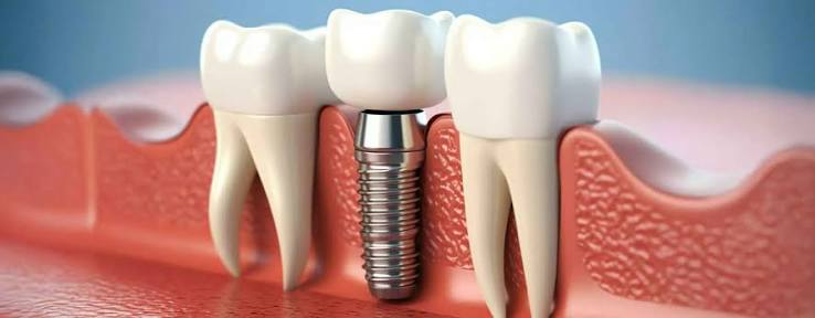
Tooth losses are anatomical conditions that deeply affect the functional mechanics of the jaw system and the preparation phase of food for digestion. The placement of titanium materials, which can biologically integrate with human bone, forms the basis of modern prosthetic approaches. The positioning of titanium screws within the jaw is part of a medical chain that consists of interconnected processes, not just a single session. The question of which stages implant treatment in Izmir involves is frequently researched by individuals looking to plan a clinical schedule.
What Stages Does Implant Treatment in Izmir Involve and How is the Initial Diagnosis Made?
Planning in medical procedures is the most critical step for the process to proceed according to clinical norms. During the initial consultation, not only the intraoral condition of the patient but also their general systemic health is recorded through detailed forms. Vital anatomical points, such as the vertical and horizontal dimensions of the bone structure, the location of the sinus cavities, and the path of the mandibular nerve canal, are examined through two-dimensional (panoramic) and three-dimensional (CBCT) radiographic scans.
After obtaining the radiological imaging, the dentist determines the diameter and length of the titanium screw to be used. The area and angle of intervention are simulated digitally. If the individual is using any medical drugs or has chronic conditions, a consultation is requested from the relevant medical branches to clarify the operation schedule.
How is the Surgical Operation Phase Carried Out in Jaw Surgery?
The operation process is carried out in sterile clinical environments where hygiene standards are maintained at the highest level. Since the procedure is performed under local anesthesia, sensitivity during the surgical intervention is controlled medically. The placement of the titanium screw into the bone takes an average of twenty to thirty minutes for a single tooth in standard cases. The time spent in the chair may vary depending on the nature and number of the procedures.
| Procedure Step | Medical Content and Purpose |
|---|---|
| Local Anesthesia | Blockage of nerve conduction in the relevant area to ensure tissue comfort during the surgical procedure. |
| Tissue Incision (Cut) | Gently removing the gum tissue over the bone with a scalpel and exposing the intervention area. |
| Cavity Preparation | Gradually opening the implant socket without generating heat in the bone, using physiological saline (cooling fluids) equivalent. |
| Implant Placement | The titanium material is positioned at a level suitable for the bone threshold, with a specific torque (compression force) value. |
What to Expect During the Titanium Screw’s Integration with Bone (Osteointegration) Phase?
Although the part of the operation that takes place in the chair is completed, the essential functioning of the body begins at this stage. Despite being a foreign material placed in the jawbone, titanium is one of the rare elements not rejected by the immune system. Thanks to its biocompatible structure, repair cells reaching the area through the bloodstream settle on the surface of the screw and begin to weave new bone tissue.
During this waiting period, it is crucial not to apply chewing force on the titanium screw. The material must remain stable (immobile) within the bone; this is the fundamental rule for the formation of hard bone tissue instead of soft tissue. Temporary prosthetic applications may be made in edentulous areas at this stage to address the aesthetic concerns of the patient, but functional loading should be avoided.
How is the Interdisciplinary Management of Implant Processes Realized Within Avrupadent?
Different disciplines of dentistry provide supportive medical data in solving complex cases. It is not enough to simply place the titanium screw where the bone is densest; it must also stop at an opening where it will have the correct occlusion relationship with the teeth that will be placed on it. Therefore, before the surgical phase, the prosthesis design is done digitally, and the screw is aligned according to this target.
What Procedures Are Added to Treatment Stages That Require Advanced Surgery?
Over long periods of dental extraction, the jawbone in that area may undergo resorption (deterioration) as it no longer functions. The stability of the titanium screw depends on the surrounding hard bone wall being at least 1.5 – 2 millimeters thick. If sufficient thickness is not detected in the tomography analysis, synthetic, rigid source, or autogenous (taken from the patient) biomaterials will be placed in that area to expand the tissue.
In areas where the upper jaw molars are located, there are anatomical air cavities referred to as maxillary sinuses. After tooth loss, these cavities sag downwards, narrowing the bone distance. In these cases, the “Sinus Lift” procedure surgically stretches the sinus membrane upwards, and the resulting cavitation is filled with bone powder. Such additional interventions can add healing times of up to six months to the total treatment duration.
When and How Do We Transition to the Prosthetic (Upper Structure) Phase in Clinics?
When biological compatibility is clinically validated to be at the desired level, the construction of the parts that will form the tooth body over the titanium root begins. The first step of this stage is the use of gum shapers referred to as “gingiva former”. A small intervention is made to access the embedded screw, and a cylinder-shaped piece is placed to create a proper emergence profile for the gum tissue.
| Prosthetic Phase | Clinical Procedure |
|---|---|
| Prosthetic Transfer | Special transfer parts are placed inside the implant to create a three-dimensional copy of the jaw. |
| Framework Trial | The passive fit of the skeletal structure made from metal or zirconium is checked inside the mouth. |
| Porcelain/Dentin Trial | The aesthetic form, color of the tooth, and the contact points for chewing with the opposing jaw are tested. |
| Fixation (Cementation) | The final prosthesis, after polishing, is permanently attached to the tooth using special medical adhesives (cement) or screw systems. |
Where Are Digital Dentistry Technologies Used in Treatment Stages?
In traditional methods, the jaw structure is observed only in two dimensions, while in digital dentistry, the anatomy is considered in all its depth. The surgical templates (guides) that assist the surgeon during the operation help isolate the openings, depth, and position of the implant from manual deviations, allowing for much smaller surgical cuts. This reduces the risk of complications and bleeding to minimum levels after the operation.
Instead of traditional dough-like materials that can trigger the gag reflex during anesthesia, pen-shaped oral scanners are preferred. These devices, which capture thousands of images of the scanned area within seconds, immediately transfer the data to a digital laboratory environment, optimizing the marginal fit of porcelain or zirconium crowns at the micron level.
What are the Anatomical and Systemic Factors Affecting the Treatment Process?
Oral tissues are an integrated structure within the body’s general circulatory and lymphatic systems. Systematic conditions present in a patient’s medical history affect the healing speed of the wound site. If blood sugar levels in diabetic individuals do not stay within reference ranges, the repair of bone cells may be compromised due to disrupted protein synthesis. Therefore, measuring HbA1c values before the operation is a medical protocol.
Heavy smoking decreases the oxygen carrying capacity in the blood by constricting peripheral (end) capillaries, reducing blood flow to the area. Healing is delayed at the non-nourished wound site, and the susceptibility to infection increases. Doctors may extend or revise the treatment schedule according to these parameters in the patient’s medical history to ensure smooth tissue repair.
What Medical Processes Should Patients Pay Attention to During the Post-Operation Healing Period?
After the surgical procedure is completed in a clinical environment, the body’s repair efforts depend on the care taken by the hospital. It is a typical physiological condition to encounter mild local swelling when anesthesia effects wear off. To maintain the integrity of the blood clot, one should avoid severe agitation in the mouth and refrain from activities that would create pressure, such as coughing, on the day of the operation.
If there are stitches (sutures) in the operation area, the cleanliness of this region should be done carefully but protected from brush injuries. Chlorhexidine-containing or saline gargles recommended by the physician help control bacterial populations during the first days when mechanical cleaning cannot be performed. In standard procedures, stitches are removed between the seventh and tenth days to confirm the epithelialization (surface closure) of the tissue.
How to Ensure Long-Term Oral Care After Completion of Treatment Stages?
Although the titanium material itself is not exposed to wear, the surrounding soft tissue and jawbone are sensitive to bacterial attacks. If food residues and plaque accumulation remain around the prosthesis for an extended period, it may lead to gum inflammation. When this inflammation progresses to the bone, it can weaken biological support, leading to long-term complications.
In summary, the surgical protocols applied for replacing missing teeth are medical processes that progress step by step while respecting the body’s biological repair limits. In a location like İzmir, which has a broad healthcare infrastructure, applying to clinically equipped institutions to create your personal anatomical map would be the most rational step in planning the suitable processes for you. A comprehensive and physician-supported diagnosis lays the groundwork for the construction of chewing functions that can be comfortably used in the years to come.