

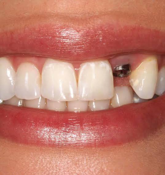
Does Every Missing Tooth Require an Implant in Izmir?
Tooth loss is an anatomical issue that disrupts the force distribution within the masticatory system (stomatognathic system), puts asymmetric loads on the temporomandibular joint (TMJ), and causes irreversible resorption over time in the jawbone. Titanium artificial root applications developed to replace lost tissues have a significant presence in today’s medicine. However, one of the main medical questions that arises in the minds of individuals who have lost multiple teeth is whether a separate surgical intervention is needed for each lost root.
Does Every Missing Tooth Require an Implant and How is Biomechanical Planning Done?
The fundamental principle in dental practice is to provide the minimum biomechanical support that can tolerate the masticatory forces (occlusal stresses) transmitted to the jawbone. Placing a single titanium material in the gap for a single missing tooth is a common clinical approach. However, in cases of extensive gaps where three or four teeth are lost side by side, placing a screw for each tooth can restrict vascularization and nutrition between cells within the bone. It is essential that there is a certain millimeter distance (typically at least 3 mm) between titanium materials to maintain the health of the bone.
When performing biomechanical planning, the force produced by the chewing muscles of the patient, the position of the opposing teeth, and the diameter of the screw to be placed are calculated. For example, two thick titanium screws placed at both ends of a gap with three missing teeth can comfortably support a porcelain or zirconium bridge that will carry the missing middle tooth as a body.
How Many Titanium Roots Are Applied in Cases of Multiple Adjacent Tooth Loss?In the case of three adjacent missing teeth, typically, two titanium roots placed at both ends of the gap provide sufficient biomechanical support. In the case of four missing teeth, either two or three materials can be planned according to the closure force of the jaw. Doctors optimize this number based on the patient’s bone density from panoramic radiography data.
The number of artificial roots to be applied is more of an engineering and physiology calculation than a mathematical one. Each region of the jawbone does not have the same density. The frontal area of the lower jaw has a very hard and dense (cortical) structure, while the back areas of the upper jaw are spongy (cancellous) and neighboring air cavities. In areas where the bone is softer, increasing the number of screws may be medically preferred to distribute the force over a wider area.Average Prosthetic Approaches in Regional Tooth Deficiencies Number and Location of Missing Teeth Proposed Number of Titanium Roots Biological Purpose of the Clinical Approach Adjacent 2 Tooth Deficiency Usually 2 pieces Each artificial root carries its independent crown, and the interface cleanliness is maintained as closely as possible.
Adjacent 3 Tooth Deficiency 2 pieces (for the beginning and end sections) A single body tooth is suspended between the two arches to construct an ‘implant-supported bridge’. This protects the central bone from trauma. Side-by-Side 4 Tooth Deficiency 2 or 3 pieces The number of supports is increased according to the chewing force of the patient (presence of bruxism/tooth clenching) to balance stress distribution.
Is a separate material positioned for each tooth in complete toothlessness (Total Edentulism) charts?
In complete toothlessness charts where all teeth are lost, no material is placed for each missing tooth. Despite the natural human dentition containing fourteen teeth in each jaw, usually four to six titanium roots are sufficient for the lower jaw, while for the upper jaw, typically six to eight titanium roots are positioned according to anatomical conditions to construct a fixed prosthetic restoration.Trying to place material in each tooth gap in complete toothlessness restorations only serves to extend surgical trauma and damage intrabony vascular structures. Each screw placed causes a certain amount of inflammation (tissue reaction) and initiates the repair process. Puncturing the jawbone like Swiss cheese can negatively affect the tissue’s self-recovery (osteointegration) capacity.
Instead, advanced surgical concepts such as ‘All-On-4’ or ‘All-On-6’ are utilized by strategically positioning implants with certain angles in the hardest and most voluminous regions of the bone. These interconnected (splinted) titanium fixtures create a common mechanical resistance that will support 12 to 14 unit hybrid, porcelain, or zirconium bridges produced in a laboratory.
Which radiological criteria are used in medical evaluations conducted within Avrupadent in Izmir?
Organizations such as Avrupadent, which provide services in İzmir localization, utilize three-dimensional dental tomography (CBCT) technologies during the planning phase. These devices map the vertical height, horizontal width, and regional hardness of the jawbone in millimeters, ensuring that the number and position of the titanium to be used are calculated specifically for the individual in a virtual environment.The clinical assessment process is not a procedure that can be completed merely by looking inside the mouth or examining two-dimensional standard X-rays. Although two-dimensional images reflect the bone structure, they do not show the thickness of the bone (the volume towards the side or tongue). In medical institutions with a multidisciplinary approach, volumetric data is used to increase the surgeon’s predictability.
Virtual Planning and Surgical Guide (Guide) Production:Tomographic data is transferred to digital software to obtain a three-dimensional copy of the patient’s skull. Doctors design the prostheses (crowns) for the missing teeth in a virtual environment and then place titanium screws in such a way that they will sit directly under these prostheses on the bone. This digital plan, where the gaps and distances between the titanium are optimally adjusted, is turned into a physical “surgical template” using 3D printers. During the operation, this template is used to minimize manual error margins.
What is an Implant Bridge (Implant Supported Prosthesis) and In Which Anatomical Conditions is it Preferred?
An implant bridge is a single-piece prosthetic restoration that is placed on titanium roots, which are situated only at specific points instead of inserting a screw for each gap in cases of wide dental gaps. This system is preferred to minimize the surgical requirements in areas where bone volume is weak.Traditional bridge prosthetics applied to natural teeth are based on the principle of reducing (cutting) the healthy teeth on the right and left of the missing tooth to serve as supporting legs. This leads to the loss of healthy enamel tissue. However, in implant-supported bridges, there is no physical intervention made on neighboring natural teeth. The supporting legs consist entirely of artificial roots within the jawbone.
Biomechanically, the rigid connection (splinting) of the titanium roots restricts the force from being directed to a single point. The chewing pressure is distributed evenly across the entire system. Especially in areas where bone resorption is very severe, and in risky anatomical areas where the sinus cavity is sagging or the inferior alveolar nerve is very close to the surface, the bridge system is established by placing the legs in safe areas.
How Does the Volume Insufficiency of the Jawbone Affect the Number of Titanium Screws?
Trying to place a large number of titanium materials in areas where the bone volume is weak, narrow, or short can disrupt the blood supply to the remaining thin tissue, leading to cell loss (necrosis). In such physiological situations, doctors plan to support the system with bridges by identifying the most voluminous areas of the bone and selecting fewer yet broader-longer materials.The stability of titanium screws in the jaw for many years depends on the presence of at least a 1.5-2 millimeter thick healthy and vascularized bone wall surrounding them. When a lot of material is placed very close together on a narrowed area of the jaw (ridge), the thin bone membrane between the two screws does not receive enough nutrients and oxygen over time, leading to degradation (resorption).
To prevent this, surgeons adopt the philosophy of “few but strategic” placement. If the bone in the area is very weak and a supporting leg is absolutely necessary for the bridge placed, biomaterials (bone powder/graft) are first applied to that area. After the volume of the area is increased with the grafting procedure, a titanium root is safely positioned in the related area following the completion of cellular maturation (approximately 4-6 months).
What Physiological Changes Are Observed When Titanium Material Is Not Placed in the Missing Tooth Area?After a tooth is removed from an area, the jawbone, deprived of the chewing force, will gradually undergo atrophy and lose its vertical and horizontal volume over time. Additionally, the neighboring teeth adjacent to the gap exhibit a tilting tendency towards this area, while the opposing teeth elongate towards the gap due to gravitational effects; this disrupts the entire closing mechanism.
The human body shapes itself according to the principle of mechanical stimulation. The gradual weakening of non-functional organs or tissues is a fundamental rule of physiology. The titanium materials placed in the jawbone send a signal to the bone cells mimicking the pressure waves transmitted by a natural root, indicating “there is a function here.” This signal keeps the osteoblastic (bone-forming) activities alive.Long-Term Physiological Consequences of Tooth Absence Timeline Clinical and Anatomical Table Functional Effects First 6 – 12 Months A noticeable narrowing (resorption) of 25% occurs in the bone width in the chewing area. Due to the embedding of the food in that area, the patient tends to chew unilaterally, and the jaw joint becomes asymmetrically enlarged. 1 – 3 Years Neighboring teeth begin to tilt into the void due to lack of support. The vertical height of the bone decreases. Gaps between the teeth open up, food accumulation increases, and the risk of new cavities and gum diseases increases. 3 Years and Above The opposing tooth in the opposing arch sags into the gap (if the upper is below, if the lower is above). The bone blade can thin as much as a sheet. The dynamics of jaw closure are disrupted (occlusal trauma), and pain occurs in the muscles. If an implant is planned in the future, advanced bone graft surgery will be needed.
What Differences Exist Between the Medical Procedures Applied for Anterior Tooth Deficiencies and Posterior Molar Tooth Deficiencies?
In anterior area surgeries, the harmony of gums and lip profile, referred to as “pink aesthetics”, is prioritized; material placement is planned with millimeter gaps according to the smile line. In the posterior molar regions, it is prioritized to use thicker-tapered titanium roots to resist chewing forces rather than aesthetics and to pay attention to limiting anatomy such as sinus cavities.The human jaw presents different biomechanical and aesthetic needs depending on the regions. In the anterior segment, which includes the aesthetic area, the jawbone is naturally thinner. Placing a screw for each missing tooth in this area may lead to the crushing of the triangular structures (papillae) between the gums and the formation of black triangular spaces. To achieve an aesthetic emergence profile in the anterior areas, dentists sometimes limit the number of screws and ensure the natural wave of the soft tissue with zirconia bridges.
In the posterior regions (molar area), the situation is entirely mechanical. These areas, where the chewing function is realized, are subjected to chewing pressures equivalent to kilograms. Therefore, dentists prefer materials with a wide diameter (for example, 4.5 mm – 5 mm) whenever possible in the back areas. A larger diameter optimizes the resistance developed against the pressure on the material by increasing the surface area in contact with the bone. The greatest anatomical risk in the posterior area is the sinuses in the upper jaw and the nerve canal in the lower jaw; these risks are isolated through virtual planning.
How Are Multiple Titanium Screws Managed in Individuals with Systemic Disorders?
In individuals with chronic systemic conditions such as diabetes, cardiovascular problems, or hypertension, placing multiple screws increases surgical trauma, so medical controls are conducted more frequently. In consultation with the relevant specialist doctors, blood values are normalized to reference ranges, and to alleviate the load on the vascular system, multiple operations can be divided into different sessions.Placing multiple titanium materials in the jawbone activates the body’s general inflammatory (repair) response. While this process acts as a normal healing response in healthy individuals, wound healing may be compromised in those with metabolic diseases.
- Diabetic Control: In individuals with unstable blood sugar, high glucose levels can disrupt protein synthesis and slow down wound healing. The HbA1c level of a diabetic patient planned for multiple surgeries should be maintained at medical standards (generally below 7%).
- Cardiovascular Diseases: The use of anticoagulants complicates bleeding control during multiple surgeries. Before the operation, drug doses are adjusted under the supervision of a cardiologist by performing coagulation (INR) tests.
- Osteoporosis: In patients using intravenous bisphosphonates for osteoporosis, multiple bone cuts may carry the risk of jaw necrosis (cell death). In these cases, the number of screws is reduced to a minimum, or alternative prosthetic methods are evaluated to ensure medical safety.
How is Tissue Sensitivity of Patients Monitored During and After Surgery?
During multiple surgical interventions, the relevant areas are anesthetized with local anesthetic agents, and nerve transmission is medically isolated, so the patient does not feel sensitivity in the chair. After the completion of the surgical session, any tissue swelling and edema that may develop in relation to cellular repair are managed at normal levels with analgesic medications prescribed by the doctor and cold compresses applied externally.There is a misconception in society that placing multiple screws will be an intense process. However, the jawbone tissue does not contain a dense nerve network by itself. Transmission mainly occurs through the periosteum covering the bone and the surrounding soft tissue. Current local anesthesia techniques used in dentistry offer a high level of comfort for both the doctor and the patient during the operation by anesthetizing large areas at once (block anesthesia).
When the effect of local anesthesia wears off, the body’s “inflammation” (repair) process begins. Blood circulation carries defense cells to the surgical area, leading to mild swelling (edema) on the surface. The larger the operation area and the more screws used, the higher the rate of edema may correspondingly increase. Following the physician’s guidance, applying regional ice therapy for the first forty-eight hours helps reduce blood flow, making the physiological process more comfortable for the patient.
How Does Multiple Titanium Implant Applications Affect the Healing Timeline?
The use of intense tobacco narrows the peripheral (distal) capillaries through the nicotine and toxins it contains; it significantly reduces the amount of blood and oxygen reaching the surgical site. In multiple titanium applications, the nutrition of cells that require more oxygen deteriorates, which disrupts the bone integration (osteointegration) cycle, increasing the likelihood of infection and complicating the healing timeline in a medical sense.For any biomaterial placed within the organism to transition into a living structure, it is possible through the formation of new blood vessels (angiogenesis) around it. Carbon monoxide in tobacco smoke inhibits the oxygen-carrying capacity of the blood, suppressing the functions of fibroblast cells, which play a key role in wound healing. Areas with reduced blood supply remain defenseless against bacterial attacks.
Especially in patients where multiple titanium implants have been placed and an immediate loading bridge is applied, there is a need for an ideal biological environment for tissue healing. In surgical procedures carried out in the İzmir location, it is a medical necessity strongly recommended by specialists for patients to restrict their tobacco consumption for the first few months after the procedure, starting at least two weeks before the operation when cellular integration is completed, to ensure the normal progression of the medical procedure.
How Should Long-Term Maintenance of Titanium Root-Based Porcelain or Zirconium Prostheses Be Carried Out?
The long-term health of bridge systems constructed on multiple titanium roots depends on the individual’s daily mechanical cleaning routine. Oral irrigators and superfloss dental threads should be used to remove plaques that may accumulate under the bridge bodies. Clinical cleaning by a specialist at six-month intervals reduces the likelihood of peri-implantitis (inflammation of surrounding tissue).While compensating for root deficiencies may seem like a finished surgical stage, the most important aspect is the maintenance of the prosthetic system over the years. Titanium materials are not exposed to tooth decay; however, the surrounding gum tissue and supporting bone structure can react with bacterial flora similarly to natural teeth. This inflammatory condition is referred to as ‘peri-implantitis.’
Special Hygiene Protocols:Normal tooth brushing techniques are insufficient for accessing the lower parts of interconnected (splinted) bridge prostheses. Therefore, patients are requested to modify their care routines:
\t- Oral Irrigator (Water Flosser): These devices, which spray water under pressure, remove food particles mechanically by entering the fine space (embrasure) between the bridge body and gum tissue.
\t
- Interdental Brushes: Using brushes of various thicknesses, bacteria plaque is eliminated before it organizes by gently cleaning between the titanium roots.
\t
- Clinical Follow-Up: The doctor examines the bone level by taking panoramic radiographs during regular check-ups and monitors the occlusion balance (bite) of the prostheses against the opposing jaw.
The number of artificial roots to be applied is more of an engineering and physiology calculation than a mathematical one. Each region of the jawbone does not have the same density. The frontal area of the lower jaw has a very hard and dense (cortical) structure, while the back areas of the upper jaw are spongy (cancellous) and neighboring air cavities. In areas where the bone is softer, increasing the number of screws may be medically preferred to distribute the force over a wider area.
| Number and Location of Missing Teeth | Proposed Number of Titanium Roots | Biological Purpose of the Clinical Approach |
|---|---|---|
| Adjacent 2 Tooth Deficiency | Usually 2 pieces | Each artificial root carries its independent crown, and the interface cleanliness is maintained as closely as possible. |
| Adjacent 3 Tooth Deficiency | 2 pieces (for the beginning and end sections) | A single body tooth is suspended between the two arches to construct an ‘implant-supported bridge’. This protects the central bone from trauma. |
| Side-by-Side 4 Tooth Deficiency | 2 or 3 pieces | The number of supports is increased according to the chewing force of the patient (presence of bruxism/tooth clenching) to balance stress distribution. |
Is a separate material positioned for each tooth in complete toothlessness (Total Edentulism) charts?
Trying to place material in each tooth gap in complete toothlessness restorations only serves to extend surgical trauma and damage intrabony vascular structures. Each screw placed causes a certain amount of inflammation (tissue reaction) and initiates the repair process. Puncturing the jawbone like Swiss cheese can negatively affect the tissue’s self-recovery (osteointegration) capacity.
Instead, advanced surgical concepts such as ‘All-On-4’ or ‘All-On-6’ are utilized by strategically positioning implants with certain angles in the hardest and most voluminous regions of the bone. These interconnected (splinted) titanium fixtures create a common mechanical resistance that will support 12 to 14 unit hybrid, porcelain, or zirconium bridges produced in a laboratory.
Which radiological criteria are used in medical evaluations conducted within Avrupadent in Izmir?
The clinical assessment process is not a procedure that can be completed merely by looking inside the mouth or examining two-dimensional standard X-rays. Although two-dimensional images reflect the bone structure, they do not show the thickness of the bone (the volume towards the side or tongue). In medical institutions with a multidisciplinary approach, volumetric data is used to increase the surgeon’s predictability.
Tomographic data is transferred to digital software to obtain a three-dimensional copy of the patient’s skull. Doctors design the prostheses (crowns) for the missing teeth in a virtual environment and then place titanium screws in such a way that they will sit directly under these prostheses on the bone. This digital plan, where the gaps and distances between the titanium are optimally adjusted, is turned into a physical “surgical template” using 3D printers. During the operation, this template is used to minimize manual error margins.
What is an Implant Bridge (Implant Supported Prosthesis) and In Which Anatomical Conditions is it Preferred?
Traditional bridge prosthetics applied to natural teeth are based on the principle of reducing (cutting) the healthy teeth on the right and left of the missing tooth to serve as supporting legs. This leads to the loss of healthy enamel tissue. However, in implant-supported bridges, there is no physical intervention made on neighboring natural teeth. The supporting legs consist entirely of artificial roots within the jawbone.
Biomechanically, the rigid connection (splinting) of the titanium roots restricts the force from being directed to a single point. The chewing pressure is distributed evenly across the entire system. Especially in areas where bone resorption is very severe, and in risky anatomical areas where the sinus cavity is sagging or the inferior alveolar nerve is very close to the surface, the bridge system is established by placing the legs in safe areas.
How Does the Volume Insufficiency of the Jawbone Affect the Number of Titanium Screws?
The stability of titanium screws in the jaw for many years depends on the presence of at least a 1.5-2 millimeter thick healthy and vascularized bone wall surrounding them. When a lot of material is placed very close together on a narrowed area of the jaw (ridge), the thin bone membrane between the two screws does not receive enough nutrients and oxygen over time, leading to degradation (resorption).
To prevent this, surgeons adopt the philosophy of “few but strategic” placement. If the bone in the area is very weak and a supporting leg is absolutely necessary for the bridge placed, biomaterials (bone powder/graft) are first applied to that area. After the volume of the area is increased with the grafting procedure, a titanium root is safely positioned in the related area following the completion of cellular maturation (approximately 4-6 months).
What Physiological Changes Are Observed When Titanium Material Is Not Placed in the Missing Tooth Area?After a tooth is removed from an area, the jawbone, deprived of the chewing force, will gradually undergo atrophy and lose its vertical and horizontal volume over time. Additionally, the neighboring teeth adjacent to the gap exhibit a tilting tendency towards this area, while the opposing teeth elongate towards the gap due to gravitational effects; this disrupts the entire closing mechanism.
The human body shapes itself according to the principle of mechanical stimulation. The gradual weakening of non-functional organs or tissues is a fundamental rule of physiology. The titanium materials placed in the jawbone send a signal to the bone cells mimicking the pressure waves transmitted by a natural root, indicating “there is a function here.” This signal keeps the osteoblastic (bone-forming) activities alive.Long-Term Physiological Consequences of Tooth Absence Timeline Clinical and Anatomical Table Functional Effects First 6 – 12 Months A noticeable narrowing (resorption) of 25% occurs in the bone width in the chewing area. Due to the embedding of the food in that area, the patient tends to chew unilaterally, and the jaw joint becomes asymmetrically enlarged. 1 – 3 Years Neighboring teeth begin to tilt into the void due to lack of support. The vertical height of the bone decreases. Gaps between the teeth open up, food accumulation increases, and the risk of new cavities and gum diseases increases. 3 Years and Above The opposing tooth in the opposing arch sags into the gap (if the upper is below, if the lower is above). The bone blade can thin as much as a sheet. The dynamics of jaw closure are disrupted (occlusal trauma), and pain occurs in the muscles. If an implant is planned in the future, advanced bone graft surgery will be needed.
What Differences Exist Between the Medical Procedures Applied for Anterior Tooth Deficiencies and Posterior Molar Tooth Deficiencies?
In anterior area surgeries, the harmony of gums and lip profile, referred to as “pink aesthetics”, is prioritized; material placement is planned with millimeter gaps according to the smile line. In the posterior molar regions, it is prioritized to use thicker-tapered titanium roots to resist chewing forces rather than aesthetics and to pay attention to limiting anatomy such as sinus cavities.The human jaw presents different biomechanical and aesthetic needs depending on the regions. In the anterior segment, which includes the aesthetic area, the jawbone is naturally thinner. Placing a screw for each missing tooth in this area may lead to the crushing of the triangular structures (papillae) between the gums and the formation of black triangular spaces. To achieve an aesthetic emergence profile in the anterior areas, dentists sometimes limit the number of screws and ensure the natural wave of the soft tissue with zirconia bridges.
In the posterior regions (molar area), the situation is entirely mechanical. These areas, where the chewing function is realized, are subjected to chewing pressures equivalent to kilograms. Therefore, dentists prefer materials with a wide diameter (for example, 4.5 mm – 5 mm) whenever possible in the back areas. A larger diameter optimizes the resistance developed against the pressure on the material by increasing the surface area in contact with the bone. The greatest anatomical risk in the posterior area is the sinuses in the upper jaw and the nerve canal in the lower jaw; these risks are isolated through virtual planning.
How Are Multiple Titanium Screws Managed in Individuals with Systemic Disorders?
In individuals with chronic systemic conditions such as diabetes, cardiovascular problems, or hypertension, placing multiple screws increases surgical trauma, so medical controls are conducted more frequently. In consultation with the relevant specialist doctors, blood values are normalized to reference ranges, and to alleviate the load on the vascular system, multiple operations can be divided into different sessions.Placing multiple titanium materials in the jawbone activates the body’s general inflammatory (repair) response. While this process acts as a normal healing response in healthy individuals, wound healing may be compromised in those with metabolic diseases.
- Diabetic Control: In individuals with unstable blood sugar, high glucose levels can disrupt protein synthesis and slow down wound healing. The HbA1c level of a diabetic patient planned for multiple surgeries should be maintained at medical standards (generally below 7%).
- Cardiovascular Diseases: The use of anticoagulants complicates bleeding control during multiple surgeries. Before the operation, drug doses are adjusted under the supervision of a cardiologist by performing coagulation (INR) tests.
- Osteoporosis: In patients using intravenous bisphosphonates for osteoporosis, multiple bone cuts may carry the risk of jaw necrosis (cell death). In these cases, the number of screws is reduced to a minimum, or alternative prosthetic methods are evaluated to ensure medical safety.
How is Tissue Sensitivity of Patients Monitored During and After Surgery?
During multiple surgical interventions, the relevant areas are anesthetized with local anesthetic agents, and nerve transmission is medically isolated, so the patient does not feel sensitivity in the chair. After the completion of the surgical session, any tissue swelling and edema that may develop in relation to cellular repair are managed at normal levels with analgesic medications prescribed by the doctor and cold compresses applied externally.There is a misconception in society that placing multiple screws will be an intense process. However, the jawbone tissue does not contain a dense nerve network by itself. Transmission mainly occurs through the periosteum covering the bone and the surrounding soft tissue. Current local anesthesia techniques used in dentistry offer a high level of comfort for both the doctor and the patient during the operation by anesthetizing large areas at once (block anesthesia).
When the effect of local anesthesia wears off, the body’s “inflammation” (repair) process begins. Blood circulation carries defense cells to the surgical area, leading to mild swelling (edema) on the surface. The larger the operation area and the more screws used, the higher the rate of edema may correspondingly increase. Following the physician’s guidance, applying regional ice therapy for the first forty-eight hours helps reduce blood flow, making the physiological process more comfortable for the patient.
How Does Multiple Titanium Implant Applications Affect the Healing Timeline?
The use of intense tobacco narrows the peripheral (distal) capillaries through the nicotine and toxins it contains; it significantly reduces the amount of blood and oxygen reaching the surgical site. In multiple titanium applications, the nutrition of cells that require more oxygen deteriorates, which disrupts the bone integration (osteointegration) cycle, increasing the likelihood of infection and complicating the healing timeline in a medical sense.For any biomaterial placed within the organism to transition into a living structure, it is possible through the formation of new blood vessels (angiogenesis) around it. Carbon monoxide in tobacco smoke inhibits the oxygen-carrying capacity of the blood, suppressing the functions of fibroblast cells, which play a key role in wound healing. Areas with reduced blood supply remain defenseless against bacterial attacks.
Especially in patients where multiple titanium implants have been placed and an immediate loading bridge is applied, there is a need for an ideal biological environment for tissue healing. In surgical procedures carried out in the İzmir location, it is a medical necessity strongly recommended by specialists for patients to restrict their tobacco consumption for the first few months after the procedure, starting at least two weeks before the operation when cellular integration is completed, to ensure the normal progression of the medical procedure.
How Should Long-Term Maintenance of Titanium Root-Based Porcelain or Zirconium Prostheses Be Carried Out?
The long-term health of bridge systems constructed on multiple titanium roots depends on the individual’s daily mechanical cleaning routine. Oral irrigators and superfloss dental threads should be used to remove plaques that may accumulate under the bridge bodies. Clinical cleaning by a specialist at six-month intervals reduces the likelihood of peri-implantitis (inflammation of surrounding tissue).While compensating for root deficiencies may seem like a finished surgical stage, the most important aspect is the maintenance of the prosthetic system over the years. Titanium materials are not exposed to tooth decay; however, the surrounding gum tissue and supporting bone structure can react with bacterial flora similarly to natural teeth. This inflammatory condition is referred to as ‘peri-implantitis.’
Special Hygiene Protocols:Normal tooth brushing techniques are insufficient for accessing the lower parts of interconnected (splinted) bridge prostheses. Therefore, patients are requested to modify their care routines:
\t- Oral Irrigator (Water Flosser): These devices, which spray water under pressure, remove food particles mechanically by entering the fine space (embrasure) between the bridge body and gum tissue.
\t
- Interdental Brushes: Using brushes of various thicknesses, bacteria plaque is eliminated before it organizes by gently cleaning between the titanium roots.
\t
- Clinical Follow-Up: The doctor examines the bone level by taking panoramic radiographs during regular check-ups and monitors the occlusion balance (bite) of the prostheses against the opposing jaw.
The human body shapes itself according to the principle of mechanical stimulation. The gradual weakening of non-functional organs or tissues is a fundamental rule of physiology. The titanium materials placed in the jawbone send a signal to the bone cells mimicking the pressure waves transmitted by a natural root, indicating “there is a function here.” This signal keeps the osteoblastic (bone-forming) activities alive.
| Timeline | Clinical and Anatomical Table | Functional Effects |
|---|---|---|
| First 6 – 12 Months | A noticeable narrowing (resorption) of 25% occurs in the bone width in the chewing area. | Due to the embedding of the food in that area, the patient tends to chew unilaterally, and the jaw joint becomes asymmetrically enlarged. |
| 1 – 3 Years | Neighboring teeth begin to tilt into the void due to lack of support. The vertical height of the bone decreases. | Gaps between the teeth open up, food accumulation increases, and the risk of new cavities and gum diseases increases. |
| 3 Years and Above | The opposing tooth in the opposing arch sags into the gap (if the upper is below, if the lower is above). The bone blade can thin as much as a sheet. | The dynamics of jaw closure are disrupted (occlusal trauma), and pain occurs in the muscles. If an implant is planned in the future, advanced bone graft surgery will be needed. |
What Differences Exist Between the Medical Procedures Applied for Anterior Tooth Deficiencies and Posterior Molar Tooth Deficiencies?
The human jaw presents different biomechanical and aesthetic needs depending on the regions. In the anterior segment, which includes the aesthetic area, the jawbone is naturally thinner. Placing a screw for each missing tooth in this area may lead to the crushing of the triangular structures (papillae) between the gums and the formation of black triangular spaces. To achieve an aesthetic emergence profile in the anterior areas, dentists sometimes limit the number of screws and ensure the natural wave of the soft tissue with zirconia bridges.
In the posterior regions (molar area), the situation is entirely mechanical. These areas, where the chewing function is realized, are subjected to chewing pressures equivalent to kilograms. Therefore, dentists prefer materials with a wide diameter (for example, 4.5 mm – 5 mm) whenever possible in the back areas. A larger diameter optimizes the resistance developed against the pressure on the material by increasing the surface area in contact with the bone. The greatest anatomical risk in the posterior area is the sinuses in the upper jaw and the nerve canal in the lower jaw; these risks are isolated through virtual planning.
How Are Multiple Titanium Screws Managed in Individuals with Systemic Disorders?
Placing multiple titanium materials in the jawbone activates the body’s general inflammatory (repair) response. While this process acts as a normal healing response in healthy individuals, wound healing may be compromised in those with metabolic diseases.
- Diabetic Control: In individuals with unstable blood sugar, high glucose levels can disrupt protein synthesis and slow down wound healing. The HbA1c level of a diabetic patient planned for multiple surgeries should be maintained at medical standards (generally below 7%).
- Cardiovascular Diseases: The use of anticoagulants complicates bleeding control during multiple surgeries. Before the operation, drug doses are adjusted under the supervision of a cardiologist by performing coagulation (INR) tests.
- Osteoporosis: In patients using intravenous bisphosphonates for osteoporosis, multiple bone cuts may carry the risk of jaw necrosis (cell death). In these cases, the number of screws is reduced to a minimum, or alternative prosthetic methods are evaluated to ensure medical safety.
How is Tissue Sensitivity of Patients Monitored During and After Surgery?
There is a misconception in society that placing multiple screws will be an intense process. However, the jawbone tissue does not contain a dense nerve network by itself. Transmission mainly occurs through the periosteum covering the bone and the surrounding soft tissue. Current local anesthesia techniques used in dentistry offer a high level of comfort for both the doctor and the patient during the operation by anesthetizing large areas at once (block anesthesia).
When the effect of local anesthesia wears off, the body’s “inflammation” (repair) process begins. Blood circulation carries defense cells to the surgical area, leading to mild swelling (edema) on the surface. The larger the operation area and the more screws used, the higher the rate of edema may correspondingly increase. Following the physician’s guidance, applying regional ice therapy for the first forty-eight hours helps reduce blood flow, making the physiological process more comfortable for the patient.
How Does Multiple Titanium Implant Applications Affect the Healing Timeline?
For any biomaterial placed within the organism to transition into a living structure, it is possible through the formation of new blood vessels (angiogenesis) around it. Carbon monoxide in tobacco smoke inhibits the oxygen-carrying capacity of the blood, suppressing the functions of fibroblast cells, which play a key role in wound healing. Areas with reduced blood supply remain defenseless against bacterial attacks.
Especially in patients where multiple titanium implants have been placed and an immediate loading bridge is applied, there is a need for an ideal biological environment for tissue healing. In surgical procedures carried out in the İzmir location, it is a medical necessity strongly recommended by specialists for patients to restrict their tobacco consumption for the first few months after the procedure, starting at least two weeks before the operation when cellular integration is completed, to ensure the normal progression of the medical procedure.
How Should Long-Term Maintenance of Titanium Root-Based Porcelain or Zirconium Prostheses Be Carried Out?
While compensating for root deficiencies may seem like a finished surgical stage, the most important aspect is the maintenance of the prosthetic system over the years. Titanium materials are not exposed to tooth decay; however, the surrounding gum tissue and supporting bone structure can react with bacterial flora similarly to natural teeth. This inflammatory condition is referred to as ‘peri-implantitis.’
Normal tooth brushing techniques are insufficient for accessing the lower parts of interconnected (splinted) bridge prostheses. Therefore, patients are requested to modify their care routines:
-
\t
- Oral Irrigator (Water Flosser): These devices, which spray water under pressure, remove food particles mechanically by entering the fine space (embrasure) between the bridge body and gum tissue.
- Interdental Brushes: Using brushes of various thicknesses, bacteria plaque is eliminated before it organizes by gently cleaning between the titanium roots.
- Clinical Follow-Up: The doctor examines the bone level by taking panoramic radiographs during regular check-ups and monitors the occlusion balance (bite) of the prostheses against the opposing jaw.
\t
\t







