Can implants be placed in both the upper and lower jaws at the same time?
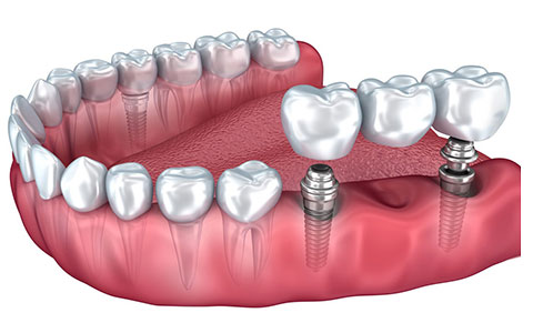
Titanium materials applied to the jawbone for the restoration of missing teeth are advanced surgical procedures that reconstruct the chewing function and the balance of the jaw joint (Temporomandibular Joint) in line with biological norms. Individuals experiencing multiple or total (complete) tooth loss in both the lower jaw (mandible) and the upper jaw (maxilla) often explore time management and surgical comfort aspects during the planning of their treatment process.
Can implants be placed in both the upper and lower jaws at the same time and how is this medical decision made?
Intervening in two different anatomical regions during the same surgical procedure in jaw surgery is quite common in the medical literature and is supported physiologically. Planning two separate surgical sessions means that the patient will undergo local anesthesia twice, initiate two separate inflammation (repair) cycles in the binding system, and spread the use of prophylactic antibiotics over two different periods. A single surgical session facilitates the body’s overall adaptation by concentrating this physical stress into a single time frame.
In making medical decisions, the medical boards (maxillofacial surgeons and prosthodontists) evaluate the physiological tolerance of the patient. Individuals with blood values within reference ranges, no clotting issues, and no medical obstacles for remaining in the surgical chair for a certain period are classified as suitable candidates for double jaw surgeries. This way, the cellular integration of the lower and upper jaws with titanium materials can be synchronized to start the production of permanent prosthetics for both jaws simultaneously.
What Anatomical Criteria is Double Jaw Planning Based on at Avrupadent Clinics?
Double jaw surgery requires much more comprehensive biomechanical engineering than just placing screws into two separate bones. The lower and upper jaws work together like two gears in a machine that must cooperate while chewing. The angle of the artificial root placed in one jaw directly affects the angle of the artificial root to be placed in the other jaw and the contact point of the porcelain tooth that will come on top.
- Virtual Occlusion Detection: In patients who are edentulous or have multiple bone deficiencies, the distance at which the lower jaw should stand in relation to the upper jaw (vertical dimension) is recalculated in digital software.
- Bone Density Analysis: The upper jaw (maxilla) is structurally spongy, while the lower jaw (mandible) is dense and hard (cortical). Doctors choose the materials appropriate for these two different densities in the diameter and groove design based on tomography.
- Surgical Template Production: To shorten the operation time and minimize tissue trauma, medical templates are produced for both jaws using 3D printers. This allows the surgeon to place screws into the bone at planned openings without experiencing millimeter deviations.
How Does Simultaneous Intervention on the Upper and Lower Jaws Affect the Healing Period?
The physiological healing (osteointegration) cycle operates according to the cellular architecture of each jawbone. The upper jawbone, being adjacent to the respiratory tract and cranial air cavities (sinuses), contains more microscopic voids in its structure (Type 3 or Type 4 bone quality). This porous structure requires a longer cellular maturation time for the hardened bone cells around the titanium material to fill in.
The alternate jaw structure is much more compact (tight) as it works against gravity, where the masticatory muscles are attached. In this area, the cells organize the titanium surface more quickly. In double jaw surgeries, even if the healing of the lower jaw is completed, the dentist usually waits for the upper jaw’s bone integration timeline to be fulfilled before transitioning to a prosthetic crown. This way, measurements for both jaws’ porcelain/zirconium are taken in the same session, and the occlusal relationship between the jaws is transmitted to the laboratory with perfect biomechanical compatibility.
How Are the Conditions Felt by the Patient During Surgical Procedures Controlled Medically?In double jaw surgeries, because there is a wide surgical area, tissue sensitivity and nerve transmission are completely isolated using specific local anesthetic agents (block anesthesia). Depending on the length of the procedure or the patient’s anxiety status, conscious sedation (semi-sleep state) medical protocols can be applied under the supervision of the dentist and anesthesiologist to maximize comfort during the operation.
Feeling anxiety in extensive jaw surgery procedures is a natural reflection of human physiology; however, the internal parts of the bone tissue (spongy area) do not contain dense nerve endings. Nerve fibers are primarily found in the mucosa of the gum tissue and the periosteum that wraps around the bone. Local anesthetic molecules block sodium channels in the cell membranes of these areas, restricting the brain’s perception of stimuli for a certain time period.In sessions where both the upper and lower jaw will be intervened, the duration for which the patient will spend in the hospital is generally longer compared to single jaw surgeries. To eliminate physiological fatigue such as the need for prolonged swallowing and the mouth being open for an extended period, sedation applied via intravenous means may be preferred in clinical environments that ensure hospital standards. Under sedation, the individual experiences a deep relaxation, does not clearly remember the procedure, and follows the doctor’s commands, significantly supporting the surgical team’s work.
How Long Does the Operation Take in Double Jaw Surgery and How is it Managed?
Depending on the number of titanium roots to be placed, the presence of additional surgical needs such as bone grafts, and the use of digital surgical templates, the surgeries intervening both the upper and lower jaws typically last between one and a half to three hours. The duration is managed within medical norms supported by the patient’s physiological rest breaks.The most critical parameter determining the operation duration is the readiness of the bone for surgical intervention. If the patient has sufficient bone thickness and vertical dimensions, it is possible to directly open drill slots without needing to make extensive incisions (flaps) using 3D templates derived from digital planning. This advanced technique known as “Flapless” (non-incision/no cut) nearly halves the time spent in the chair and minimizes the potential amount of swelling that may occur post-operation.
Medical organizations follow an ergonomic discipline in such extensive operations. Typically, surgery begins with either the lower jaw or the upper jaw, and once one area is completed, the other area is addressed; thus, local anesthesia doses are administered to the body in a balanced manner. Medical teams ensure that there is no fatigue buildup in the patient’s jaw muscles (trismus) by providing special apparatus support during the operation or by taking medical breaks to maintain the natural flow of blood.
Can Augmentation (Bone Graft) Be Applied to Both Jaws Simultaneously in Individuals with Insufficient Bone Volume?
When multiple bone deficiencies are detected, such as horizontal thinning in the lower jaw and sinus sagging in the upper jaw, graft (bone powder) and membrane applications can be performed on both jaws in the same surgical session. These advanced surgical protocols stimulate the formation of new tissue by directing the body’s own bone repair cells simultaneously to both areas based on medical standards.Individuals who have used removable palate prostheses for many years experience serious volumetric resorption (atrophy) in their jaw bones due to mechanical pressure and functional deficiencies. In this anatomical situation, the maxillary sinus air cavities located in the upper jaw sag downwards, reducing the bone distance to millimeter levels. In the lower jaw, the bone surface transforms into a sharp form like a blade.
Surgeons use “Sinus Lifting” (elevation of the sinus membrane) techniques in the upper jaw and “Guided Bone Regeneration” techniques in the lower jaw to overcome these limitations. Biomaterials made of sterile calcium phosphate particles are placed in the relevant areas. Adding bone powder to both jaws in the same session simultaneously triggers the body’s vascularization response. Once the months-long repair period is completed, both jaws transform into a resilient human bone capable of supporting titanium implants.
How Are All-On-4 or All-On-6 Systems Projected for Both Upper and Lower Jaws Simultaneously?
In cases of complete tooth loss, All-On-4 or All-On-6 systems can be projected simultaneously for the upper and lower jaws. Titanium materials placed at angles of 30-45 degrees to avoid anatomical voids are installed in the anterior areas of the jaws flat and in the posterior areas. When they provide primary stability (compression force), it allows for the application of stable temporary bridges for both jaws on the same day.In complete tooth loss restorations, the “All-on” concepts aim to establish a wide biomechanical bridge while avoiding contact with the sinuses and nerve channels due to bone resorption in the posterior areas. Including both the upper and lower jaws in this system at the same time allows for the reconstruction of the occlusal relationship (vertical dimension) from zero and in a flawless manner.
Clinical Advantages of Double Jaw All-On Systems Physiological / Biomechanical Parameters Clinical Function and Effect Avoidance of Voluminous Grafting Surgery With the open placement, the need for sinus lifting in the upper jaw and vertical augmentation in the lower jaw is significantly limited. Immediate Function (Immediate Loading) The high torque compression of titanium materials supports the screwing of stable acrylic temporary teeth for both jaws on the same day. Reassessment of Vertical Closure The distance between the upper and lower jaws (VDO) is brought to the most ideal physiological ratio by the dentist to correct any sagging in the facial muscles.
Can Individuals with Systemic Diseases be Included in Double Jaw Implant Surgery?
Individuals with diabetes, hypertension, or cardiovascular disorders can be included in double jaw surgery, provided that their blood values (e.g., HbA1c) are kept stable within reference ranges, in consultation with the relevant specialist doctors. When the systemic condition is medically controlled, the body’s tissue repair capacity can operate within physiological limits.Making surgical incisions on both jaws at once initiates a widespread inflammatory (repair) cycle in the body. This situation requires intensive effort from the vascular and circulatory systems. Chronic metabolic diseases are parameters that directly affect tissue repair speed. In particular, in patients with uncontrolled Type 2 diabetes, high circulating glucose in the blood damages cell walls, disrupting the vascularization (angiogenesis) of the tissue around titanium.
For this reason, equipped clinical organizations in the İzmir region request an extensive blood test (hemogram, coagulation profile, fasting blood sugar, etc.) from the patient before the operation. For heart patients using anticoagulants, medication doses are optimized under physician supervision. Procedures carried out after obtaining medical approval progress similarly to a healthy individual’s repair timeline. A systemic disease is not an obstacle before treatment, but a detail in medical planning that requires extra attention.
What Anatomical Data Does Radiological Imaging (3D Tomography) Provide in Double Jaw Operations?
Three-dimensional dental computed tomography (CBCT) measures the vertical and horizontal volume of both jawbones in millimeters; it maps the sagging rate of maxillary sinuses in the upper jaw and follows the path of the mental foramen and the mandibular nerve canal in the lower jaw in three-dimensional space. This radiological data prepares the ground for the secure placement of titanium roots in a virtual environment without contacting anatomical cavities.In operations involving complex and wide areas such as the double jaw, relying solely on two-dimensional panoramic films means ignoring the thickness of the bone towards the lips and tongue. A finely contoured lower jaw, like a knife’s edge, can present misleading images that appear thick and sufficient on panoramic radiographs. Three-dimensional volumetric scans allow physicians to examine the jaw slice by slice (in sections).
Additionally, the bone density scale known as Hounsfield Unit (HU) is determined before the procedure thanks to tomography software. Knowing in advance how the titanium material to be placed will encounter resistance in the bone, the physician modifies the milling (socket preparation) technique according to this hardness level. These radiological diagnostic tools are objective medical technologies that leave no room for chance in surgical success.
How Should Nutritional Routine Be Planned After Surgery in Upper and Lower Jaw Surgery Patients?
Since surgical intervention is performed on both jaws, it is medically necessary for patients to consume soft, liquid, and puree consistency foods at room temperature for the first few weeks. To avoid disrupting the stabilization of titanium roots (primary retention) within the bone, all mechanical forces created by hard, fibrous, or crusty foods should be completely avoided to limit trauma to the wound areas.When a single jaw is intervened in, the patient can usually perform partial chewing using the other jaw. However, in double jaw surgeries, since the entire chewing system undergoes reconstruction, the nutritional physiology should be revised. The newly placed titanium materials should be kept away from micro-movements (vibration) until they are surrounded by bone cells (osteointegration phase).
Medical Nutrition and Protection Protocol:- Temperature Control: During the first forty-eight hours after the operation, very hot drinks and foods should be avoided to prevent vasodilation that could trigger bleeding.
- Pressure Management: Even if fixed temporary teeth are placed on the same day, these teeth serve aesthetic and slight functional purposes. Chewing on foods like hazelnuts, walnuts, or tough meats can lead to rotation of the material inside the bone (loss of stabilization).
- Fluid Intake: Using a straw creates negative pressure that can dislodge the clot in the wound inside the mouth. Fluids should be sipped directly from a glass.
Can Elderly Patients Tolerate Procedures Applied to Both the Upper and Lower Jaw Simultaneously?
Individuals in the geriatric (elderly) group can tolerate double jaw surgery as long as their cellular ages and overall conditions meet medical standards, independent of their chronological ages. By using minimally invasive (non-sutured) techniques and modifying anesthesia protocols to be tailored to the individual, the physiological wear caused by these individuals remaining edentulous for many years can be compensated with a comfortable medical intervention.Old age is not a direct contraindication in medical procedures. On the contrary, the impacts of dentures, nutritional deficiencies, and related gastrointestinal (digestive) system discomforts can significantly reduce the quality of life for elderly individuals. The restoration of a stable chewing system provides substantial positive momentum for their systemic health.
Doctors know that elderly patients’ bone metabolism tends to work a bit slower compared to younger individuals. To balance this situation, trauma applied to the bone during surgical procedures is minimized, wound edges are meticulously closed, and the biological healing (waiting) timeline can be kept a few months longer than standard patients. Thus, tissues find the opportunity to grasp the titanium structure at their physiological speeds.
How is Aesthetic and Closure Biomechanics (Occlusion) Designed After Double Jaw Surgeries?
Simultaneously designing the prostheses for the upper and lower jaws gives the doctor the ability to reconstruct the lost vertical closure dimension, support of facial muscles, and aesthetic smile line freely (occlusal rehabilitation). Zirconium or porcelain blocks prepared in the laboratory using digital measurements and facial scans are produced to exhibit anatomical compatibility with lower jaw movements.When a prosthesis is made for only one jaw, the doctor must comply with the position and shape of the existing teeth on the opposing jaw. However, when both jaws are restored simultaneously, the dentist works as if they have a pure white canvas. Due to long-term edentulousness, the nose and chin may approach each other, and the lips may recede (elderly facial expression). The distance between the upper and lower jaws (vertical dimension) can be elevated with prosthetic restorations, creating a natural botox effect on the facial tissues.
Besides aesthetics, function is the core of this design. During chewing, the prosthetics must not interfere with each other in the movements of the jaw going right, left, and forward (preventing early contacts). Otherwise, lateral forces are applied to the titanium roots, and this stress can lead to bone damage over time. Prosthetics designed using specialized articulators (jaw joint simulators) show perfect medical compatibility with the biomechanics of the temporomandibular joint (TMJ).
How Should Oral Hygiene and Medical Maintenance Be Ensured After Surgery?
To protect the gum tissue around the prosthetics anchored to titanium roots from microbial attacks, interdental brushes, superfloss threads, and oral irrigators should be used to clean the niches beneath the prosthetics daily. Clinical tartar cleaning performed by specialists every six months preserves the long-term function of the system by limiting the development of peri-implantitis (inflammation of the surrounding tissue).Repairing the entire jaw system with artificial materials does not reduce the importance of daily cleaning rituals; on the contrary, it increases them. Bacteria that decay natural teeth cannot decompose inorganic structures (zirconium, porcelain); however, they can inflame the biological limit between titanium and bone by migrating through the neck region of the prosthetic. This insidious tissue deterioration, known as peri-implantitis, is the greatest medical enemy of extensive restorations.
In double jaw restorations, the bridge bodies are longer and have wide anatomical spaces for food accumulation underneath. The bristles of standard toothbrushes cannot enter these horizontal tunnels. Oral irrigators that shoot pressurized water help reduce the adherence of anaerobic (oxygen-free) bacteria in these areas. During clinical maintenance sessions, doctors ensure periodic medical support by removing accumulated mineral plaques with special Teflon instruments that do not scratch the titanium surfaces.
How Does Smoking Alter Cellular Repair in Double Jaw Implant Operations?
The nicotine present in cigarette smoke causes vasoconstriction by constricting the capillaries within the mucosa, slowing the delivery of repair cells and oxygen to the surgical site. In double jaw operations where multiple titanium materials are expected to integrate, reduced blood flow interrupts the osteointegration cycle, increasing the risk of infection; therefore, it is essential to limit these habits during the medical process.Any biomaterial placed in the jawbone requires a significant blood flow (vascularization) from the living tissue to surround it. In procedures involving broad wound surfaces like double jaw surgery, the body’s demand for oxygen is at its peak. When carbon monoxide mixes with the blood, it replaces the oxygen that cells need and suppresses the fibroblast cells responsible for repair.
Moreover, smoking alters the response of the gums related to periodontal health. In smokers, the gums do not bleed (giving no warning) despite plaque accumulation, allowing inflammation to progress stealthily into the bone depths. Oral surgeons typically map out a medical guideline to restrict these habits weeks before surgery and throughout the biological waiting period to ensure long-term structural stability.
Feeling anxiety in extensive jaw surgery procedures is a natural reflection of human physiology; however, the internal parts of the bone tissue (spongy area) do not contain dense nerve endings. Nerve fibers are primarily found in the mucosa of the gum tissue and the periosteum that wraps around the bone. Local anesthetic molecules block sodium channels in the cell membranes of these areas, restricting the brain’s perception of stimuli for a certain time period.
In sessions where both the upper and lower jaw will be intervened, the duration for which the patient will spend in the hospital is generally longer compared to single jaw surgeries. To eliminate physiological fatigue such as the need for prolonged swallowing and the mouth being open for an extended period, sedation applied via intravenous means may be preferred in clinical environments that ensure hospital standards. Under sedation, the individual experiences a deep relaxation, does not clearly remember the procedure, and follows the doctor’s commands, significantly supporting the surgical team’s work.
How Long Does the Operation Take in Double Jaw Surgery and How is it Managed?
The most critical parameter determining the operation duration is the readiness of the bone for surgical intervention. If the patient has sufficient bone thickness and vertical dimensions, it is possible to directly open drill slots without needing to make extensive incisions (flaps) using 3D templates derived from digital planning. This advanced technique known as “Flapless” (non-incision/no cut) nearly halves the time spent in the chair and minimizes the potential amount of swelling that may occur post-operation.
Medical organizations follow an ergonomic discipline in such extensive operations. Typically, surgery begins with either the lower jaw or the upper jaw, and once one area is completed, the other area is addressed; thus, local anesthesia doses are administered to the body in a balanced manner. Medical teams ensure that there is no fatigue buildup in the patient’s jaw muscles (trismus) by providing special apparatus support during the operation or by taking medical breaks to maintain the natural flow of blood.
Can Augmentation (Bone Graft) Be Applied to Both Jaws Simultaneously in Individuals with Insufficient Bone Volume?
Individuals who have used removable palate prostheses for many years experience serious volumetric resorption (atrophy) in their jaw bones due to mechanical pressure and functional deficiencies. In this anatomical situation, the maxillary sinus air cavities located in the upper jaw sag downwards, reducing the bone distance to millimeter levels. In the lower jaw, the bone surface transforms into a sharp form like a blade.
Surgeons use “Sinus Lifting” (elevation of the sinus membrane) techniques in the upper jaw and “Guided Bone Regeneration” techniques in the lower jaw to overcome these limitations. Biomaterials made of sterile calcium phosphate particles are placed in the relevant areas. Adding bone powder to both jaws in the same session simultaneously triggers the body’s vascularization response. Once the months-long repair period is completed, both jaws transform into a resilient human bone capable of supporting titanium implants.
How Are All-On-4 or All-On-6 Systems Projected for Both Upper and Lower Jaws Simultaneously?
In complete tooth loss restorations, the “All-on” concepts aim to establish a wide biomechanical bridge while avoiding contact with the sinuses and nerve channels due to bone resorption in the posterior areas. Including both the upper and lower jaws in this system at the same time allows for the reconstruction of the occlusal relationship (vertical dimension) from zero and in a flawless manner.
| Physiological / Biomechanical Parameters | Clinical Function and Effect |
|---|---|
| Avoidance of Voluminous Grafting Surgery | With the open placement, the need for sinus lifting in the upper jaw and vertical augmentation in the lower jaw is significantly limited. |
| Immediate Function (Immediate Loading) | The high torque compression of titanium materials supports the screwing of stable acrylic temporary teeth for both jaws on the same day. |
| Reassessment of Vertical Closure | The distance between the upper and lower jaws (VDO) is brought to the most ideal physiological ratio by the dentist to correct any sagging in the facial muscles. |
Can Individuals with Systemic Diseases be Included in Double Jaw Implant Surgery?
Making surgical incisions on both jaws at once initiates a widespread inflammatory (repair) cycle in the body. This situation requires intensive effort from the vascular and circulatory systems. Chronic metabolic diseases are parameters that directly affect tissue repair speed. In particular, in patients with uncontrolled Type 2 diabetes, high circulating glucose in the blood damages cell walls, disrupting the vascularization (angiogenesis) of the tissue around titanium.
For this reason, equipped clinical organizations in the İzmir region request an extensive blood test (hemogram, coagulation profile, fasting blood sugar, etc.) from the patient before the operation. For heart patients using anticoagulants, medication doses are optimized under physician supervision. Procedures carried out after obtaining medical approval progress similarly to a healthy individual’s repair timeline. A systemic disease is not an obstacle before treatment, but a detail in medical planning that requires extra attention.
What Anatomical Data Does Radiological Imaging (3D Tomography) Provide in Double Jaw Operations?
In operations involving complex and wide areas such as the double jaw, relying solely on two-dimensional panoramic films means ignoring the thickness of the bone towards the lips and tongue. A finely contoured lower jaw, like a knife’s edge, can present misleading images that appear thick and sufficient on panoramic radiographs. Three-dimensional volumetric scans allow physicians to examine the jaw slice by slice (in sections).
Additionally, the bone density scale known as Hounsfield Unit (HU) is determined before the procedure thanks to tomography software. Knowing in advance how the titanium material to be placed will encounter resistance in the bone, the physician modifies the milling (socket preparation) technique according to this hardness level. These radiological diagnostic tools are objective medical technologies that leave no room for chance in surgical success.
How Should Nutritional Routine Be Planned After Surgery in Upper and Lower Jaw Surgery Patients?
When a single jaw is intervened in, the patient can usually perform partial chewing using the other jaw. However, in double jaw surgeries, since the entire chewing system undergoes reconstruction, the nutritional physiology should be revised. The newly placed titanium materials should be kept away from micro-movements (vibration) until they are surrounded by bone cells (osteointegration phase).
- Temperature Control: During the first forty-eight hours after the operation, very hot drinks and foods should be avoided to prevent vasodilation that could trigger bleeding.
- Pressure Management: Even if fixed temporary teeth are placed on the same day, these teeth serve aesthetic and slight functional purposes. Chewing on foods like hazelnuts, walnuts, or tough meats can lead to rotation of the material inside the bone (loss of stabilization).
- Fluid Intake: Using a straw creates negative pressure that can dislodge the clot in the wound inside the mouth. Fluids should be sipped directly from a glass.
Can Elderly Patients Tolerate Procedures Applied to Both the Upper and Lower Jaw Simultaneously?
Old age is not a direct contraindication in medical procedures. On the contrary, the impacts of dentures, nutritional deficiencies, and related gastrointestinal (digestive) system discomforts can significantly reduce the quality of life for elderly individuals. The restoration of a stable chewing system provides substantial positive momentum for their systemic health.
Doctors know that elderly patients’ bone metabolism tends to work a bit slower compared to younger individuals. To balance this situation, trauma applied to the bone during surgical procedures is minimized, wound edges are meticulously closed, and the biological healing (waiting) timeline can be kept a few months longer than standard patients. Thus, tissues find the opportunity to grasp the titanium structure at their physiological speeds.
How is Aesthetic and Closure Biomechanics (Occlusion) Designed After Double Jaw Surgeries?
When a prosthesis is made for only one jaw, the doctor must comply with the position and shape of the existing teeth on the opposing jaw. However, when both jaws are restored simultaneously, the dentist works as if they have a pure white canvas. Due to long-term edentulousness, the nose and chin may approach each other, and the lips may recede (elderly facial expression). The distance between the upper and lower jaws (vertical dimension) can be elevated with prosthetic restorations, creating a natural botox effect on the facial tissues.
Besides aesthetics, function is the core of this design. During chewing, the prosthetics must not interfere with each other in the movements of the jaw going right, left, and forward (preventing early contacts). Otherwise, lateral forces are applied to the titanium roots, and this stress can lead to bone damage over time. Prosthetics designed using specialized articulators (jaw joint simulators) show perfect medical compatibility with the biomechanics of the temporomandibular joint (TMJ).
How Should Oral Hygiene and Medical Maintenance Be Ensured After Surgery?
Repairing the entire jaw system with artificial materials does not reduce the importance of daily cleaning rituals; on the contrary, it increases them. Bacteria that decay natural teeth cannot decompose inorganic structures (zirconium, porcelain); however, they can inflame the biological limit between titanium and bone by migrating through the neck region of the prosthetic. This insidious tissue deterioration, known as peri-implantitis, is the greatest medical enemy of extensive restorations.
In double jaw restorations, the bridge bodies are longer and have wide anatomical spaces for food accumulation underneath. The bristles of standard toothbrushes cannot enter these horizontal tunnels. Oral irrigators that shoot pressurized water help reduce the adherence of anaerobic (oxygen-free) bacteria in these areas. During clinical maintenance sessions, doctors ensure periodic medical support by removing accumulated mineral plaques with special Teflon instruments that do not scratch the titanium surfaces.
How Does Smoking Alter Cellular Repair in Double Jaw Implant Operations?
Any biomaterial placed in the jawbone requires a significant blood flow (vascularization) from the living tissue to surround it. In procedures involving broad wound surfaces like double jaw surgery, the body’s demand for oxygen is at its peak. When carbon monoxide mixes with the blood, it replaces the oxygen that cells need and suppresses the fibroblast cells responsible for repair.
Moreover, smoking alters the response of the gums related to periodontal health. In smokers, the gums do not bleed (giving no warning) despite plaque accumulation, allowing inflammation to progress stealthily into the bone depths. Oral surgeons typically map out a medical guideline to restrict these habits weeks before surgery and throughout the biological waiting period to ensure long-term structural stability.