

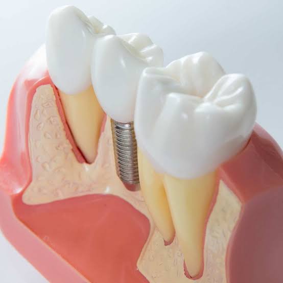
In which situations is a Short Implant used in Izmir?
In the process following tooth loss, the lack of mechanical stimulation to the jawbone leads to a cellular level resorption (absorption) of the tissue. This physiological shrinkage, which lasts for years, causes a significant reduction in the vertical distance of the jawbone (from top to bottom). The traditional approach in dental practice suggests that the titanium roots to be placed instead of missing teeth must have a certain length; however, modern medical technologies have shifted towards less traumatic and tissue-preserving protocols. In cases where the vertical bone distance is limited and is very close to the anatomical nerve canals or sinus cavities, the concept of “Short Implant” enters the stage.
In which situations is a Short Implant used and what is its medical definition?
In modern dental practices, it was once thought that a titanium root needed to be at least 10 to 12 millimeters long to remain stable in the jawbone. However, biomechanical research and advancements in surface technologies have proven that the retention in bone (osteointegration) is dependent on the total surface area in contact with the bone (Bone-to-Implant Contact – BIC) rather than the length of the material.
Even if the jawbone is shortened vertically (longitudinally), if it maintains a certain thickness horizontally (laterally), short materials come into play. These materials are designed to compensate for the deficiency in lengths with the width of their diameters. During the surgical procedure, a prototypical support point is formed by utilizing the volume of the existing surface bone without having to access anatomically risky areas (nerve networks or air pockets).
Why Are Short Implant Applications Preferred for the Mandible?
In the human skeletal structure, the mandible contains a vital nerve line that runs longitudinally like a tunnel. In normal anatomical conditions, the tooth roots are quite far from this tunnel. However, as teeth are lost and bone resorbs, the crest of the jaw erodes downwards towards this tunnel. Eventually, there comes a point where only a thin band of bone, about 5 or 6 millimeters, remains between the gum tissue and the nerve canal.
In this anatomical narrowing scenario, the physician has two surgical options: The first is to perform vertical bone grafting (vertical augmentation) to restore the bone to its former height, which places the patient through a repair process lasting several months. The second, and more tissue-friendly option, is to place a 4-millimeter short titanium root implant that is appropriate for the remaining 5-6 millimeters of bone. The second method minimizes the risk of nerve damage while also limiting local tissue trauma.
How Do Short Materials Offer an Alternative in the Presence of Upper Jaw Sinus Cavities?
The upper jaw’s molar region in the skull skeleton is directly adjacent to the sinus cavities (spaces), which are part of the respiratory system. When a tooth is extracted, the air pressure inside the sinus cavity expands toward the underlying bone (pneumatization), thinning the bone from the inside out. Using a standard titanium screw in this area causes the material to spill into the air cavity (inside the sinus).
| Medical Parameter | Sinus Lifting (Advanced Surgery) | Short Implant Application |
|---|---|---|
| Procedure Duration and Trauma | The gum is opened widely, and the membrane is stretched upward to pack bone graft. The operational area is extensive. | The procedure is limited locally by opening only a socket that fits the existing bone. |
| Cellular Maturation (Waiting) | The transition of added grafts to living bone creates an average waiting period of 6 to 8 months. | The process is completed in the bone’s own healing cycle (3-4 months), and the prosthetic stage is initiated. |
| Complication Management | There is a risk of tearing the sinus membrane or infection of the added particles. | No intervention has been made to sinus malfunction, so respiratory tract related reactions are restricted. |
How is the Biomechanical Resistance of Short Implants Against Biting Forces?
The rules of physics and engineering also apply in jaw surgery. The force applied to a titanium root works to move (bend) that root within the bone. The moment of bending of a long screw inside the bone (lever effect) is actually greater than that of a short screw. Clinical studies indicate that the biting force is most intense in the 3-4 millimeter area of the first neck (crest) where the material meets the bone. The end parts of the screw remain more passive in terms of stress absorption.
In the light of this medical data, a short material that is only 4 millimeters long but has a diameter of 5 millimeters has almost the same (or greater) surface retention as a much longer but thinner material when surface area is calculated. This biomechanical design allows the safe transmission of intense biting forces in the posterior molar regions to the jawbone.
How is Short Implant Planning Conducted in İzmir Avrupadent Clinics?
In the treatment protocols adopted by corporate organizations, no anatomical measurement is left to assumptions. Although two-dimensional standard X-rays may indicate how much height the bone retains, they do not reveal the thickness of the bone (the width in the lip-tongue direction). However, if a short titanium root is to be used, the horizontal thickness where that root will be placed must be quite healthy and broad according to medical norms.
-
\t
- Dimension Detection: In tomographic sections, the safe limit between the nerve canal or sinus membrane and the crest (bone peak) is measured millimetrically, and material length is selected leaving a safety margin of 2 mm.
- Crown-Root Ratio Calculation: If the length of the porcelain tooth (crown) to be placed is significantly longer than that of the short screw within the bone, it poses a risk of mechanical tipping (lever). This ratio is analyzed in digital software to revise the prosthesis design.
- Splinting (Connection) Plan: In cases of multiple tooth loss, the prostheses placed over adjacent short materials are designed to be interconnected (splinted) to support the toleration of mechanical forces on a common surface.
\t
\t
What are the Effects of Short Implant Application on the Operation and Healing Process?
Traditional advanced surgical (bone powder) operations create a large wound area in the body. Closing this wound area, producing new blood vessels, and converting foreign materials into bone tissue is a quite labor-intensive inflammatory process for the connective tissue system. In short implant procedures, however, the body does not struggle to convert extra materials into bone; it focuses solely on repairing the small cavity opened in the existing living bone.
Opening only a short cavity of 4 or 5 millimeters during the operation means that the intra-bone blood vessels and cells sustain less damage. Tissues whose blood supply has not been disrupted exhibit a much faster cellular recovery during the post-operative period. When the effect of local anesthesia wears off, the physiological exudate encountered is managed within normal limits through analgesic medications (pain relievers) and the application of cold compresses.
How Does a Short Implant Function Medically in Patients with Osteoporosis?
Systemic bone loss (osteoporosis) is a condition characterized by a reduction in calcium density in the skeletal system. Many of these patients use bisphosphonate-type medications that slow down bone loss. While these medications strengthen the bone, they also suppress its ability to regenerate. In dentistry, performing extensive surgical interventions (advanced grafting) on such bone can lead to non-healing in that area and tissue death.
Oral surgeons are responsible for choosing the least traumatic (minimally invasive) approach possible in osteoporosis patients. Short titanium materials precisely meet this medical need. These structures, which only grip the outer (cortical) hard layer of the bone and provide stabilization without penetrating deeper, help reconstruct the chewing function without overburdening the patient’s existing tissue. The surgical schedule is carefully managed by modifying the medications used by the patient under the physician’s supervision.
What Physiological Processes Are Expected During the Healing (Osteointegration) Period After Surgery?The implanted short titanium root is enveloped at the cellular level by jawbone cells (osteoblasts) to fuse (osteointegration), which takes an average of two to three months in the lower jaw and three to four months in the upper jaw. During this waiting period, it is essential that no movement or pressure is applied to the material so that hard bone tissue can form instead of soft tissue.
Regardless of how short the dimensions may be, the biological communication established by titanium materials with bone is the same as that of standard materials. The screw placed in the socket opened in the jawbone, thanks to its hydrophilic (water-attracting) micro-pores on its surface, attracts proteins and repair cells from the blood. In the first weeks, tissues bound to the material through a blood clot reach mechanical stability by hardening with calcium accumulation over the months.During this physiological adaptation period, it is crucial that no pressure is applied (to avoid vibration) on the short material, as it relies on the bone surface for mechanical support. If the patient attempts to chew tough foods in that area or if a temporary prosthesis is in contact with the region, instead of bone cells, flexible connective tissue cells will surround the screw. This erroneous repair known as fibrointegration may lead to the loss of the material’s stability, making it medically necessary for the patient to adhere to the dietary recommendations provided by the doctor during the waiting period.
How is the Prosthetic Design on Short Implants Optimized?
The sizes of prosthetic structures (crowns) that will come over short materials are specially designed to limit the tipping (lever) moment that occurs during chewing. The chewing surface of the prosthesis (occlusal table) is produced in a slightly narrower form than a natural tooth, allowing mechanical stresses in both horizontal and vertical directions to be dissipated evenly over the titanium collar region.Dentistry is a discipline that not only concerns biological tissues but also the physical forces (biomechanics) acting on those tissues. If a very tall and wide porcelain tooth is built on a short root (disruption of the crown/root ratio), the pressure applied to the abutment of the tooth during the chewing of food generates a force (torque) aimed at dislodging the underlying short root.
Stress-Reducing Medical Strategies in Prosthetic Design Design Modification Mechanical and Physiological Purpose Narrowing the Chewing Surface The area that comes into contact with food is minimized, reducing the total load transmitted to the titanium material to a minimal level. Leveling of Tubercle (Pulp) Edges The bumps on the surface of porcelain crowns are flattened, limiting the transmission of lateral (horizontal) forces to the bone. Splinting (Multiple Connection) The joining of prosthetics made of short materials applied side by side ensures that the force is distributed to the entire block instead of a single screw.
Is There an Advantage of Applying Short Implants in Patients with Systemic Diseases?
In individuals with diabetes (sugar disease) or cardiovascular (heart and vascular) disorders, advanced surgical (bone dust) procedures may carry a high risk of infection and bleeding. The short implant procedure is a more advantageous medical approach that allows individuals with systemic diseases to benefit from surgical treatment without risking their overall health status since it narrows the surgical area and shortens tissue repair time.As the diameter of the surgical procedure increases, the need for cellular defense and blood flow that the body must direct to that area also increases proportionally. In uncontrolled diabetic cases, due to weakened microcirculation, large tissue sections and added external biomaterials may not receive adequate nourishment; this increases the potential for tissue inflammation (necrosis).
The placement of short titanium materials only requires the creation of a small millimetric socket (cavity) in the jawbone, which creates minimal stress on the body’s repair mechanism. Additionally, the short duration of the operation helps to control life-threatening fluctuations that can develop in hypertensive patients or individuals with heart disorders that are dependent on anesthesia stress and the duration of stay in the chair. Of course, obtaining medical approval through consultations with the relevant branch physicians is an indispensable medical protocol.
How Should Long-Term Care and Maintenance of Prosthetics on Short Implants Be Managed?
As is the case with all prosthetic restorations, it is essential to adhere to daily care routines (such as using an interdental brush and super floss) to maintain the longevity of shorter materials. Clinical plaque cleaning and radiological bone monitoring sessions conducted under the supervision of a specialist every six months preserve the biological integrity of the system by limiting the development of peri-implantitis (inflammation of surrounding tissues).Short titanium materials derive their stability from limited but dense bone support at their surfaces. If bacterial plaque accumulates in the neck area due to the patient’s insufficient oral hygiene, and this plaque leads to tooth decay, the inflammation (peri-implantitis) beginning in the gum tissue progresses toward the bone. While 2 millimeters of bone loss can be tolerated in a longer material, experiencing 2 millimeters of bone resorption in a short material, which is already 4 or 5 millimeters long, means losing half of the support of the screw.
This physiological sensitivity requires patients with short materials to exceed standard oral hygiene practices. The use of water flossers limits the proliferation of harmful bacteria by flushing out oxygen-deprived environments beneath prosthetics. During routine medical check-ups, the closure relationships of the prosthesis are reassessed, as asymmetrical deviations that may occur over time in chewing balances (occlusion) can lead to excessive load on short material. Small adjustments made by the doctor eliminate this mechanical stress.
Regardless of how short the dimensions may be, the biological communication established by titanium materials with bone is the same as that of standard materials. The screw placed in the socket opened in the jawbone, thanks to its hydrophilic (water-attracting) micro-pores on its surface, attracts proteins and repair cells from the blood. In the first weeks, tissues bound to the material through a blood clot reach mechanical stability by hardening with calcium accumulation over the months.
During this physiological adaptation period, it is crucial that no pressure is applied (to avoid vibration) on the short material, as it relies on the bone surface for mechanical support. If the patient attempts to chew tough foods in that area or if a temporary prosthesis is in contact with the region, instead of bone cells, flexible connective tissue cells will surround the screw. This erroneous repair known as fibrointegration may lead to the loss of the material’s stability, making it medically necessary for the patient to adhere to the dietary recommendations provided by the doctor during the waiting period.
How is the Prosthetic Design on Short Implants Optimized?
Dentistry is a discipline that not only concerns biological tissues but also the physical forces (biomechanics) acting on those tissues. If a very tall and wide porcelain tooth is built on a short root (disruption of the crown/root ratio), the pressure applied to the abutment of the tooth during the chewing of food generates a force (torque) aimed at dislodging the underlying short root.
| Design Modification | Mechanical and Physiological Purpose |
|---|---|
| Narrowing the Chewing Surface | The area that comes into contact with food is minimized, reducing the total load transmitted to the titanium material to a minimal level. |
| Leveling of Tubercle (Pulp) Edges | The bumps on the surface of porcelain crowns are flattened, limiting the transmission of lateral (horizontal) forces to the bone. |
| Splinting (Multiple Connection) | The joining of prosthetics made of short materials applied side by side ensures that the force is distributed to the entire block instead of a single screw. |
Is There an Advantage of Applying Short Implants in Patients with Systemic Diseases?
As the diameter of the surgical procedure increases, the need for cellular defense and blood flow that the body must direct to that area also increases proportionally. In uncontrolled diabetic cases, due to weakened microcirculation, large tissue sections and added external biomaterials may not receive adequate nourishment; this increases the potential for tissue inflammation (necrosis).
The placement of short titanium materials only requires the creation of a small millimetric socket (cavity) in the jawbone, which creates minimal stress on the body’s repair mechanism. Additionally, the short duration of the operation helps to control life-threatening fluctuations that can develop in hypertensive patients or individuals with heart disorders that are dependent on anesthesia stress and the duration of stay in the chair. Of course, obtaining medical approval through consultations with the relevant branch physicians is an indispensable medical protocol.
How Should Long-Term Care and Maintenance of Prosthetics on Short Implants Be Managed?
Short titanium materials derive their stability from limited but dense bone support at their surfaces. If bacterial plaque accumulates in the neck area due to the patient’s insufficient oral hygiene, and this plaque leads to tooth decay, the inflammation (peri-implantitis) beginning in the gum tissue progresses toward the bone. While 2 millimeters of bone loss can be tolerated in a longer material, experiencing 2 millimeters of bone resorption in a short material, which is already 4 or 5 millimeters long, means losing half of the support of the screw.
This physiological sensitivity requires patients with short materials to exceed standard oral hygiene practices. The use of water flossers limits the proliferation of harmful bacteria by flushing out oxygen-deprived environments beneath prosthetics. During routine medical check-ups, the closure relationships of the prosthesis are reassessed, as asymmetrical deviations that may occur over time in chewing balances (occlusion) can lead to excessive load on short material. Small adjustments made by the doctor eliminate this mechanical stress.







