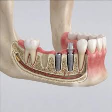
Is an implant placed in the jawbone for narrow jaw?
Titanium materials used for compensating dental deficiencies need to be surrounded by a minimum thickness and density of a bone wall. Human anatomy has a dynamic cellular cycle that exhibits resorption of non-functional tissues. During the process following tooth loss, the jawbone, deprived of masticatory forces, can shrink to a form as thin as a knife’s edge, especially in the horizontal axis (in the cheek and tongue direction). This anatomical narrowing creates a barrier that limits the feasibility of standard jaw surgery procedures.
Is an implant placed in the jawbone for narrow jaw, and how is it biologically possible?
The long-term physiological stability of any titanium screw placed inside the jawbone depends on being surrounded by a healthy bone tissue of at least 1.5 – 2 millimeters in thickness. If the threads of the screw protrude out of the bone or are covered by a very thin bony membrane, the tissue in that area risks necrosis over time due to insufficient blood supply, which may lead to material loss.
Advanced surgical branch of dentistry aims to overcome such anatomical deficiencies by utilizing the body’s self-repair capacity. When inorganic bone powders (grafts) are placed in the narrowing bone crest (top), the body uses these materials as a scaffold. Thanks to the blood circulation, osteoblast (bone-forming) cells that reach the area infiltrate into these powders over a period of months, transforming them into living, hard, and voluminous human bone. This newly formed biological volume provides a suitable ground for the placement of artificial roots.
What Causes Anatomical Narrowing in the Jawbone?
The human skeletal system is a reactive structure under mechanical stress (Wolff’s Law). Our natural teeth transmit the force generated while chewing food to the jawbone, ensuring that bone cells remain continuously active and maintain their mineral density. When a tooth is extracted, this mechanical stimulus signal is completely interrupted. The body starts to resorb the calcium and minerals in this bone tissue, which has been coded as no longer functional, back into the bloodstream.
This resorption process does not progress uniformly everywhere. In the upper and lower ridges, particularly in the bone layer facing the cheeks (buccal side), the cortical plate is structurally quite thin. Within the first year following extraction, this outer bone layer rapidly resorbs, allowing for inward collapse. The width of the jawbone decreases, and the bone takes on a shape resembling a sharp blade edge or a thin triangle. Even if the vertical height appears sufficient, this serious loss of tissue in the horizontal direction eliminates the width that titanium roots could support.
What Surgical Methods Are Used to Expand the Narrowing Jawbone?
The most suitable surgical method is chosen by the physicians according to the anatomical map of the bone (how much it has thinned and how flexible its structure is). All these methods integrate the principles of tissue engineering into clinical practice.
| Surgical Method | Clinical Operation | Medical Application Area |
|---|---|---|
| Guided Bone Regeneration (GBR) | Calcium particles (graft) are placed on the outer surface of the narrowed bone, covered with biodegradable/non-biodegradable membranes. | It is used to thicken the tissue outwardly in mild to moderate levels of horizontal narrowing. |
| Crest Expansion (Split Crest) | Placement of material by stretching the top point of the bone in the blade ridge form with micro instruments. | The bone is planned in specific anatomical crest areas that have a certain flexibility but are too thin. |
| Autogenous Block Graft | Transplantation of a small bone block taken from the tip of the patient’s jaw or the back molar region to the narrow site with micro-screws. | In cases of severe horizontal bone loss, it is preferred to obtain the highest cellular response. |
How is Crest Expansion (Split Crest) Operation Performed?
This medical approach is based on the philosophy of expanding the bone from within rather than thickening it from the outside. However, the presence of a spongy and flexible layer called “calcellus” in the middle of the bone is a prerequisite for this procedure to be performed. If the bone is fully hardened (cortical), there is a risk of fracture when trying to stretch it.
During surgical procedures, piezo-surgery devices, which use sound waves and cut the bone at the micron level, are generally preferred over traditional rotary drills. An incision is made in a fine line from the top of the bone, and then special expanding hand tools (wedge-shaped instruments) are used to gradually and millimetrically expand the bone plate facing the cheek outward. A titanium screw is placed in the center of this newly created volume. Bone dust is inserted into the voids remaining at the edges to support the body’s blood clot and cells, after which sutures are applied.
What Medical Stages Do Bone Dust (Graft) and Membrane Applications Include?
The basis of this procedure, known as Guided Bone Regeneration (GBR), relies on managing cellular competition. Gum cells are soft tissue cells and multiply very quickly; bone cells (osteoblasts), on the other hand, reproduce much more slowly. If you simply place bone dust on a narrow bone and cover it, the rapidly multiplying gum cells will seep between those dust particles, limiting osseointegration and turning that area into connective tissue.
To manage this cellular competition, doctors place special medical membranes on top of bone powders. These membranes serve as a barrier. They physically restrict the rapid proliferation of soft tissue cells from invading the spaces of the bone powders (creating a barrier). Thus, they provide the isolated biological environment and time necessary for the slow-growing bone cells to envelop the particles and convert them into living tissue (through the osteochondral process).
How are Narrow Jaw Cases Evaluated at Avrupadent Clinics in Izmir?
In surgical interventions where there are anatomical constraints, there is no room for estimates or two-dimensional incomplete data. Although standard panoramic X-rays show how much length of the bone remains, they do not reflect the thickness of the bone in the direction of the lip and tongue (the third dimension). A very thin bone protrusion, reminiscent of paper, is a result of the inadequacy of two-dimensional films when the gums of a patient are exposed.
In institutional medical organizations, the process begins with radiological volumetric scans. Every millimeter of the jawbone is examined in slices in a computer environment. The thickness of the bone is measured digitally, indicating how many millimeters it is, and how much space the cortical (hard) layer occupies. Based on this objective data, medical boards decide before the operation which bone augmentation method (split crest, block graft, or YKR) will provide the most suitable (least traumatic) response for the patient’s physiology.
Can Titanium Roots Be Placed in the Same Session as Bone Augmentation Procedure?
In the medical literature, this situation is classified as “Simultaneous (Single-Stage)” and “Sequential (Two-Stage)” surgical approaches. For a titanium root to complete the osteointegration (bonding with the bone) cycle, it is a fundamental medical rule that it must not move at all (should not show micro-movement) right after it is placed in the bone.
When the doctor expands the jaw, if the remaining original bone material can withstand a rotational force (torque) of about 35-40 Ncm, both procedures can be completed in the same session, limiting the number of surgical sessions for the patient. However, if the jawbone is so thin that the material shows wobbling inside the bone after expansion, the chance of that material bonding with the bone is close to zero. In this physiological scenario, the doctor will, sticking to medical principles, only close the area with grafts and stitches. The body will convert those powders into a living and healthy bone block in approximately six months. In a second session, titanium screws can be securely positioned in this newly formed and expanded foundation.
How Long Does Bone Integration (Consolidation) Last After Surgery?
Volume enhancement operations performed by cutting the bone or adding powders demand a significant cellular workload from the body. The added biomaterials are initially inert structures. For the body to accept these materials as its own bone, they need to establish new capillary networks through a process known as “angiogenesis.” Repair cells (osteoblasts) cannot migrate to the area until blood vessels reach that location.
While the fusion of titanium in a standard bone structure takes two to three months, the timeline for an externally added structure to become vascularized and evolve into a living tissue can extend over the course of months. To ensure that this healing stage remains uninterrupted, it is essential that a temporary movable prosthesis does not exert pressure on the surgical area. A graft site under constant pressure will lead to the production of flexible connective tissue instead of bone cells, jeopardizing the medical stability of the procedure.
Do Age Factors Affect Medical Decisions in Narrow Jaw Surgeries?
There is a common misconception that cellular aging in society completely prevents bone surgery. Human bone has the capacity to repair fractures and remodel itself throughout all stages of life. Individuals in advanced age who have been edentulous for years are the group that experiences anatomical narrowing the most. Achieving a healthy chewing function for these individuals is crucial for the preservation of their gastrointestinal (digestive) systems.
The clinical approach of physicians towards elderly patients focuses on expanding the repair timeline and minimizing surgical stress. Additionally, the bisphosphonate-type drugs frequently used by elderly individuals due to osteoporosis may suppress the bone’s self-renewal mechanism. If these types of medications are being used, consultations with relevant medical doctors are conducted to make medication adjustments, and the surgical planning is revised according to the patient’s medical history.
Can Those with Systemic Diseases Undergo Bone Augmentation?
Advanced surgical interventions such as bone expansion involve the widening of the periodontal tissue and the manipulation of the underlying bone. This situation initiates a widespread inflammatory (repair) cycle in the organism. Chronic metabolic diseases directly affect the wound healing speed and the responsiveness of the connective tissue system.
Especially in patients with Type 2 diabetes, a high glucose level in the blood disrupts protein synthesis, complicating the closure of wounds. Due to weakened microvascular circulation, the added bone powders cannot reach sufficient oxygen, resulting in the risk of necrosis (cell death). When the patient’s HbA1c level is maintained within medical standards, the body’s physiology approaches its normal limits, and the surgical intervention achieves the planned repair goals.
How Does Tobacco Usage Affect Narrow Jaw Surgery?
The absolute success criterion for augmentation (bone grafting) procedures is the ability to form new blood vessels in that area. In smokers, carbon monoxide enters the bloodstream, displacing the oxygen that cells need. The activity of cells known as fibroblasts (wound healing cells) and osteoblasts (bone-forming cells) is severely suppressed in an oxygen-deprived environment.
Additionally, the sutured gum tissue, when exposed to the heat and toxins generated by tobacco products, loses its tension and shows a tendency to open. This opening means that the underlying bone powders come into contact with bacteria in the surrounding environment, which leads to inflammation of the material and its expulsion. Jaw surgeons establish clinical guidelines to restrict smoking for individuals undergoing bone expansion treatment in the weeks and months leading up to the surgery.
What Medical Precautions Should Be Taken to Prevent Jaw Bone Narrowing?
The greatest challenges faced in clinics arise from patients neglecting the area for many years after tooth extraction. There is an approach in modern dentistry known as ‘socket preservation.’ If the patient who has had a tooth extracted does not have the means for a prosthetic treatment at that moment, the doctor can slow down the collapse of that area using medical methods by placing some bone powder into the extraction site.
However, the most physiological way to preserve bone volume is to timely place a system (titanium root) that transmits chewing pressure to the bone in that area. The titanium root sends a signal to the surrounding bone cells indicating ‘there is mechanical function here,’ stopping the cells from reabsorbing calcium and preserving the anatomical form of the area for many years.