

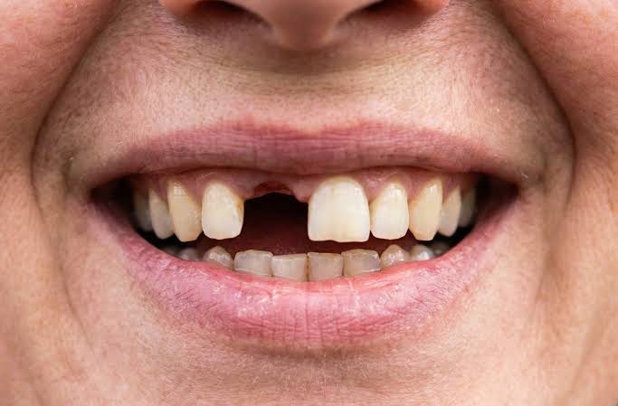
Can implants be placed for congenitally missing teeth?
Structural dental deficiencies arising from the failure of dental germs (buds) to form during embryological development are referred to in the medical literature as “dental agenesis”. This anatomical condition, often genetically inherited, not only affects the masticatory mechanism of individuals but also leads to insufficient growth of the jawbone in that area, tipping of adjacent teeth into the gap, and restrictions in speech (phonation) functions. In dental practice, there are fundamental physiological and biomechanical differences between placing an artificial root for a tooth that has been extracted later and compensating for a tooth that has never existed.
Can implants be placed for congenitally missing teeth and what are the causes of this medical condition?Implants (titanium artificial roots) can be applied for congenitally missing teeth (dental agenesis) provided that there is adequate volumetric width of the jawbone, which can be achieved medically after the skeletal bone development of the individual is fully completed. This condition, resulting from genetic mutations, developmental disorders in the embryological stage, or syndromic presentations leading to the complete absence of the dental germ (bud), is permanently restored through joint clinical planning of oral and maxillofacial surgery and orthodontics.
The formation of teeth in human anatomy begins in the first weeks of the embryological period in the womb and is the result of a highly complex cellular arrangement that continues for years. Dental germs form when ectodermal tissues migrate into the jawbone. If a mutation occurs in certain genes during this cellular migration (for example, in the MSX1 and PAX9 genes), the dental bud will not exist in that area. In the literature of dentistry, this condition is classified as hypodontia (1 to 5 missing teeth), oligodontia (6 or more missing teeth), or anodontia (no teeth at all) depending on the number of missing teeth.Prosthetic restorations applied for congenital tooth loss require a more comprehensive biological infrastructure than procedures performed to replace a tooth that has been extracted later. This is because when a tooth is extracted, there is already a formed jawbone (alveolar ridge) in that area. However, in areas where there are no teeth since birth, the bone remains quite thin (hypoplastic) both vertically and horizontally, as it has not been stimulated. Titanium root applications are placed within the bone to take on the biomechanical function of the missing root after this thin anatomy has been reshaped using tissue engineering methods.
What Radiological Methods Are Used to Diagnose Hypodontia (Congenital Tooth Absence) in Izmir?
In the equipped dental clinics in Izmir, the diagnosis of congenital tooth loss is made by screening the dental germs within the bone using panoramic radiographs and three-dimensional dental tomography (CBCT) devices. These radiological data objectively map whether the tooth remains buried in the jawbone and provide millimetric volume measurements of the bone for treatment planning.The presence of a tooth not being visible in the oral cavity (as a result of an eruption) does not mean that the tooth is congenitally absent. Sometimes, a tooth bud has formed but has remained buried within the jawbone due to its incorrect positioning, pressure from neighboring teeth, or a thick tissue barrier in the eruption path (e.g., buried canine teeth). To diagnose a true tooth deficiency, observing the oral cavity is insufficient; it requires scanning the inside of the bone with medical devices.
In clinical evaluation processes, two-dimensional panoramic films are used as the first screening tool. If no tooth bud or root structure is found in that area on the panoramic film, the diagnosis of congenital absence (agenesis) is confirmed. After this stage, three-dimensional volumetric tomography is utilized to determine whether a titanium material can be placed in that space in the future. Tomography provides objective data to medical boards by digitally transferring the thickness of the bone in the area to software, including the angulation of any neighboring tooth roots, at a millimetric level.
How Do Surgical and Prosthetic Procedures Progress in Cases of Non-eruption of Primary Teeth?
In cases where the erupting permanent tooth is congenitally absent, the primary tooth can remain in the mouth for years without being lost because a physiological pressure that would dissolve the root of the primary tooth does not occur. The medical approach is to keep this healthy primary tooth in the arch for as many years as possible; however, when mobility begins due to root resorption, the tooth is extracted, and a titanium artificial root is planned for the area.The biological mechanism of the shedding of primary teeth at a certain age is based on the physical pressure applied to the roots of the primary tooth as the permanent teeth growing beneath begin to erupt. This pressure activates cells known as “osteoclasts,” which gradually dissolve (resorb) the root of the primary tooth. When the root is gone, the primary tooth becomes loose and falls out. If there isn’t a permanent tooth bud developing underneath, this resorption mechanism does not begin at all.
In this anatomical chart, there are individuals who continue to have their primary teeth in their mouths in a healthy manner until their thirties or forties. The clinical approach of dentists here is to preserve the existing natural tissue. As long as the primary tooth remains in the mouth, the surrounding jawbone does not resorb in order to support that tooth; it retains its shape. However, the roots of primary teeth are much shorter and weaker compared to permanent teeth; they may begin to resorb or fracture over the years due to the cumulative fatigue of the chewing forces. At the medical stage where this tissue loss is experienced, the primary tooth is removed and a titanium material is placed that will serve in the long term.
Why Is Titanium Root Application Not Done Before Bone Development Is Completed?
In individuals in the growth and development stage, the jawbones continue to expand and grow in three dimensions. In this stage, the titanium material placed in the bone will remain stable (ankylosed) by fusing with the bone; as surrounding tissues grow, the prosthetic tooth will be left below, leading to impaction (infraocclusion); since this situation can lead to serious jaw asymmetries and irreversible orthodontic disorders, these procedures are medically postponed until skeletal development is completed.Natural teeth adapt to the growth pattern of the jawbone through the periodontal connective tissue fibers around them. During growth in childhood or adolescence, natural teeth maintain their positions in the chewing plane by moving upwards (or downwards) in coordination with the jawbone, aided by these flexible connections. In contrast, titanium artificial roots used in jaw surgery integrate directly with bone cells; they lack any connective tissue that would provide flexibility.
Assuming a titanium implant was placed for a missing tooth at an early age (for example, at 14 years old), the patient’s jawbone will grow a few millimeters further downwards (or forwards) over the next five years. While the adjacent natural teeth move with the jaw, the titanium root will remain fixed in its initial position. As a result, the artificial tooth will remain above the level of the neighboring teeth (buried), creating asymmetry in the gum tissue boundaries and disrupting the overall chewing balance. Due to this physiological limitation, doctors do not proceed to the surgical phase without confirming that skeletal growth has completely finished (usually at 18 for girls and 20-21 for boys) through wrist X-rays or cephalometric films.
What Temporary Prosthetic Procedures Are Planned While Waiting for Bone Development in Young Individuals?
In young individuals who have not yet completed their skeletal growth but are experiencing aesthetic and psychological issues due to congenital tooth loss, Maryland bridges with wings, removable clear aligners (Essix), or space-maintaining appliances are used to close the gaps without damaging the dental tissues until the growth phase is complete, ensuring the temporary maintenance of function.Especially the lack of upper jaw lateral teeth (bicuspids) from birth can negatively affect the social lives and self-esteem development of adolescents. The inability to perform surgical intervention during this process does not mean that the patient must leave that area empty for years. Doctors develop conservative (protective) solutions that do not disrupt growth dynamics, do not penetrate the bone, but ensure visual integrity.
Temporary Prosthetic Options:- Maryland (Winged) Bridge: A body tooth made of composite or porcelain is placed in the position of the missing tooth. This tooth is bonded to the inner (tongue-facing) surfaces of the adjacent natural teeth with special medical cements, using metal or fiber wings at the back, without cutting the tooth. It can be easily removed when growth is complete.
- Essix Retainer (Tooth-Colored Clear Plate): Acrylic tooth is placed in the part of these clear plates, used at the end of orthodontic treatments, that corresponds to the gap. When the patient wears the plate, the missing area gets closed. The plate can be removed while sleeping at night or during cleaning.
- Removable Space Maintainers: Devices that support from the palate and hold an artificial tooth in the missing tooth area prevent the neighboring teeth from closing the gap and support aesthetic illusion.
Which Advanced Surgical Methods Are Used If There Is Insufficient Jaw Bone Volume Due to Congenital Tooth Missing?
In areas where the dental follicle is formed, the jawbone (alveolar crest) cannot develop physiologically and remains very thin (hypoplastic). To enable titanium implants to be placed in these narrow bony areas, techniques such as guided bone regeneration (bone dust and barrier membrane applications) or autogenous bone transfer are applied to provide the region with medical volume both horizontally and vertically.The part of the jawbone surrounding the tooth (alveolar process) is a structure that exists as “tooth-dependent” during embryological development. If a tooth does not develop in a region, the jawbone in that area cannot reach the thickness and height that would carry that tooth. Therefore, the bone structure encountered in regions with congenital tooth absence is much narrower, resembling the thinness of paper (ridge width) compared to the bone of an individual who had teeth extracted later on.
To ensure the stability of titanium materials in that area, the surgeon must firstly thicken that bone. Sterile biomaterials containing calcium phosphate (bone dust) are filled into the narrow area. The surfaces of these particles are covered with special collagen membranes that block the infiltration of soft gum cells. The body vascularizes these particles within an approximately six-month consolidation (ossification) cycle, transforming them into its own dense bone. The newly obtained broad anatomical substrate creates a suitable ground for the titanium implant.
Why is Orthodontic Treatment (Brackets/Plates) Planned as a Preparation for Titanium Implant Placement?
When the adjacent teeth shift or tilt over time in edentulous areas, they narrow the surgical field. It is a medical necessity to push the roots of the adjacent teeth with orthodontic appliances in order to place titanium material in the ideal axis (parallel) to the jawbone and to be able to create an aesthetic/functional crown on it, reaching the area with minimal biomechanical millimeters.In dental practice, compensating for missing tissue depends on the correct positioning of surrounding tissues. When the teeth in a human mouth find spaces next to them, they lose their mechanical support and slowly lean toward that space (mesial drift) under the influence of chewing forces. If a region is vacated from birth for twenty years, the roots of the teeth on the right and left of the gap may close off the bone beneath that area like a pair of scissors.
Attempting to forcibly place a titanium screw into this narrowed bony corridor means piercing or damaging the healthy roots of adjacent teeth. Additionally, if the screw has to be placed at an awkward angle, the load placed on the future tooth will reflect as a diagonal stress on the jawbone, leading to bone resorption. Orthodontists bring these tilted teeth back to their physiological positions (upright) with wire or clear plate systems. The opened clean and flat bony corridor leaves a secure workspace for the oral surgeon to operate according to medical standards.
How is a Multidisciplinary Approach Applied in the Management of Congenitally Missing Tooth Cases within Avrupadent?
In İzmir, in medical organizations similar to Avrupadent that provide corporate services, cases of dental agenesis are evaluated by medical boards consisting of specialists in orthodontics, jaw surgery, and prosthetic dental treatments. Treatment steps of each branch are synchronized by collaboratively designing the movement path of the teeth, the need for bone augmentation, and the final position of the porcelain tooth in a digital environment using three-dimensional radiological mappings.The treatment of complex anatomical anomalies includes medical dynamics that cannot be summarized by the knowledge of a single discipline. Particularly in cases of multiple tooth loss, the patient’s mechanical closure (occlusion) can be completely disrupted. The medical boards plan the entire journey of the patient from the day they start treatment to the day they finish using a philosophy of “backward planning.”
Using digital software, optical measurements taken from the patient’s mouth are manipulated on a computer screen. The orthodontist determines the space that will be created by virtually moving the teeth. The prosthodontist designs the width and length of the porcelain tooth that will fill that space. The jaw surgeon places the titanium screw in the virtual bone in a way that it will align perfectly with the center of the designed porcelain tooth. If the screw is to be placed externally, it is noted in the report that bone powder will be added. Once all branches reach an agreement on this digital project, the patient’s medical process in the chair is initiated.
How is Aesthetic (Pink and White) Integrity Ensured in Anterior Tooth Gap?
It is not enough to simply provide white aesthetics (zirconium/porcelain crowns) for deficiencies originating from the anterior region of the smile line; the goal is to mimic the natural form, volume, and triangular structures known as papillae of the gum tissue (pink aesthetics). For this purpose, soft tissue grafts (gingival grafts from the palate) and medical devices for shaping the gums are utilized to biologically support the crown’s emergence like a natural root from the gum.The congenital absence of upper incisor teeth (lateral teeth) is a genetic condition frequently encountered in the community. This area is the direct reflection area of the smile when a person smiles or speaks. Because the tooth bud did not develop in that area, the gum tissue (mucosa) covering the bone remains thin and has a flat structure.
Medical Procedures in Anterior Region Aesthetics Aesthetic Parameter Clinical Procedure and Biological Purpose Gum Thickness Augmentation (Biotype Modification) The thick connective tissue taken from the upper palate is transplanted under the thin gum using micro-surgery. The reflection of titanium’s gray color from the outside is restricted. Emergence Profile Formation Pressure is applied to the gum tissue using temporary crowns or special shapers; it is ensured that the tissue wraps the porcelain in a natural wave. Use of Zirconium Abutments Instead of metal parts, white/transparent zirconium connecting pieces are used, aligning the light reflection (translucency) at the gum level with natural enamel.
Why Are Zirconium Crowns Applied on Titanium Roots Preferred from a Biomechanical Perspective?
Zirconium crowns do not contain metal alloys due to their inorganic structures, and they replicate the optical depth of natural enamel tissue directly thanks to their light transmissibility. Additionally, the pure surface form of zirconium establishes a high cellular compatibility (biocompatibility) with the surrounding mucosa by making it difficult for bacterial plaque to adhere; this contributes to the medical dimension of limiting gum retraction and infection risk.In past years, porcelain teeth with metal substructures, despite being durable, did not allow light to pass through and gave a matte and dull appearance due to their internal metal skeletons. Moreover, over time, metal alloys could oxidize with the saliva of the gums, leading to purplish-gray tattoo-like stains at the gum edges. In restorations performed at an early age (in the early twenties) for treating congenital missing teeth, the compatibility of the material with the tissue (being non-reactive) is of critical medical importance.
Zirconium (zirconium dioxide) is a white crystal that, despite its ceramic structure, can compete with metals in terms of resistance to cracking and breaking. When coated with a glass-like glaze, it has no microscopic pores on its surface. Gum cells (fibroblasts) respond physiologically favorably to this poreless surface by closely surrounding zirconium. This anatomical shield, which limits plaque accumulation, ensures long-term isolation from the risk of “peri-implantitis” (surrounding bone inflammation) posed by titanium roots anchored in the jawbone.
How Long Does the Surgical Operation Process and Healing Period Take in Cases of Congenitally Missing Teeth?
In standard cases where bone grafting (augmentation) is not needed for the region with tooth deficiency, the surgical procedure is completed in a single session, and the biological bone healing phase lasts 2 to 3 months. In patients who require advanced surgery (grafting) due to bone narrowing, a minimum additional physiological waiting schedule of 4 to 6 months is implemented for the added material to cellularly rejuvenate and harden.The rate of repair for an intervention made to the jawbone depends on the size of the procedure and the existing viable bone surface. If the physician has been able to place titanium material onto the existing thin bone with the help of orthodontic forces by slightly stretching it, the cells directly envelop this material. In this case, the process of osteointegration (fusion) progresses rapidly within the limits of what is possible.
However, if the bone is so thin that it needs to be thickened by adding calcium phosphate powders from the outside, the physiological waiting time required from the body is much greater. The process of new capillary vessels (angiogenesis) being formed by the body, transporting repair cells with these vessels, and the powders transforming into viable bone blocks (consolidation) is like a laboratory activity that takes months. If this repair phase, which operates at the body’s own pace, is rushed, the tissues cannot bear the load and material loss occurs. During these biological months, patients use temporary restorations that preserve external aesthetic appearance but do not apply pressure to the region.
How are Complete Prosthetic Designs Optimized in Genetic Oligodontia with Multiple Tooth Deficiencies?
In cases of oligodontia or anodontia, where six or more teeth are genetically missing, the individual’s occlusal height and jaw development are severely impaired. Medical boards ensure mechanical rehabilitation by balancing force distribution through long-stem bridges splinted (connected) over 4 to 6 titanium artificial roots strategically placed according to the strength of the jawbone.Oligodontia cases are one of the most complex anatomical puzzles in dentistry. In these individuals’ mouths, there may be one single tooth, with distorted forms, pointed or short milk and permanent teeth present together. Due to the significant number of missing teeth, the jawbone cannot rise vertically. The lower third of the face (the distance between the tip of the nose and the tip of the jaw) remains short, the lips recede inward, and the patient exhibits a facial profile that is much older than their chronological age.
In these medical tables, the goal for doctors is not merely to place a titanium screw for every missing tooth; as excessively drilling into a thin jawbone disrupts blood supply and creates a risk of cell death (necrosis). Instead, strategic supports are placed in the densest areas of the jaw following biomechanical principles. The prosthetics to be made will elevate the patient’s vertical dimension of occlusion (VDO) and will be produced in sufficient volume to support the facial muscles. Thus, not only teeth but also a physiological recovery reflected in the entire facial skeleton is achieved.
Why are Long-term Oral Hygiene and Clinical Follow-up Appointments After Surgery So Important?
The artificial systems integrated into the jawbone must be used with stability for many years, and this is crucial for limiting microbial inflammation (peri-implantitis) that may develop in the surrounding tissues. In addition to daily brushing routines, the use of interdental brushes and mouth rinses; performing professional plaque removal by specialists every six months and conducting radiological bone measurements is a medical maintenance requirement.Titanium materials and zirconium teeth are inorganic, so they do not dissolve in acids. However, the surrounding gums and bone are living biological tissues. When bacteria from the oral flora settle in the neck area between the prosthesis and the gum, the body sends inflammatory cells to that area to fight these bacteria. Prolonged inflammation can lead to the resorption of the bone that holds the titanium root.
Individuals receiving treatment for congenital missing teeth usually complete this surgical process in their early twenties. The aim is for these artificial roots to remain in the patient’s mouth for fifty years or more. Maintaining such a long physiological lifespan requires a strict maintenance discipline, not just the comfort of saying ‘treatment is complete.’ Doctors check the potential asymmetries in the occlusion of the prostheses during clinical appointments, detect screw loosening, and remove dental calculus (tartar) from unseen cavities using special instruments that do not scratch the titanium surface. The medical insurance of the system covers these periodic checks.
The formation of teeth in human anatomy begins in the first weeks of the embryological period in the womb and is the result of a highly complex cellular arrangement that continues for years. Dental germs form when ectodermal tissues migrate into the jawbone. If a mutation occurs in certain genes during this cellular migration (for example, in the MSX1 and PAX9 genes), the dental bud will not exist in that area. In the literature of dentistry, this condition is classified as hypodontia (1 to 5 missing teeth), oligodontia (6 or more missing teeth), or anodontia (no teeth at all) depending on the number of missing teeth.
Prosthetic restorations applied for congenital tooth loss require a more comprehensive biological infrastructure than procedures performed to replace a tooth that has been extracted later. This is because when a tooth is extracted, there is already a formed jawbone (alveolar ridge) in that area. However, in areas where there are no teeth since birth, the bone remains quite thin (hypoplastic) both vertically and horizontally, as it has not been stimulated. Titanium root applications are placed within the bone to take on the biomechanical function of the missing root after this thin anatomy has been reshaped using tissue engineering methods.
What Radiological Methods Are Used to Diagnose Hypodontia (Congenital Tooth Absence) in Izmir?
The presence of a tooth not being visible in the oral cavity (as a result of an eruption) does not mean that the tooth is congenitally absent. Sometimes, a tooth bud has formed but has remained buried within the jawbone due to its incorrect positioning, pressure from neighboring teeth, or a thick tissue barrier in the eruption path (e.g., buried canine teeth). To diagnose a true tooth deficiency, observing the oral cavity is insufficient; it requires scanning the inside of the bone with medical devices.
In clinical evaluation processes, two-dimensional panoramic films are used as the first screening tool. If no tooth bud or root structure is found in that area on the panoramic film, the diagnosis of congenital absence (agenesis) is confirmed. After this stage, three-dimensional volumetric tomography is utilized to determine whether a titanium material can be placed in that space in the future. Tomography provides objective data to medical boards by digitally transferring the thickness of the bone in the area to software, including the angulation of any neighboring tooth roots, at a millimetric level.
How Do Surgical and Prosthetic Procedures Progress in Cases of Non-eruption of Primary Teeth?
The biological mechanism of the shedding of primary teeth at a certain age is based on the physical pressure applied to the roots of the primary tooth as the permanent teeth growing beneath begin to erupt. This pressure activates cells known as “osteoclasts,” which gradually dissolve (resorb) the root of the primary tooth. When the root is gone, the primary tooth becomes loose and falls out. If there isn’t a permanent tooth bud developing underneath, this resorption mechanism does not begin at all.
In this anatomical chart, there are individuals who continue to have their primary teeth in their mouths in a healthy manner until their thirties or forties. The clinical approach of dentists here is to preserve the existing natural tissue. As long as the primary tooth remains in the mouth, the surrounding jawbone does not resorb in order to support that tooth; it retains its shape. However, the roots of primary teeth are much shorter and weaker compared to permanent teeth; they may begin to resorb or fracture over the years due to the cumulative fatigue of the chewing forces. At the medical stage where this tissue loss is experienced, the primary tooth is removed and a titanium material is placed that will serve in the long term.
Why Is Titanium Root Application Not Done Before Bone Development Is Completed?
Natural teeth adapt to the growth pattern of the jawbone through the periodontal connective tissue fibers around them. During growth in childhood or adolescence, natural teeth maintain their positions in the chewing plane by moving upwards (or downwards) in coordination with the jawbone, aided by these flexible connections. In contrast, titanium artificial roots used in jaw surgery integrate directly with bone cells; they lack any connective tissue that would provide flexibility.
Assuming a titanium implant was placed for a missing tooth at an early age (for example, at 14 years old), the patient’s jawbone will grow a few millimeters further downwards (or forwards) over the next five years. While the adjacent natural teeth move with the jaw, the titanium root will remain fixed in its initial position. As a result, the artificial tooth will remain above the level of the neighboring teeth (buried), creating asymmetry in the gum tissue boundaries and disrupting the overall chewing balance. Due to this physiological limitation, doctors do not proceed to the surgical phase without confirming that skeletal growth has completely finished (usually at 18 for girls and 20-21 for boys) through wrist X-rays or cephalometric films.
What Temporary Prosthetic Procedures Are Planned While Waiting for Bone Development in Young Individuals?
Especially the lack of upper jaw lateral teeth (bicuspids) from birth can negatively affect the social lives and self-esteem development of adolescents. The inability to perform surgical intervention during this process does not mean that the patient must leave that area empty for years. Doctors develop conservative (protective) solutions that do not disrupt growth dynamics, do not penetrate the bone, but ensure visual integrity.
- Maryland (Winged) Bridge: A body tooth made of composite or porcelain is placed in the position of the missing tooth. This tooth is bonded to the inner (tongue-facing) surfaces of the adjacent natural teeth with special medical cements, using metal or fiber wings at the back, without cutting the tooth. It can be easily removed when growth is complete.
- Essix Retainer (Tooth-Colored Clear Plate): Acrylic tooth is placed in the part of these clear plates, used at the end of orthodontic treatments, that corresponds to the gap. When the patient wears the plate, the missing area gets closed. The plate can be removed while sleeping at night or during cleaning.
- Removable Space Maintainers: Devices that support from the palate and hold an artificial tooth in the missing tooth area prevent the neighboring teeth from closing the gap and support aesthetic illusion.
Which Advanced Surgical Methods Are Used If There Is Insufficient Jaw Bone Volume Due to Congenital Tooth Missing?
The part of the jawbone surrounding the tooth (alveolar process) is a structure that exists as “tooth-dependent” during embryological development. If a tooth does not develop in a region, the jawbone in that area cannot reach the thickness and height that would carry that tooth. Therefore, the bone structure encountered in regions with congenital tooth absence is much narrower, resembling the thinness of paper (ridge width) compared to the bone of an individual who had teeth extracted later on.
To ensure the stability of titanium materials in that area, the surgeon must firstly thicken that bone. Sterile biomaterials containing calcium phosphate (bone dust) are filled into the narrow area. The surfaces of these particles are covered with special collagen membranes that block the infiltration of soft gum cells. The body vascularizes these particles within an approximately six-month consolidation (ossification) cycle, transforming them into its own dense bone. The newly obtained broad anatomical substrate creates a suitable ground for the titanium implant.
Why is Orthodontic Treatment (Brackets/Plates) Planned as a Preparation for Titanium Implant Placement?
In dental practice, compensating for missing tissue depends on the correct positioning of surrounding tissues. When the teeth in a human mouth find spaces next to them, they lose their mechanical support and slowly lean toward that space (mesial drift) under the influence of chewing forces. If a region is vacated from birth for twenty years, the roots of the teeth on the right and left of the gap may close off the bone beneath that area like a pair of scissors.
Attempting to forcibly place a titanium screw into this narrowed bony corridor means piercing or damaging the healthy roots of adjacent teeth. Additionally, if the screw has to be placed at an awkward angle, the load placed on the future tooth will reflect as a diagonal stress on the jawbone, leading to bone resorption. Orthodontists bring these tilted teeth back to their physiological positions (upright) with wire or clear plate systems. The opened clean and flat bony corridor leaves a secure workspace for the oral surgeon to operate according to medical standards.
How is a Multidisciplinary Approach Applied in the Management of Congenitally Missing Tooth Cases within Avrupadent?
The treatment of complex anatomical anomalies includes medical dynamics that cannot be summarized by the knowledge of a single discipline. Particularly in cases of multiple tooth loss, the patient’s mechanical closure (occlusion) can be completely disrupted. The medical boards plan the entire journey of the patient from the day they start treatment to the day they finish using a philosophy of “backward planning.”
Using digital software, optical measurements taken from the patient’s mouth are manipulated on a computer screen. The orthodontist determines the space that will be created by virtually moving the teeth. The prosthodontist designs the width and length of the porcelain tooth that will fill that space. The jaw surgeon places the titanium screw in the virtual bone in a way that it will align perfectly with the center of the designed porcelain tooth. If the screw is to be placed externally, it is noted in the report that bone powder will be added. Once all branches reach an agreement on this digital project, the patient’s medical process in the chair is initiated.
How is Aesthetic (Pink and White) Integrity Ensured in Anterior Tooth Gap?
The congenital absence of upper incisor teeth (lateral teeth) is a genetic condition frequently encountered in the community. This area is the direct reflection area of the smile when a person smiles or speaks. Because the tooth bud did not develop in that area, the gum tissue (mucosa) covering the bone remains thin and has a flat structure.
| Aesthetic Parameter | Clinical Procedure and Biological Purpose |
|---|---|
| Gum Thickness Augmentation (Biotype Modification) | The thick connective tissue taken from the upper palate is transplanted under the thin gum using micro-surgery. The reflection of titanium’s gray color from the outside is restricted. |
| Emergence Profile Formation | Pressure is applied to the gum tissue using temporary crowns or special shapers; it is ensured that the tissue wraps the porcelain in a natural wave. |
| Use of Zirconium Abutments | Instead of metal parts, white/transparent zirconium connecting pieces are used, aligning the light reflection (translucency) at the gum level with natural enamel. |
Why Are Zirconium Crowns Applied on Titanium Roots Preferred from a Biomechanical Perspective?
In past years, porcelain teeth with metal substructures, despite being durable, did not allow light to pass through and gave a matte and dull appearance due to their internal metal skeletons. Moreover, over time, metal alloys could oxidize with the saliva of the gums, leading to purplish-gray tattoo-like stains at the gum edges. In restorations performed at an early age (in the early twenties) for treating congenital missing teeth, the compatibility of the material with the tissue (being non-reactive) is of critical medical importance.
Zirconium (zirconium dioxide) is a white crystal that, despite its ceramic structure, can compete with metals in terms of resistance to cracking and breaking. When coated with a glass-like glaze, it has no microscopic pores on its surface. Gum cells (fibroblasts) respond physiologically favorably to this poreless surface by closely surrounding zirconium. This anatomical shield, which limits plaque accumulation, ensures long-term isolation from the risk of “peri-implantitis” (surrounding bone inflammation) posed by titanium roots anchored in the jawbone.
How Long Does the Surgical Operation Process and Healing Period Take in Cases of Congenitally Missing Teeth?
The rate of repair for an intervention made to the jawbone depends on the size of the procedure and the existing viable bone surface. If the physician has been able to place titanium material onto the existing thin bone with the help of orthodontic forces by slightly stretching it, the cells directly envelop this material. In this case, the process of osteointegration (fusion) progresses rapidly within the limits of what is possible.
However, if the bone is so thin that it needs to be thickened by adding calcium phosphate powders from the outside, the physiological waiting time required from the body is much greater. The process of new capillary vessels (angiogenesis) being formed by the body, transporting repair cells with these vessels, and the powders transforming into viable bone blocks (consolidation) is like a laboratory activity that takes months. If this repair phase, which operates at the body’s own pace, is rushed, the tissues cannot bear the load and material loss occurs. During these biological months, patients use temporary restorations that preserve external aesthetic appearance but do not apply pressure to the region.
How are Complete Prosthetic Designs Optimized in Genetic Oligodontia with Multiple Tooth Deficiencies?
Oligodontia cases are one of the most complex anatomical puzzles in dentistry. In these individuals’ mouths, there may be one single tooth, with distorted forms, pointed or short milk and permanent teeth present together. Due to the significant number of missing teeth, the jawbone cannot rise vertically. The lower third of the face (the distance between the tip of the nose and the tip of the jaw) remains short, the lips recede inward, and the patient exhibits a facial profile that is much older than their chronological age.
In these medical tables, the goal for doctors is not merely to place a titanium screw for every missing tooth; as excessively drilling into a thin jawbone disrupts blood supply and creates a risk of cell death (necrosis). Instead, strategic supports are placed in the densest areas of the jaw following biomechanical principles. The prosthetics to be made will elevate the patient’s vertical dimension of occlusion (VDO) and will be produced in sufficient volume to support the facial muscles. Thus, not only teeth but also a physiological recovery reflected in the entire facial skeleton is achieved.
Why are Long-term Oral Hygiene and Clinical Follow-up Appointments After Surgery So Important?
Titanium materials and zirconium teeth are inorganic, so they do not dissolve in acids. However, the surrounding gums and bone are living biological tissues. When bacteria from the oral flora settle in the neck area between the prosthesis and the gum, the body sends inflammatory cells to that area to fight these bacteria. Prolonged inflammation can lead to the resorption of the bone that holds the titanium root.
Individuals receiving treatment for congenital missing teeth usually complete this surgical process in their early twenties. The aim is for these artificial roots to remain in the patient’s mouth for fifty years or more. Maintaining such a long physiological lifespan requires a strict maintenance discipline, not just the comfort of saying ‘treatment is complete.’ Doctors check the potential asymmetries in the occlusion of the prostheses during clinical appointments, detect screw loosening, and remove dental calculus (tartar) from unseen cavities using special instruments that do not scratch the titanium surface. The medical insurance of the system covers these periodic checks.







