

Is There an Age Limit for Implant Treatment? İzmir Clinic Procedures
Medical procedures applied for cellular and mechanical compensation of dental deficiencies focus more on the biological developmental stages and systemic health profiles of patients rather than their demographic characteristics. In medical literature, the integration of biocompatible titanium screws into the jawbone (osseointegration) is a planned surgical stage in individuals who have reached specific bone maturity. This topic addressed within the framework of the Ministry of Health’s patient information guidelines highlights the importance of both skeletal age and existing bone volume rather than just chronological age. Avrupadent, which provides medical services in İzmir, evaluates the age factor within scientific parameters by managing treatment protocols in the light of multidisciplinary clinical analyses and radiological findings.
Why Does Bone Development Create a Lower Age Criteria for Implant Treatment?
In individuals undergoing skeletal development, the jawbone continues to anatomically grow, expand, and change shape. For titanium screws to bond with the bone on a cellular basis, the position of the implant should remain fixed, which may lead to orthodontic mismatches by revealing level differences compared to neighboring natural teeth.
In the human body, growth and development continue actively until the closure of growth plates (epiphyseal cartilages) located at the ends of the bones. Skeletal growth in the jaw-face (maxillofacial) region follows a slightly different chronological course compared to overall body growth. The lower jaw (mandible) and upper jaw (maxilla) bones expand three-dimensionally until the end of puberty, gaining length and vertical volume. Natural teeth adapt to this growth process by moving and erupting in conjunction with the bone, thanks to the periodontal attachments (ligaments) surrounding them.
In contrast, dental implants connect directly to the jawbone at a microscopic level through a titanium oxide layer on their surfaces (ankylosis condition). Since there is no periodontal connective tissue around them, their positions within the bone remain fixed, and they cannot adapt to the growth vectors. When this procedure is applied to an individual in the developmental stage (generally under 18), the jawbone continues to grow while the artificial root remains in its original position. Over the years, as natural teeth grow vertically, the prosthetic restoration may remain buried below the level (infraocclusion), leading to significant occlusal disorders, damage to neighboring tooth roots, and aesthetic deformities. In clinical practices in Izmir, Avrupadent physicians do not plan this surgical procedure without clinically confirming the end of growth spurts through wrist radiographs or cephalometric X-rays in young patients.
What Medical Criteria Does Implant Treatment Depend on in Older Age Groups?
Old age is not an anatomical or cellular barrier that restricts implant surgery; the main determinants are the individual’s overall systemic health profile, healing capacity, and the existing volume of jawbone. If chronic illnesses are kept under medical control, planning can be made for any adult age group.
Geriatric (elderly) dental cases frequently encountered in geriatric dentistry are physiological conditions that directly affect patients’ chewing mechanics, digestive quality, and psychosocial status. Medical research indicates that in patients over 70 or 80 years old, the rate of osseointegration (titanium surface attachment) of bone cells (osteoblasts) may show marginal slowing compared to younger individuals, but it also demonstrates that the capacity for cellular integration remains. This physiological slowdown in the rate of cell renewal can add a few weeks to the stabilization process of the bone tissue around the artificial root; however, it does not eliminate the feasibility of the operation.
The focus here is on the patient’s biological age and the condition of systemic organ functions rather than their chronological age. Cardiovascular (heart-vessel) diseases, endocrine issues, and immunological (immune system) variations that emerge with aging are key factors affecting local anesthesia tolerance and surgical wound healing. In assessments conducted within Avrupadent, anticoagulant (blood-thinning) medications, blood pressure regulators, and blood sugar levels used by patients in the advanced age group are noted before the operation, and consultations for surgical intervention are requested from the relevant medical branches (cardiology, internal medicine) when necessary. This multidisciplinary medical approach is essential for ensuring that the process proceeds in accordance with clinical standards.
| Age Group | Biological and Anatomical Condition | Clinical Assessment Procedure |
|---|---|---|
| Under 18 Years Old (Adolescent Period) | Bone growth, jaw width expansion, and vertical bone development are actively ongoing. | Treatment is postponed until the healing is completed. Temporary prosthetic solutions or space maintainers are applied to preserve the vacancy. |
| 18 – 65 Years (Adulthood Period) | Bone development is completed, the wound healing mechanism and cellular regeneration are at physiological rates. | Standard radiological mapping and medical history ([öykü]) are obtained to proceed with routine clinical planning steps. |
| Above 65 Years (Elderly Period) | Osseointegration rate may slow down somewhat. The likelihood of systemic diseases is high. | Multidisciplinary consultations, bone density analyses, and the suitability of systemic medications for the operation are monitored. |
How Do Systemic Diseases Affect the Implant Treatment Process?
Systemic diseases such as type 2 diabetes, hypertension, and osteoporosis are factors that directly alter microcirculation, tissue perfusion, and cellular healing physiology. If these discomforts are maintained within reference ranges with pharmacological treatments prescribed by specialists, surgical procedures can be managed.
Chronic diseases affecting the overall functions of an organization directly shape the inflammatory response of oral tissues to surgical traumas. For example, in cases of diabetes (sugar disease), the chronic elevation of blood glucose levels leads to alterations in capillary structures (microangiopathy). This damage to capillaries hinders the adequate transport of oxygen and defense cells to the surgically treated area, delaying wound healing and increasing the risk of microbial infections. In diabetic patients, it is required that the HbA1c value, which shows the average blood sugar level over the last three months, is within specific reference ranges (generally below 7). When these values are maintained, diabetic patients can also have implant prostheses in accordance with medical protocols.
Another important systemic parameter is the discomforts affecting bone metabolism. In patients diagnosed with osteoporosis (bone loss) who use “bisphosphonate” group drugs (which inhibit bone resorption), the blood flow dynamics of the jawbone change. In cases of intensive intravenous use of these drugs, there is a risk of impaired bone healing and tissue necrosis (osteonecrosis) in the area after surgical interventions performed on the jawbone. In patients with hypertension, appropriate solutions are preferred by calculating the effects of vasoconstrictor components in local anesthetics on blood pressure. Avrupadent, located in the Izmir region, analyzes patients’ medical histories within the framework of these biological reactions and creates personalized risk maps.
- Endocrine Control: Examination of HbA1c tests in diabetic patients before the operation and obtaining physician approval.
- Cardiovascular Evaluation: Organizing the use of anticoagulant medications a few days before surgery in consultation with cardiology.
- Bone Metabolism: Determining surgical constraints based on the type of pharmacological agents used in individuals receiving treatment for osteoporosis (oral or intravenous).
- Oncological History: In patients who have received radiation therapy (radiation treatment) in the head and neck region, managing the process by communicating with radiation oncologists, as tissue perfusion is reduced.
How Does Advanced Age Influence Surgical Planning for Mandibular Insufficiency?
As age progresses, bone resorption (loss) caused by prolonged toothlessness or trauma physically complicates the retention of titanium screws. The volume losses identified through advanced radiological examinations are compensated through reconstructive procedures using biological bone grafts (bone powder).
In the process following tooth extraction, the alveolar crests in the relevant region of the jawbone are deprived of masticatory forces. According to the skeletal rule known as Wolff’s Law, bone tissue that is not mechanically stimulated loses its volume and mineral density over time (resorption). Particularly in the elderly population who have used removable (palate) prostheses for many years, the pressure exerted by the prosthesis on the mucosa further accelerates this bone loss. As a result, there may be insufficient height or width of an anatomical base in the region where the implant will be placed. It is a medical necessity to have a healthy bone wall of millimeter thickness around the cavity that will be opened surgically to ensure vascularization and cellular integration.
To overcome such anatomical limitations, “bone augmentation” techniques are employed. In the surgical area of the hospital, alloplastic materials derived from inorganic bovine sources or synthetic calcium phosphate, or autogenous bone blocks taken from another region of the patient’s own jaw are placed. In the upper jaw posterior (molar) areas, the sagging of anatomical voids, such as maxillary sinuses, is a frequently occurring issue. In this case, the sinus membrane (Schneiderian membrane) is carefully lifted upward with special instruments (Sinus Lifting) to position graft particles in the newly formed void. During the procedures at Izmir Avrupadent, biological tissue healing periods are meticulously awaited for the integration and vascularization of the added particles with the patient’s own living bone (angiogenesis). These interventions can add months to the surgical schedule according to the anatomical situation, but they form the medical foundation of the operation.
| Type of Graft Material | Source and Processing Time | Biological Function |
|---|---|---|
| Autogenous Graft | Live bone taken from the person’s own body (usually the chin tip or the area of the 20-year molar). | Provides the highest biological compatibility and organization because it contains live bone cells. |
| Xenograft | Bone reduced to an inorganic form in laboratories from animal sources (usually bovine or porcine). | Functions as a structural scaffold that can be integrated into our own living bone cells and harden. |
| Allograft | Bone structures obtained from human tissue banks, sterilized and with antigenic properties removed. | Contributes to the bone remodeling process, creating a mechanical support area. |
| Alloplastic Graft | Inorganic materials synthesized entirely in a laboratory environment, such as hydroxyapatite or tricalcium phosphate derivatives. | Synthetic filling materials designed to protect bone volume without carrying infection risk. |
Why is Radiological Examination Important During Implant Treatment?
Two-dimensional and three-dimensional radiological scans map out the width and length of the jawbone, the volume of the sinus cavities, and the path of the nerve canals running beneath the lower jaw in millimeters. These data form the basis for creating patient-specific surgical guides and planning for the accurate anatomical placement of the screw.
During clinical examination, the physician can only observe the soft tissues in the mouth and the thickness of the mucosa. The alveolar bone architecture beneath the mucosa and adjacent anatomical formations become visible only through radiological investigations. Two-dimensional panoramic X-rays provide general preliminary information about the jaw structure, while three-dimensional dental volumetric tomography (CBCT – Cone Beam Computed Tomography) allows for examination of the bone in slices. Analyses conducted on tomographic data also numerically calculate the density of the bone’s trabecular (spongy) structure in Hounsfield units (HU).
One of the most critical reference points when planning an operation in alternatives is the bundle of nerves called the “inferior alveolar nerve,” which provides sensation to the lip and chin areas. The location of this nerve canal determines the selection of the vertical length of the implant to be placed. The millimetric safety margin that must be left between the nerve and the tip of the implant is clarified through tomography measurements. During the stages of treatment in the clinics of İzmir Avrupadent, the obtained 3D data is transferred to software applications to implement backward planning. Initially, the ideal position of the porcelain or zirconium tooth that will function in the patient’s mouth is digitally modeled, and then a virtual implant is placed at an appropriate angle towards the bone from the center axis of this tooth. This way, a clinical route is determined that allows the chewing forces to be transmitted to the jawbone at the most balanced angle.
What Are the Functional Benefits of Dental Implants in Older Individuals?Prosthetic impacts, intraoral movements, and retention issues commonly encountered by individuals using removable dentures are minimized with implant-supported systems. The chewing force is transmitted directly to the jawbone, which improves the quality of food mastication, thereby contributing to the normal digestive process of the stomach and intestinal system.
Prosthetic impacts, intraoral movements, and retention issues commonly encountered by individuals using removable dentures are minimized with implant-supported systems. The chewing force is transmitted directly to the jawbone, which improves the quality of food mastication, thereby contributing to the normal digestive process of the stomach and intestinal system.
Most patients in the older age group face the condition of total edentulism (loss of all teeth), where all teeth are lost. Traditionally applied complete (denture) prosthetics rely on the vacuum effect created by adhesion to the tissue and the existing ridge elevations of the jawbone. However, especially in the lower jaw, the continuous movement of the tongue and the effects of gravity biomechanically force the denture to remain stable. This mobility of the prosthetic during the function in the mouth can lead to tissue irritations related to friction in the gums and painful aphthous lesions.
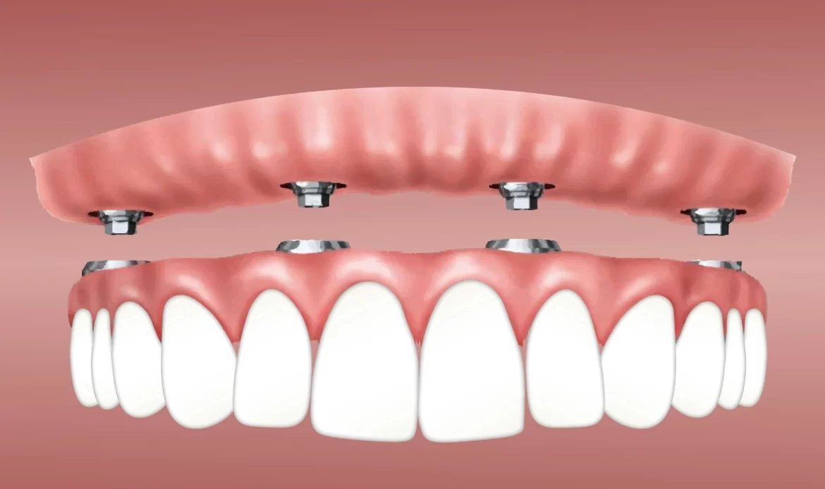
Implant surgery is a medical alternative that structurally eliminates these physiological handicaps. In movable implant prostheses called “Overdenture,” two to four titanium screws positioned in certain areas of the jaw (typically in canine tooth formations) secure the prosthesis with precise retention systems (snap-on or bar-type). This locking mechanism significantly limits the positional changes of the prosthesis during eating and speaking. In fixed concepts referred to as “All-on-4” or “All-on-6,” the prosthesis is made non-removable by being directly connected to the implants via screws. The forces generated during the chewing action are transferred to the bone tissue through the titanium roots, rather than applying pressure to the mucosa, allowing individuals to chew fibrous and hard foods (meat, vegetables, apples, etc.) with a function close to that of natural teeth. Since digestive enzymes become active in the mouth along with chewing, the effective chewing of foods is clinically important for preserving gastrointestinal (stomach-intestinal) system health.
Does Smoking Affect the Implant Process Independently of Age?
The carbon monoxide and nicotine molecules found in cigarette smoke constrict peripheral (micro) blood vessels, reducing tissue oxygenation in the operation area. Independently of age, this situation delays the healing of the wound, disrupts the function of defense cells, and structurally increases the likelihood of infection.
The use of tobacco products is one of the most significant biological risk factors encountered during the cellular healing process of oral surgical interventions. When tobacco is smoked, the oxygen-carrying capacity of hemoglobin in the blood decreases. For the surgical wound that forms in the area where the titanium screw is placed in the jawbone to heal, a substantial amount of oxygen and nutrients must be delivered to the region for osteoblasts (bone-forming cells) to synthesize new bone tissue. The vasoconstrictor effect created by nicotine hampers this cellular nutrition.
However, tobacco consumption prepares the ground for the proliferation of anaerobic (living in an oxygen-free environment) bacteria by altering the bacterial flora in the oral cavity. A bacterial infection that may develop in the area during the healing period can lead to the displacement of bone tissue from the implant surface and can impair osseointegration. Regardless of age, it is recommended in the medical protocols practiced by the Avrupadent physicians in İzmir that patients limit or cease the use of tobacco products at specific intervals before and after the operation. This recommendation is a physiological precaution taken to ensure the surgical procedure concludes appropriately according to the clinical timeline.
Does Post-Implant Care Habits Vary by Age?
Regardless of the individual’s age, the clinical lifespan of implant-supported restorations depends on individual mechanical oral hygiene practices. Brushing with fluoride toothpaste twice a day, using interdental brushes or oral irrigators, and professional dental cleaning performed every six months are standard medical procedures.
Since the titanium infrastructure and porcelain/zirconium superstructures of dental implants are inorganic materials, they are not affected by acids that cause tooth decay. However, the biological tissues surrounding the artificial roots, which are the gums (peri-implant mucosa) and jawbone, are extremely sensitive to microbial dental plaque. Periodontal connections that slow down infections reaching the bone in natural teeth are absent around implants. This anatomical difference facilitates the progression of inflammation caused by accumulated bacterial plaque (peri-mucositis) to the bone tissue, leading to bone loss (peri-implantitis).
Especially in older individuals, the physiological slowdown of salivary gland functions or the occurrence of “xerostomia” (dry mouth) due to side effects of systemic medications is frequently observed. As the natural healing and antibacterial effects of saliva in the mouth decrease, the accumulation of food debris on porcelain surfaces and gum borders accelerates. This situation further emphasizes the importance of daily mechanical cleaning. After prosthetic restorations performed at İzmir Avrupadent clinics, patients are informed about the use of special thick dental threads called “superfloss” to clean porcelain bridges, in addition to standard brushing, along with the use of oral irrigators that provide hydrodynamic cleaning with pressurized water. Radiological bone level measurements and ultrasonic tartar cleanings performed by the dentist at six or eight-month routine intervals are necessary clinical procedures for maintaining functions over many years, regardless of age.
What Protocols Are Applied in Implant Planning at Avrupadent?
The İzmir Avrupadent clinics operate with a comprehensive system that includes the collection of detailed medical history, anatomical analysis via 3D tomography, surgical procedures under aseptic conditions, and the design of personalized prosthetic superstructures using digital systems (CAD/CAM) following bone healing.
The initial phase of the treatment organization is the assessment and data collection process. In this process, which combines the principles of jaw surgery, periodontology, and prosthetic dental treatment, the panoramic X-rays and volumetric tomographies (CBCT) of the patient are examined. The bone quality in the region where the implant will be placed is evaluated according to a scale ranging from D1 to D4, which classifies bone density. The doctor digitally selects screws of appropriate diameter and length suitable for the patient’s anatomical structure before the operation. Planning may also include the production of surgical guide plates using 3D printers; these plates ensure that the angle of the screw is controlled with millimeter precision during the operation.







