

What Are the Application Areas of Single Tooth Implant Treatment? Missing Tooth Treatment in Izmir
Tooth loss is an anatomical condition that arises in human physiology due to pathological processes such as trauma, fracture (caries), or periodontal (gum) diseases, which directly affect chewing mechanics. Dental implants, which are among the restorative methods applied in medicine to compensate for these losses, work on the principle of surgically integrating titanium alloy materials into the jawbone (alveolar crest). Avrupadent, which provides medical services in Izmir, addresses cases of single tooth loss through a multidisciplinary diagnosis process; the width, height of the jawbone, and the patient’s systemic medical history are mapped with three-dimensional radiological analyses.
What is Single Tooth Implant Treatment and When is it Applied?
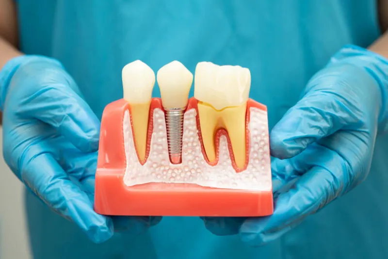
Single tooth implant treatment is a procedure in which an artificial root made of titanium alloy is placed in the gap where a single tooth has been lost due to trauma, infection, or deep fracture, and a prosthetic crown is placed on top. This medical application is planned to provide localized restoration without the need to affect the adjacent teeth.
The jawbone (maxilla and mandible), part of the human skeletal system, is a biological tissue that maintains its volume through mechanical stimuli provided by the tooth roots it holds. The extraction of a single tooth, leaving its function aside, creates an isolated tissue loss in that area. If the enamel tissues of the teeth adjacent to the area of deficiency are in healthy form, their support for traditional restorations means a loss of adjacent tissue in medical practice. Threaded screws made of titanium are placed directly into this missing socket to mimic the functions of the natural tooth root at a cellular level.
The medical indications of the application (requirements) include; congenital tooth deficiencies (agenesis), failure of primary teeth to fall out during childhood and the absence of the permanent tooth from below, trauma-related root fractures, and cystic lesions that cannot be restored with root canal treatment. If there is no active acute infection in the relevant area and the bone density meets certain millimetric standards, this surgical intervention is performed according to medical protocols. In the procedures at Avrupadent located in Izmir, these types of pathologies are analyzed through panoramic or three-dimensional tomography, and the operation schedule is designed according to the hospital’s cellular healing capacity.
Why is a Single Tooth Deficiency Anatomically Important?
The absence of a single tooth disrupts the distribution of chewing forces over time, leading to the neighboring teeth tilting towards the gap and allowing the opposing tooth to elongate in the vertical axis. These structural changes alter the occlusal contact mechanics and cause the bone tissue in the extraction area to enter a volumetric resorption process due to lack of stimulation.
The dental arch in the oral cavity, like interlocking stones supporting each other, creates contact points side by side, balancing the chewing load. The removal of a tooth eliminates this locking mechanism. Over the following years, the teeth at the back of the gap (usually molars) tend to tilt into the gap (mesially). This tilt prepares the ground for the formation of pockets and food accumulation by disturbing the attachment of the tooth roots to the bone through periodontal fibers.
On the other hand, the opposing (antagonist) tooth in the arch will perform a vertical movement directed towards the issue in question due to the influence of gravity. According to Wolff’s Law of the skeletal system, the bone cells in the area of the jawbone (alveolar ridge) lose their mineral density due to the lack of mechanical stimulation, triggering the process of resorption. In the first six-month period after extraction, a significant part of the alveolar bone width physiologically narrows. Compensating for these deficiencies medically is a clinical necessity to restore functional balance, maintain the alignment of teeth, and ensure the process of food particle breakdown, which is the beginning of the digestive system, continues normally.
How is Single Tooth Implant Treatment Planned in İzmir Avrupadent Clinics?
In the İzmir location of Avrupadent clinics providing medical services, the planning involves measuring the width and height of the jawbone through three-dimensional dental tomographies, calculating the distance to the nerve canals, and examining the patient’s medical history. Based on the obtained data, surgical guides are formed to create an objective clinical map.
The assessment and planning phase is the most critical stage that determines the biological and mechanical limits of the operation. This process, which begins when the patient sits in the dental chair, deepens with the collection of medical anamnesis (health history). The presence of systemic diseases such as diabetes (sugar disease), hypertension, and osteoporosis is recorded, as well as any anticoagulants (blood thinners) or immunosuppressive medications the patient is taking. It is requested by the physician that the blood sugar level (HbA1c) falls within certain reference ranges for the procedure to be carried out.
After the physical oral examination, a 3D CBCT (Cone Beam Computed Tomography) scan is taken of the localized missing area. On the computer screen, the proximity to the ‘nervus alveolaris inferior’ canal in the lower jaw, which carries the sensation of the lip, and the membrane of the ‘maxillary sinus’ in the upper jaw is calculated over the millimetric sections. By classifying the density of the patient’s jawbone between D1 (very hard) and D4 (spongy), the length and thread thickness of the screw to be used are selected. With the backward planning concept, the porcelain crown to be made in the mouth is virtually designed, and then the implant angle most suitable for the central axis of this crown is determined computationally, and the procedure is shaped.
| Anatomical Area / Measurement | Clinical Evaluation Purpose | Effect on Surgery |
|---|---|---|
| Bone Width (Bucco-Lingual) | Determining the existing thickness to prevent the screw from protruding out of the bone. | Determines the diameter of the implant (for example, 3.5mm or 4.5mm) and whether bone powder is needed. |
| Bone Height (Vertical) | Calculation of the millimetric safety distance to the lower jaw nerve and the upper jaw sinuses. | Determines the vertical length of the titanium screw to be used (for example, 8mm or 10mm). |
| Bone Density (Trabecular Structure) | Numerical analysis in Hounsfield Unit (HU) terms of the hardness degree. | Guides the drilling techniques and thread design to be used during the operation. |
| Mesio-Distal Distance | The horizontal measurement between the two adjacent teeth of the missing space. | The prosthetic crown determines the anatomical volume of the external ridge in the initial stage. |
How is Implant Application Conducted in Anterior Tooth Deficiencies?
In applications in the anterior incisor region, aesthetic parameters such as the volume of bone, thickness of the gingiva, and light permeability guide clinical planning. Following the placement of the artificial root, zirconium-based crowns are often preferred to avoid metal reflection under porcelain, aimed at achieving a restoration that is compatible with the natural tooth physiognomy.
The anterior region of the edentulous space is defined as the aesthetic zone where lip support is provided and speech articulation is managed. The tooth losses that occur in this area affect the patient’s facial profile. The anterior mandible is quite thin anatomically (it can be in the shape of a knife blade). When surgical intervention is performed, the sensitivity of the outer (buccal) wall of the alveolar crest to resorption is taken into account. The three-dimensional position of the titanium screw within the bone directly determines the millimetric shape of the gingiva (pink aesthetic).
If the patient’s gingiva biotype is thin (translucent), the titanium material, which has a metallic color, may produce a grayish reflection beneath the gingiva. To overcome this optical handicap, during the evaluation stage, a thin connective tissue graft (piece) is taken from the patient’s palate area to thicken the relevant gingiva (soft tissue surgery). When transitioning to the prosthetic stage, the connecting piece (abutment) and the upper crown placed on the implant are processed in a laboratory setting from zirconium or full ceramic materials that can transmit light as natural enamel does, thus providing a natural harmony.
What Medical Criteria Are Considered in the Implantation of Posterior Molars (Molar Region)?
In the back molar regions, wide-diameter titanium materials with biomechanical resistance are chosen because intense chewing and crushing forces (vertical loads) emerge. The maxillary sinus distance in the upper jaw and the proximity to the mandibular nerve canal in the lower jaw are calculated radiologically, and the medical boundaries of the operation are determined at a millimeter level.
The posterior region of oral anatomy is where the food that goes into the digestive system is crushed and chewed. As a result of the contraction of the masseter and temporal muscles, hundreds of Newtons of vertical and horizontal pressure are exerted onto each chewing cycle on a molar tooth. The artificial root to be applied in the case of a single molar tooth loss must be able to absorb this enormous force into the surrounding bone. Using a narrow-diameter screw in the relevant area may lead to material fatigue and breakage of the abutment screw in the long term. Due to these biomechanical needs, the widest diameter implant platforms that the anatomical volume allows (for example, above 4.5 mm or 5.0 mm) are planned for molar region restorations.
Anatomically, the maxillary molar region is subject to the drooping of the maxillary sinuses (pneumatization) after tooth loss. The bony layer between the two organs can reduce to the thinness of a paper. Since extending titanium directly into the sinus cavity would cause a medical complication, ‘Sinus Lifting’ surgeries come into play, where the sinus membrane is pushed up and bone grafts (particles) are placed in between. In the lower jaw, the location of the thick nerve network called ‘nervus alveolaris inferior’ is scanned millimetrically; it’s a non-negotiable rule of the operation that the screw must end at least two millimeters away from this nerve. In the European Dent procedures carried out in İzmir, the laboratory occlusal designs of upper porcelain restorations to withstand the mechanical load of the region are also made by calculating these vector forces.
How Does a Single Tooth Implant Differ from Traditional Bridge Prostheses?
Bridge prosthetics require the reduction of the enamel of healthy teeth on either side of the gap, whereas implant applications only involve surgical intervention in the socket where the missing tooth is. This method preserves the anatomical integrity of adjacent teeth and slows down the process of local bone resorption by transmitting chewing force to the jawbone.
In dental practice, there have been two primary prosthetic approaches for single tooth deficiencies for decades: tooth-supported fixed bridges and implant-supported crowns. In conventional bridge restoration, healthy teeth that will serve as supports (abutments) are reduced with specific millimeter drills to close the gap (preparation process). The prepared three-unit porcelain block is constructed using medical agents on top of these supporting teeth. The forces that arise during the chewing process are distributed to these reduced teeth through the body of the bridge.
Implant surgery, on the other hand, adopts a conservative (protective) medical approach. No mechanical drilling is performed on the enamel tissues or roots of adjacent healthy teeth; only a procedure is applied under local anesthesia to the area where the deficiency exists. The most significant biomechanical difference is observed in the bone tissue. The bone tissue remaining under the bridge bodies gradually loses volume (resorption) over time due to not receiving functional pull or pressure stimulation, leading to gaps forming between the porcelain and the gum. Implants transmit chewing pressure directly to the alveolar bone like a natural root, stimulating cells, supporting local blood flow, and controlling the bone resorption process at the cellular level. In terms of hygiene practices, while the areas beneath bridge bodies require specific threads for cleaning, the interface cleaning of a single artificial root is maintained with natural dental floss routine.
| Medical Parameter | Single Tooth Implant Application | Traditional Bridge Prosthesis |
|---|---|---|
| Effect on Adjacent Tooth Tissue | Adjacent teeth anatomically preserve their integrity, no abrasion occurs. | It is necessary to reduce the size of the two adjacent healthy teeth to support them. |
| Effect on Jawbone Volume | It stimulates osteoblasts by transferring force to the bone, slowing down bone loss. | No stimulation reaches the bone tissue; the area underneath the body gradually loses volume over time. |
| Chewing Mechanics (Vectorial Load) | The biting load is transmitted from its own screw structure directly to the bone underneath. | The single tooth biting load is also applied onto the roots of the supporting (abutment) teeth. |
| Oral Hygiene Routine | Standard brushing and standard dental floss are used for intermediate cleaning. | The gaps underneath the body (pontic) need to be cleaned with special (superfloss) threads or oral irrigators. |
How Long Does the Osseointegration Process of the Implant Take?
The fusion of titanium material with the jawbone cells (osteoblasts) is a cellular process that lasts on average two to three months, depending on the structural density in the lower jaw. Because the upper jaw has more spongy tissue, this biological integration process takes a time period ranging from three to six months.
Osseointegration is the process where an inorganic material (titanium oxide surface) forms a structural and functional unity at the microscopic level with living bone tissue. During the surgical procedure, when the implant is placed in the cavity created by drills, the clinician calculates the compression force applied by the screw to the bone (primary stability) in Newton-centimeters (Ncm). Initial retention is a mechanical concept. Within the first few hours after the operation, blood flows between the screw and the bone walls, forming a fibrin (clot) mesh here.
In the past weeks, the body’s connective and repair cells migrate over this fibrin mesh and embed into the porous microstructure of the titanium surface. Bone-forming osteoblast cells synthesize calcium and phosphate minerals in the area, creating new living bone matrix; this phase is called cellular bonding (secondary stability). The mandible has an extremely thick and dense cortical structure, which allows this cellular structuring process to take shape more quickly in clinical standards. In contrast, the maxilla is a structure that is quite trabecular, with a lot of space and fine bone trabeculae. The cells filling this spongy structure and wrapping around the screw occur over a broader timeline due to anatomical reasons. This waiting period is the biological repair physiology of the organism and cannot be accelerated by external intervention; in the Izmir Avrupadent protocols, porcelain dental crowns are not placed until this maturation phase of the bone cells is completed.
How is Anesthesia Management Carried Out in Single Tooth Implant Surgery?
The surgical procedure is performed while the application area is anesthetized with local anesthetic agents. As the nerve transmission in the relevant area is pharmacologically blocked, the patient remains conscious but can only perceive pressure and vibration during the intervention. As a result of medical consultations, the procedure is completed in a comfort zone according to clinical protocols.
Surgical incisions made to the intraoral tissues (mucosa and periosteum) require a pharmacological pain control system due to the dense nerve networks they contain. In dental practice, local anesthesia is applied by the practitioner injecting the anesthetic solution (usually with active ingredients of articaine or lidocaine) around the nerve endings with a needle. This solution temporarily suspends the transmission of pain signals from the surgical area to the brain by blocking sodium channels. An operation aimed at a single tooth loss is generally prolonged between 20-30 minutes by anesthetizing just that area (infiltration or regional block).
During the operation, the physician intervenes with devices called micromotors on the bone. These devices create a vibration and a slight mechanical pressure sensation while continuously cooling the area with physiological serum (to prevent bone overheating). The anesthesia used blocks the perception of pain but does not completely shut off the thick nerve fibers carrying the sensation of pressure; thus, it is commonplace for the patient to perceive the procedure as a pressure sensation. In patients with severe dental phobia (fear of the dentist), those with very strong nausea reflexes, or those who cannot tolerate stress systematically, an anesthesiology specialist consultation is performed, and “conscious sedation” (the administration of a sedative through the vascular route) methods are employed to ensure that the patient’s vital signs are monitored in the procedural environment.
What Medical Methods Are Used When There Is Insufficient Volume of the Jawbone?
When bone narrowing or height loss is detected in tomographic sections, reconstructive bone grafts and barrier membrane applications are used. Through these biomaterials, the volume of the jawbone is increased, and when necessary, the sinus membrane is elevated (sinus lifting) to create an anatomical foundation for the artificial root.
It is essential that the amount of bone does not fall within the three-dimensional limits that would support the length and diameter of the implant; this brings forth advanced surgical techniques known as “augmentation”. Long-term tooth loss, severe periodontal infections, or a history of cystic operations can create serious cavities in the vestibular (outer) or lingual (inner) walls of the jawbone. It is a medical rule that the threads of the titanium screw must not be exposed to the outside environment in any way.
To restore these anatomical limitations, xenografts (sheep/pork sourced), alloplastic (synthetic), or autogenous (bone taken from the patient’s own jaw) bone blocks/particles are used. The graft material is mixed with the patient’s blood and placed into the deficient area, and it is covered with a medical collagen or PTFE membrane (overlay) that limits the seepage of connective tissue cells into the area, and the gum is sewn. Biological particles gradually turn into living bone tissue within months by providing a skeletal groundwork for the body’s own repair cells (osseointegration). In the back regions of the upper jaw (maxilla), the sagging sinus floor is elevated with special surgical lifts (open or closed sinus lifting), and vertical height is achieved by placing the same particles into the opened space.
What Issues Should Be Considered During the Healing Period After Surgery?It is a medical guideline to use the specific dosages of antibiotics and mouthwashes prescribed by the doctor to support tissue healing after surgery. In the first two days, applying cold compresses helps control the possible swelling; avoiding extreme physical exertion, hot consumables, and tobacco products is essential for wound closure.
It is a medical guideline to use the specific dosages of antibiotics and mouthwashes prescribed by the doctor to support tissue healing after surgery. In the first two days, applying cold compresses helps control the possible swelling; avoiding extreme physical exertion, hot consumables, and tobacco products is essential for wound closure.
As is the case after every surgical intervention, the oral mucosa and jawbone also develop an inflammatory response to trauma. This response manifests as fluid accumulation (edema) in the tissues and dilation of the capillaries. Immediately after the procedure, the sterile gauze placed by the physician in the area should be kept warm by the patient for half an hour to initiate the clotting mechanism. Periodically applying ice or cold gel compresses from the outside (the cheek) helps physiologically limit edema and potential minor bruising (ecchymosis) by causing vasoconstriction.
Regarding eating habits, for the first few days, soft, seedless, and warm foods should be consumed to prevent the lips of the wound from opening and to keep the stitches stable. Hot beverages and acidic foods can disrupt the clot structure, leading to bleeding as drainage at the wound site. The patient is expected to gently clean the operation site with chemical mouthwashes containing chlorhexidine as recommended by the physician. Smoking, the external factor that most affects the healing dynamics, reduces oxygenation by constricting blood vessels, cutting off cell nutrition; thus, refraining from tobacco during this process prevents medical complications. In the protocols of İzmir Avrupadent clinic, the patient is asked to return for a check-up seven to ten days post-operation, where the stitches (sutures) in the area are removed by the physician, and the healing tissue (epithelization) is examined.
How is the Care and Maintenance of a Single Tooth Implant Carried Out?
The long-term functional stabilization of dental implants is ensured through mechanical hygiene performed using toothbrushes and dental floss, in addition to regular check-ups every six months to prevent inflammation of the surrounding gum tissue (peri-mucositis). Although titanium is an inorganic material that does not corrode, regular clinical controls are a medical maintenance rule.
A crown supported by an implant provides a biomechanical function that is aesthetically and functionally very close to a natural tooth, but the defense mechanism of the surrounding tissues against infection is different from that of natural teeth. The roots of natural teeth are surrounded by periodontal fiber systems, which physiologically slow down the penetration of bacterial plaque (tartar) that accumulates in the gums into the bone tissue. However, titanium surfaces are in direct contact with bone cells (ankylosis) and have no connective tissue fibers around them. This anatomical deficiency can lead to the rapid access of bacterial plaque from the gums to the jawbone (peri-implantitis) in the presence of poor oral hygiene.
The only way to prevent the development of this medical condition is through individual mechanical hygiene. In addition to standard fluoride tooth brushing performed twice a day, thick dental floss (superfloss) that reaches the edges of porcelain restorations and pressure water jet oral irrigators (water flosser) should be used to clean food residues in micro-spaces. Tartar formed by the deposition of calcium ions within the entire area cannot be removed by the patient with a toothbrush. Therefore, it is a clinical requirement to visit Izmir Avrupadent clinic for professional periodontal cleaning with ultrasonic devices at least twice a year and to measure the surrounding alveolar bone level of the implant through radiological exams (to check for any crestal defects).







