

How is Implant Treatment Applied in Izmir?
Tissue losses occurring in oral anatomy lead to extensive cellular changes ranging from the chewing function (mastication) in human physiology to the digestive mechanism and the cellular structure of the jawbone. One of the biological restoration methods frequently applied in modern medical literature to compensate for tooth loss is the integration of artificial roots made of titanium or zirconium alloys into the jawbone (alveolar ridge) through surgical protocols. Implant treatment in Izmir is a multi-step procedure in which the specific anatomical and systemic data of patients are measured with objective medical devices.
What is Implant Treatment in Izmir and what are its Biological Foundations?
Implant treatment in Izmir is the process of placing biocompatible titanium screws into the jawbone for the functional restoration of missing teeth, and the osseointegration of this inorganic material with bone cells at the microscopic level. The application aims to restore the anatomical structures to their normal physiology by ensuring that chewing forces are transmitted to the jawbone.
The maxilla (upper jaw) and mandible (lower jaw) bones, which are essential components of the human skeletal system, maintain their volume through mechanical pressures transmitted by the natural tooth roots present in them. When tooth loss occurs, the alveolar bone in the related area withdraws mineral support at the cellular level due to a lack of mechanical stimulation. The activity of osteoclasts (bone-resorbing cells) increases, leading to significant tissue resorption in the area. The biological basis of implant application relies on controlling this resorption process at the cellular level. The titanium alloy placed in the bone during the operation is not perceived as a foreign body by the organism.
Titanium material forms a thin layer of titanium oxide on its surface when it comes into contact with oxygen. This chemical property (biocompatibility) limits the rejection reaction of the binding system. The fibroblast and osteoblast (bone-forming) cells within the clot that forms in the operational area adhere to the specifically refined micro-porous surface of the screw. In the following months, these cells synthesize calcium and phosphate minerals in the area, establishing a cellular bridge (ankylosis) between the inert metal and living bone tissue. In the Avrupadent clinics operating in Izmir, this biological bonding process is closely monitored through periodic X-ray examinations to determine the extent of tissue integration according to medical norms.
How is the Clinical Evaluation Process for Implant Treatment Conducted in Izmir?
The clinical evaluation process consists of taking the patient’s medical history and mapping the vertical and horizontal volume of the jawbone in millimeters using three-dimensional dental volumetric tomography (CBCT). Based on this data, the physician objectively determines the surgical procedure trajectory, sizes of the materials to be used, and anatomical proximity.
In the general medical management of surgical interventions, the diagnostic (diagnosis) phase carried out before rather than during the operation determines the course of the procedures. This process, which starts as the patient sits in the dental chair, is deepened by obtaining a systemic anamnesis (patient history). The patient’s general health status is evaluated according to the physical risk classification of the American Society of Anesthesiologists (ASA). When chronic conditions affecting systemic circulation or cellular healing are detected, medical consultations are requested from the relevant specialty physicians.
After the physical examination, the radiological imaging phase is entered. Two-dimensional panoramic films show the overall curvature of the jaw, while Cone Beam Computed Tomography (CBCT) technology is used to measure the millimetric thickness of the area where the implant will be placed and the density of its spongy tissue. With these three-dimensional software, the distance between the ‘inferior alveolar nerve’ canal, which carries the sensation in the lower jaw, and the ‘maxillary sinus’ air cavities, which contribute to sound resonance in the upper jaw, is presented with numerical data. The physician determines whether the bone is cortical (hard) or trabecular (soft) by looking at the Hounsfield Unit (HU) values in the tomography slices. In the clinical protocols at İzmir Avrupadent, planning any surgical invasive procedure without this anatomical data is considered contrary to medical standards.
What Are the Stages of Implant Treatment at İzmir Avrupadent?
The treatment stages include; planning based on radiological diagnosis, opening a cavity under local anesthesia to place the titanium screw into the bone, waiting for the biological tissue healing (osseointegration) period, and taking measurements to adapt the prosthetic superstructure (porcelain or zirconium crown) into the mouth.
On the day of the operation, the clinical settings ensure asepsis and antisepsis standards to minimize cross-infection risks. After applying local anesthesia to the patient, the physician makes an incision in the gum tissue (mucosa) at the operation site and lifts the mucosal flap to expose the underlying bone tissue. Following the millimetric measurements determined on the CBCT, a gradual drilling (osteotomy) procedure is performed on the alveolar crest using special surgical micromotors that cool with sterile physiological serum. During the drilling of the bone, if the tissue temperature exceeds 47 degrees Celsius, it may lead to bone cell death (necrosis), making the cooling function of the devices critical from a clinical perspective.
Once the osteotomy is completed, the titanium implant body, taken out from its special sterile package, is placed into the socket using low-speed torque devices. The pressure exerted by the screw on the bone (primary stability) is measured in Newton-centimeters (Ncm). After placing a cover screw inside the implant, the lifted gum flap is closed with surgical threads (suture). Following this initial surgical session, post-operative care instructions are provided to the patient, and a prescription medication is arranged for the normal healing of the wound site. Cellular organization continues within the tissue during the months following the removal of stitches. In the second surgical stage, the cover screw is removed, and healing abutments (gingiva former) that prepare the gum for prosthetic components are placed. In the European clinical pathway applied in Izmir, no transition to measurement and restorative stages (third stage) is made without radiological approval of bone density.
| Procedure Stage | Medical Procedure Performed | Recommended Timeline |
|---|---|---|
| 1. Surgical Stage (Placement) | Flap opening, osteotomy, and placement of the screw into the bone for suturing (stitching) under local anesthesia. | 20 – 45 minutes per tooth. |
| 2. Biological Healing (Osseointegration) | The bone tissue enveloping the titanium surface at the cellular level and creating mechanical resistance. | Lower Jaw: 2-3 Months |
| 3. Mucosal Shaping | Opening of the gum and the placement of the healing cap, as well as the formation of the mucosal emergence profile (pink aesthetics). | 7 – 14 Days |
| 4. Prosthetic Measurement and Delivery | Taking digital (CAD/CAM) or conventional measurements, manufacturing crowns in the laboratory, and making occlusal adjustments. | 10 – 20 Days |
Which Medical Procedures are Followed for Jaw Bone Insufficiency in Izmir?
If it is determined through radiological examinations that the volume of alveolar bone is insufficient in the vertical or horizontal direction, augmentation (bone grafting) techniques are applied. Volume is supported by placing allograft, synthetic, or autogenous bone grafts (powders) in the patient; in the upper jaw, structural areas are created by lifting the sinus membrane (sinus lifting).
The stability of titanium material over the years depends on the thickness of the surrounding healthy bone wall. A minimum of 1.5 – 2 millimeters of bone tissue thicker than the implant diameter is required. If there have been long-term tooth losses, it may create significant defects in the cortical walls of the jawbone due to cystic pathologies or traumatic extractions. In the presence of these defects, when a surgical gap is opened, the threads of the screw may remain exposed; the exposed threads can lead to infection accumulation in the gum pocket. To compensate for this anatomical issue, reconstructive surgical interventions are performed.
In the augmentation procedure, graft particles that serve as a biological scaffold are placed in the deficient area. These particles can be xenografts (inorganic cortical bone), allografts (human donor-derived), or alloplastic (synthetic calcium phosphate-derived) structures. The body’s own osteoblasts gradually progress over this scaffold over a period of months, transforming the area into a living bone matrix. The maxillary sinus membrane (Schneiderian membrane), which sags down due to toothlessness in the posterior regions of the upper jaw, is carefully pushed upwards with special hand tools (Sinus Lifting), and graft materials fill the newly formed physiological area. The physicians at Avrupadent in Izmir prepare the tissue physiology, performing these advanced surgical applications either during the same session as the implant placement or about six months prior to the implantation procedure, depending on the condition of the surgical area.
How Does Implant Treatment Differ in Cases of Single Tooth Loss and Complete Toothlessness?In cases of single tooth deficiency, a localized cavity is opened to place a single implant while preserving the integrity of adjacent teeth. In cases of complete edentulism, 2 to 6 implants are strategically placed in the jaw to create a biomechanical support mechanism that evenly transmits the mechanical forces of removable or fixed prostheses to the bone.
In cases of single tooth deficiency, a localized cavity is opened to place a single implant while preserving the integrity of adjacent teeth. In cases of complete edentulism, 2 to 6 implants are strategically placed in the jaw to create a biomechanical support mechanism that evenly transmits the mechanical forces of removable or fixed prostheses to the bone.
The lack of teeth in the patient’s mouth anatomy modifies the scope of the surgical concept applied. In the case of a single tooth loss (for example, central incisor or first molar), intervention is carried out in that area under local anesthesia. As in traditional bridge prostheses, the enamel (tissues) of the healthy teeth next to the missing space is not ground down. The placed single implant transmits the occlusal (chewing) force to the underlying alveolar bone along its long axis.
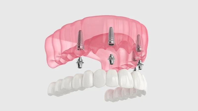
In cases of complete edentulism, the oral flora, muscle dynamics, and force distribution present a completely different scenario. Traditional complete (plate) prostheses can move during chewing, as they are supported by soft tissue, which may cause tissue irritation. In complete edentulism cases, if the patient’s bone structure is suitable, a mechanical support structure is created by placing at least 2 or 4 implants in the lower jaw and at least 4 or 6 implants in the upper jaw. In fixed prosthesis concepts referred to as “All-on-4” or “All-on-6”, implants placed at specific angles in the jaw are connected with a rigid bridge. This way, the chewing force is distributed throughout the body, limiting excessive load on a single screw (overloading). In removable “Overdenture” systems, the prosthesis is secured using bars or precision retainers (clip-on) mounted on the screws. In the planning at Avrupadent in Izmir, these biomechanical force analyses are projected by considering Newton values through digital software.
How is the Osseointegration (Bone Healing) Process Monitored in Izmir?
The microscopic bonding process known as osseointegration between the inorganic titanium surface and living bone cells lasts an average of two to three months in the lower jaw and three to six months in the upper jaw. This healing stage is monitored through routine clinical examinations and periodic local radiographic images, in which tissue density is assessed by the physician.
Immediately after the surgical placement procedure, the physical compression (primary stability) decreases gradually over time, while the cellular retention formed by cell proliferation (secondary stability) increases. Initial hemostasis (bleeding control) in the operative area and the formation of fibrin clots pave the way for bone cells to migrate to the area. The inflammation and proliferation phases, which are the normal wound healing mechanisms of human physiology, are followed by bone mineralization.
The maxilla (upper jaw) bone has a more porous structure with finer trabeculae due to the nose and sinus cavities. The placement of cells into this spongy tissue and the resulting calcification (hardening) requires a longer timeline compared to the denser mandibula (lower jaw) bone. The healing period is a specific biological clock that cannot be shortened by vitamins or medications provided externally, being dependent on the organism’s own metabolism. According to European dental clinical protocols, patients are invited for X-ray evaluations at certain intervals to check for radiolucency around the implant. An implant that has no soft tissue thickening around it and establishes clear radiographic contact with the bone is considered ready for functional loading (prosthesis implantation).
How is Anesthesia Management Performed During Implant Surgery in Izmir?
The area where the procedure will be performed is anesthetized locally using pharmacological agents that block nerve transmission (such as articaine or lidocaine solutions). The patient remains conscious under anesthesia; due to medical protocols that completely disrupt pain transmission, only pressure from the bone and device vibrations are perceived during the procedure.
Pain management in maxillofacial (jaw and face) surgery is based on the mechanism of temporarily blocking peripheral (surrounding) nerve fibers. Local anesthetic solutions used in dentistry disrupt the permeability of sodium channels in the nerve cell membrane, interrupting the transmission of electrochemical pain signals (impulses) from the surgical site to the brain. Under this pharmacological effect, making a mucosal incision or drilling (boring) on the bone does not create a sensation of biological trauma.
During the operation, the doctor uses solutions containing epinephrine (adrenaline) with vasoconstrictive effects to prolong the duration of local anesthesia and control bleeding (hemostasis) at the surgical site. However, in patients with systemic hypertension (high blood pressure) or severe cardiovascular (heart) arrhythmias, solutions without adrenaline are preferred, as the epinephrine that enters the bloodstream can cause an acute increase in blood pressure. While the micromotor device opens a cavity in the bone, it performs intensive cooling with physiological serum and creates a slight vibration (tremor) in the bone. While local anesthesia blocks thin pain fibers, thick pressure fibers are not blocked to the same extent; therefore, it is an anatomical fact that the patient feels pressure on their jaw.
How is the Prosthetic (Crown) Phase in Implant Treatment Progressed in Izmir?
Once the osseointegration phase is confirmed radiologically, measurements are taken using digital or conventional methods with the help of pieces shaped by healing abutments from the gum tissue.
When the cellular adaptation of the surgical stage is completed, prosthetic rehabilitation, the second and final major step of the treatment, begins. First, under local anesthesia (or without the need for anesthesia), the gum (mucosa) is opened over the cover screw, and healing abutments, referred to as ‘gingiva formers’, are attached. These abutments provide a cylindrical form to the gum over a period of 7 to 14 days to ensure the porcelain tooth looks natural as if it’s emerging from the bone (pink aesthetics).
Following the shaping of the mucosal profile, transfer pieces known as ‘measurement posts’ are screwed into the inner body of the implant. High-precision silicone measurement materials or intraoral (in-mouth) optical scanners are used to replicate the patient’s jaw closure relations and the three-dimensional position of the space in the laboratory. In restorative planning within the Izmir Avrupadent framework, the contact points of the lower and upper jaws (occlusal points) are taken into consideration. Metal-supported ceramics with high mechanical fracture resistance are used in the posterior molar areas, while zirconia full ceramic materials resembling natural tooth enamel in light fracture index are used in the anterior incisive areas. These crowns are integrated into chewing function by being screwed (screw system) or attached with medical agents (cemented system) via an intermediary piece (abutment) to the implant body.
| Material Classification | Structural Property and Strength | Anatomical Indication Area |
|---|---|---|
| Metal Supported Ceramic Crown | Internally made with a chrome-cobalt alloy, externally it contains aesthetic porcelain. Fracture resistance is at the highest level. | The area of the Back Molars exposed to high masticatory (chewing) force. |
| Zirconium-Based Crown | Provides high biocompatibility and light permeability with a white zirconium oxide base. Does not reflect metallic light. | The region of the Front and Small Molars where aesthetic expectations and the smile line are present. |
| Monolithic Zirconium | Obtained by milling from a single block of zirconium. Since there is no porcelain coating on it, ceramic chipping does not occur. | All jaw areas in individuals with severe teeth grinding (Bruxism) discomfort. |
How is Implant Treatment Planned for Individuals with Systemic Diseases in Izmir?
In patients with diabetes, hypertension, osteoporosis, or cardiovascular disorders, blood values and regularly used medical medications are consulted with relevant specialists. When medical values (HbA1c, INR) are regulated to reference ranges, surgical procedures are carried out with attention to infection and bleeding control protocols.
Implant surgery, which is a dental procedure, is an integral part of the body’s overall physiology, along with tissue blood supply and the vascular system. The healing of the surgical site and bone synthesis is directly affected by the patient’s systemic condition (ASA risk group). In cases of uncontrolled diabetes (diabetes mellitus), high levels of glucose in the blood can lead to blockages in peripheral capillaries (microangiopathy). This situation reduces the quantity of oxygen, nutrients, and defense cells (leukocytes) that can reach the surgical area, slowing down tissue repair and increasing the potential for infection. In medical evaluations conducted in Izmir, recent HbA1c tests are requested from diabetic patients, and it is required that the average value remains within medical limits (around 7%).
In patients using anticoagulants (blood thinners) due to cardiovascular issues or atrial fibrillation, the clotting mechanism (cascade) has been pharmacologically slowed down. To manage the increased risk of prolonged bleeding when an incision is made, temporary adjustments to the medication dosage (bridging therapy) are made in consultation with a cardiologist. Additionally, in individuals receiving intravenous (intra-venous) bisphosphonates for osteoporosis, there is a risk of necrosis in the operation area due to the cessation of the bone’s cellular renewal cycle (Bisphosphonate-Related Jaw Osteonecrosis). In this regard, all systemic diseases in the Avrupadent processes are analyzed individually, and the timing and scope of the surgery are shaped according to these concrete findings.
What Considerations Should Be Made During the Healing Period After Implant Treatment?
To control the swelling associated with the expansion of capillaries in the surgical area after the operation, cold compresses (ice) should be applied from the outside for the first 48 hours. To ensure that healing is not interrupted, it is medically necessary to use the prescribed medications precisely as directed and to consume soft foods in the initial days.
Every incision and osteotomy performed in the tissues inside the mouth initiates a biological repair (inflammatory) response in the body. Symptoms of this response include slight swelling (edema) and, rarely, capillary bruising (ecchymosis) on the outer skin, which are physiological findings commonly encountered in the early days of wound healing. The cold press application, which the patient will apply to the operation area (from outside the cheek) at 15-minute intervals, constricts the blood vessels (vasoconstriction), limiting fluid accumulation and possible slight bleeding seepage.
It is essential to pay attention to the dietary routine to keep the wound lips stable and to prevent the disruption of the clot structure in the bone. During the first two days, one should avoid very hot beverages, acidic and spicy foods, as well as hard foods such as nuts that could cause mechanical damage to the wound area; a diet should be followed that has a pureed consistency at room temperature. One of the leading factors that severely disrupt physiological wound healing is tobacco (smoking). The nicotine and carbon monoxide in tobacco smoke dramatically reduce tissue oxygenation, jeopardizing the ossification phase. In the clinical protocols of İzmir Avrupadent, post-operative care instructions are given to patients in writing, aimed at maintaining the individual compliance with the surgical success rate (osseointegration stability).
How Should Long-Term Care for Implants Applied in İzmir Be?
The functional maintenance of implant-supported restorations is dependent on the mechanical limitation of tartar formation using dental floss and interface brushes, in addition to routine tooth brushing practices. To prevent peri-implantitis (inflammation around the implant), clinical examinations every six or eight months are a long-term clinical protocol.
The inorganic structure of titanium alloys and the porcelain crowns over them makes it impossible for cavities to form in these areas. However, this should not create a medical misconception that prostheses can be neglected. The mucosa (gum) surrounding the implant body and the underlying alveolar bone is much more vulnerable to microbial dental plaque in the mouth compared to natural teeth. The periodontal fiber bundles (ligaments) that slow down the penetration of bacteria into the bone found at the roots of natural teeth are not present around titanium surfaces (direct contact – ankylosis).
Due to this anatomical difference, the microbial bacterial plaque that accumulates in the groove between the gum and the porcelain initially causes inflammation in the soft tissue (peri-mucositis). If mechanical hygiene is not maintained, bacteria can quickly infiltrate the bone tissue, leading to irreversible bone resorption around the titanium (peri-implantitis). The care routines emphasized by the experts at Izmir Avrupadent for patients involve cleaning under porcelain bridges with specially designed threads (superfloss) or pressure water jet dental flossers (water flosser). Since hardened tartar cannot be removed with a toothbrush, periodic clinical visits twice a year for the radiological analysis of bone levels by the physician are an integral part of medical maintenance.







